

Gramicidin, a Bactericidal Antibiotic, Is an Antiproliferative Agent for Ovarian Cancer Cells
- PMID: 38138162
- PMCID: PMC10744341
- DOI: 10.3390/medicina59122059
Gramicidin, a Bactericidal Antibiotic, Is an Antiproliferative Agent for Ovarian Cancer Cells
Abstract
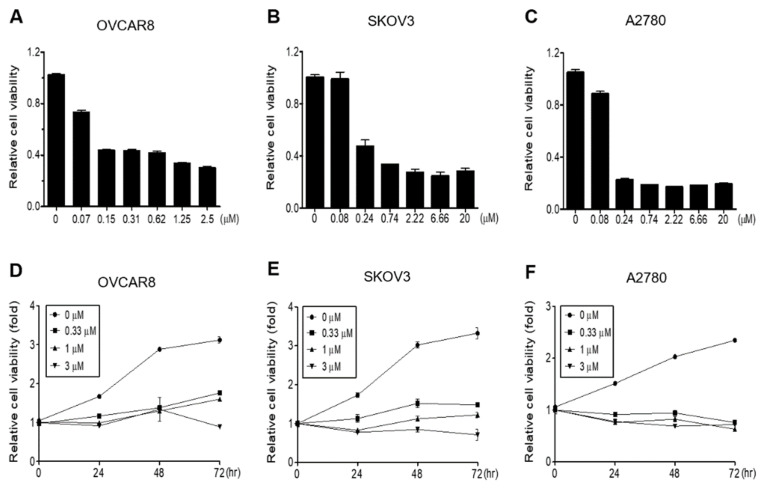
Background and Objectives: Gramicidin, a bactericidal antibiotic used in dermatology and ophthalmology, has recently garnered attention for its inhibitory actions against cancer cell growth. However, the effects of gramicidin on ovarian cancer cells and the underlying mechanisms are still poorly understood. We aimed to elucidate the anticancer efficacy of gramicidin against ovarian cancer cells. Materials and Methods: The anticancer effect of gramicidin was investigated through an in vitro experiment. We analyzed cell proliferation, DNA fragmentation, cell cycle arrest and apoptosis in ovarian cancer cells using WST-1 assay, terminal deoxynucleotidyl transferase dUTP nick and labeling (TUNEL), DNA agarose gel electrophoresis, flow cytometry and western blot. Results: Gramicidin treatment induces dose- and time-dependent decreases in OVCAR8, SKOV3, and A2780 ovarian cancer cell proliferation. TUNEL assay and DNA agarose gel electrophoresis showed that gramicidin caused DNA fragmentation in ovarian cancer cells. Flow cytometry demonstrated that gramicidin induced cell cycle arrest. Furthermore, we confirmed via Western blot that gramicidin triggered apoptosis in ovarian cancer cells. Conclusions: Our results strongly suggest that gramicidin exerts its inhibitory effect on cancer cell growth by triggering apoptosis. Conclusively, this study provides new insights into the previously unexplored anticancer properties of gramicidin against ovarian cancer cells.
Keywords: apoptosis; drug repositioning; gramicidin; ovarian cancer.
Conflict of interest statement
The authors declare no conflict of interest. The funders have no role in the design of the study; the collection, analyses, or interpretation of data; the writing of the manuscript; or the decision to publish the results.
Figures


Similar articles
-
Repurposing Valrubicin as a Potent Inhibitor of Ovarian Cancer Cell Growth.Anticancer Res. 2024 Oct;44(10):4301-4307. doi: 10.21873/anticanres.17259. Anticancer Res. 2024. PMID: 39348966
-
Effect of combined treatment with progesterone and tamoxifen on the growth and apoptosis of human ovarian cancer cells.Oncol Rep. 2012 Jan;27(1):87-93. doi: 10.3892/or.2011.1460. Epub 2011 Sep 14. Oncol Rep. 2012. PMID: 21922150
-
Fostamatinib Inhibits the Proliferation of Ovarian Cancer Cells Through Apoptosis Induction.Anticancer Res. 2024 Nov;44(11):4895-4903. doi: 10.21873/anticanres.17315. Anticancer Res. 2024. PMID: 39477304
-
The anti-tumor effect of OP-B on ovarian cancer in vitro and in vivo, and its mechanism: An investigation using network pharmacology-based analysis.J Ethnopharmacol. 2022 Jan 30;283:114706. doi: 10.1016/j.jep.2021.114706. Epub 2021 Oct 3. J Ethnopharmacol. 2022. PMID: 34614446
-
Gentian Violet Inhibits Cell Proliferation through Induction of Apoptosis in Ovarian Cancer Cells.Biomedicines. 2023 Jun 7;11(6):1657. doi: 10.3390/biomedicines11061657. Biomedicines. 2023. PMID: 37371752 Free PMC article.
Cited by
-
Identification of novel Gramicidin S analogs from Aneurinibacillus aneurinilyticus isolated from corn steep liquor with potential antifungal activity.Microb Cell Fact. 2025 Sep 2;24(1):199. doi: 10.1186/s12934-025-02835-5. Microb Cell Fact. 2025. PMID: 40890732 Free PMC article.
-
Insights and therapeutic advances in pancreatic cancer: the role of electron microscopy in decoding the tumor microenvironment.Front Cell Dev Biol. 2024 Dec 18;12:1460544. doi: 10.3389/fcell.2024.1460544. eCollection 2024. Front Cell Dev Biol. 2024. PMID: 39744013 Free PMC article. Review.
-
Designed gramicidin-inspired stabilized peptide-based therapeutics to potentiate immunotherapy against aggressive kidney cancer.Biomater Sci. 2025 Aug 19;13(17):4681-4705. doi: 10.1039/d5bm00109a. Biomater Sci. 2025. PMID: 40485488 Free PMC article.
-
Colloidal Dispersions of Gramicidin D in Water: Preparation, Characterization, and Differential Cytotoxicity.ACS Omega. 2025 Feb 24;10(8):8611-8618. doi: 10.1021/acsomega.4c11133. eCollection 2025 Mar 4. ACS Omega. 2025. PMID: 40060847 Free PMC article.
-
CPNE7 promotes colorectal tumorigenesis by interacting with NONO to initiate ZFP42 transcription.Cell Death Dis. 2024 Dec 18;15(12):896. doi: 10.1038/s41419-024-07288-z. Cell Death Dis. 2024. PMID: 39695095 Free PMC article.
References
MeSH terms
Substances
LinkOut - more resources
Full Text Sources
Medical
