. 2023 Dec 8;28(24):8006.
doi: 10.3390/molecules28248006.

Shock-Induced Degradation of Guanosine and Uridine Promoted by Nickel and Carbonate: Potential Applications

Affiliations

Shock-Induced Degradation of Guanosine and Uridine Promoted by Nickel and Carbonate: Potential Applications

Gustavo P Maia et al. Molecules. .

Abstract

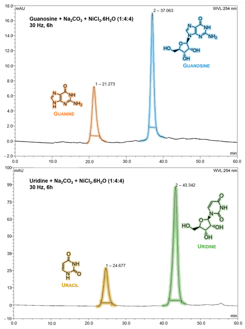
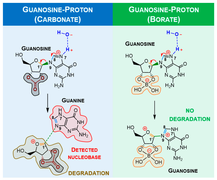
Experimental studies of the degradation of two ribonucleosides (guanosine and uridine) were carried out by making use of mechanochemistry. Mechanochemical experiments reveal the decomposition of guanosine and uridine, promoted by nickel(II) and carbonate ions, into guanine and uracil, respectively. These nucleobases were identified by HPLC and 1H NMR spectroscopy (this applied only to uracil). Additionally, density-functional theory (DFT) methodologies were used to probe the energetic viability of several degradation pathways, including in the presence of the abovementioned ions. Three mechanisms were analysed via ribose ring-opening: dry, single-molecule water-assisted, and metal-assisted, wherein the last two mechanisms confirmed the mechanochemical degradation of both ribonucleosides into respective nucleobase moieties. These results can contribute to an astrobiological interpretation of the extraterrestrial sample's contents.

Keywords: DFT; asteroids; degradation reactions; mechanochemistry; meteorites.


Conflict of interest statement

The authors declare no conflict of interest.

Figures

Figure 1
Figure 1
Ribonucleoside-Na2CO3-NiCl2·6H2O samples before and after mechanochemical treatment (30 Hz, 6 h): (a,b) A uridine sample before and after mechanochemical treatment, respectively; (c,d) A guanosine sample before and after mechanochemical treatment, respectively. Despite the low quality of the pictures, the abrupt change of colours is clear after mechanochemical experiments. These pictures only intend to serve as initial evidence of the reactivity of ribonucleosides.
Figure 2
Figure 2
HPLC chromatogram from guanosine and uridine ribonucleosides after mechanochemical treatment (30 Hz, 6 h) with Na2CO3 and NiCl2·6H2O on a ribonucleoside-Na2CO3-NiCl2·6H2O ratio of 1:4:4. Above: guanine (~21 min, A = 246 and 274 nm) and guanosine (~37 min, A = 253 nm). Below: uracil (~25 min, A = 259 nm) and uridine (~43 min, A = 262 nm). HPLC Run 1 and Run 2 conditions were applied for guanosine- and uridine-treated samples, respectively. Standard nucleobase/nucleoside solutions were previously run to identify their retention times.
Figure 3
Figure 3
1H NMR from a sample containing uridine in H2O/D2O (9:1) after mechanochemical treatment (30 Hz, 6 h) with Na2CO3 and NiCl2·6H2O, in a ratio of 1:4:4 of uridine-Na2CO3-NiCl2·6H2O. From the top: a standard of uridine 1H NMR spectra in H2O/D2O (9:1), a standard of uracil 1H NMR in H2O/D2O (9:1), and a treated sample of uridine. Yellow arrows represent the presence of uracil.
Figure 4
Figure 4
HPLC chromatogram from guanosine and uridine ribonucleosides after mechanochemical treatment (30 Hz, 6 h) with NaCl for a ribonucleoside—NaCl ratio of 1:4. Above: guanine (~21 min, A = 246 and 274 nm) and guanosine (~37 min, A = 253 nm). Below: uracil (~25 min, A = 259 nm) and uridine (~43 min, A = 262 nm). HPLC Run 1 and Run 2 conditions were applied for guanosine- and uridine-treated samples, respectively. Standard nucleobase/nucleoside solutions were previously run to identify their retention times.
Figure 5
Figure 5
X-ray conformers for guanosine (left,middle). The definition of the twist angle used for DFT calculations (right) based on X-ray experimental twist = 300° (left) and twist = 17° (middle).
Figure 6
Figure 6
Based on X-ray data, the geometries of the two conformers are π-π-stacked, forming a van der Walls dimer.
Figure 7
Figure 7
DFT calculations of the most stable conformers of guanosine. A twist angle of 71° enables 2′-hydroxyl and N3 H-bonding (a), while a twist angle of 183° enables 5′-hydroxyl and N3 H-bonding (b). In the presence of carbonate/borate, the 2′-hydroxyl H-bond is eliminated (c,d). Grey and orange moieties represent carbonate and borate, respectively.
Figure 8
Figure 8
Unsuccessfully proposed guanosine β elimination mechanism with nucleobase formation and proton transfer saddle point optimised at HF (Hartree-Fock) level.
Figure 9
Figure 9
Proposed mechanisms for guanosine degradation and labilisation in the presence of carbonate (blue) or borate ions (green), respectively. In the presence of a proton, the nucleobase moiety is eliminated, forming a double bond in the ribose moiety (highly reactive, which will induce ose degradation). The labilisation of structures promoting nucleobase elimination is lower in ribonucleoside-borate structures. Colours highlight the studied bonds.
Figure 10
Figure 10
Proposed mechanism for mechanochemical degradation of guanosine, promoted by nickel (II). In the presence of a proton/metal, the nucleobase moiety is eliminated, forming a double bond in the ribose moiety (highly reactive, which will induce ose degradation). However, this mechanism suggests that the metal ion could catalyse two ribonucleosides simultaneously, being more effective than the proton. Carbonate acts as the “activator” for ribonucleoside degradation.
Figure 11
Figure 11
DFT calculated neutrally optimised conformers of uridine. Similar to guanosine, 5′-hydroxyl and O2 H-bonding are the predominant H-bond stabilisations (blue). The carbonate moiety is highlighted in grey.
Figure 12
Figure 12
Proposed mechanism for mechanochemical degradation of uridine, promoted by nickel (II). In the presence of a proton/metal, the nucleobase moiety is eliminated, forming a double bond in the ribose moiety (highly reactive, which will induce ose degradation). Similar to guanosine degradation, the metal ion contribution is more effective than proton catalysis. Carbonate acts as the “activator” for ribonucleoside degradation.

Similar articles

References

    1. André V., Duarte M.T., Gomes C.S.B., Sarraguça M.C. Mechanochemistry in Portugal-A Step towards Sustainable Chemical Synthesis. Molecules. 2021;27:241. doi: 10.3390/molecules27010241. - DOI - PMC - PubMed
    1. Liu X., Li Y., Zeng L., Li X., Chen N., Bai S., He H., Wang Q., Zhang C. A Review on Mechanochemistry: Ap-proaching Advanced Energy Materials with Greener Force. Adv. Mater. 2022;34:2108327. doi: 10.1002/adma.202108327. - DOI - PubMed
    1. Pagola S. Outstanding Advantages, Current Drawbacks, and Significant Recent Developments in Mechanochemistry: A Perspective View. Crystals. 2023;13:124. doi: 10.3390/cryst13010124. - DOI
    1. Zhou J., Hsu T.-G., Wang J. Mechanochemical Degradation and Recycling of Synthetic Polymers. Angew. Chem. Int. Ed. 2023;62:e202300768. doi: 10.1002/anie.202300768. - DOI - PubMed
    1. Kaupp G.J.C. Mechanochemistry: The varied applications of mechanical bond-breaking. CrystEngComm. 2009;11:388–403. doi: 10.1039/B810822F. - DOI

LinkOut - more resources