. 2023 Dec 5;16(24):7508.
doi: 10.3390/ma16247508.

In Vitro Investigation of Gelatin/Polycaprolactone Nanofibers in Modulating Human Gingival Mesenchymal Stromal Cells

Affiliations

In Vitro Investigation of Gelatin/Polycaprolactone Nanofibers in Modulating Human Gingival Mesenchymal Stromal Cells

Zhiwei Tian et al. Materials (Basel). .

Abstract

The aesthetic constancy and functional stability of periodontium largely depend on the presence of healthy mucogingival tissue. Soft tissue management is crucial to the success of periodontal surgery. Recently, synthetic substitute materials have been proposed to be used for soft tissue augmentation, but the tissue compatibility of these materials needs to be further investigated. This study aims to assess the in vitro responses of human gingival mesenchymal stromal cells (hG-MSCs) cultured on a Gelatin/Polycaprolactone prototype (GPP) and volume-stable collagen matrix (VSCM). hG-MSCs were cultured onto the GPP, VSCM, or plastic for 3, 7, and 14 days. The proliferation and/or viability were measured by cell counting kit-8 assay and resazurin-based toxicity assay. Cell morphology and adhesion were evaluated by microscopy. The gene expression of collagen type I, alpha1 (COL1A1), α-smooth muscle actin (α-SMA), fibroblast growth factor (FGF-2), vascular endothelial growth factor A (VEGF-A), transforming growth factor beta-1 (TGF-β1), focal adhesion kinase (FAK), integrin beta-1 (ITG-β1), and interleukin 8 (IL-8) was investigated by RT-qPCR. The levels of VEGF-A, TGF-β1, and IL-8 proteins in conditioned media were tested by ELISA. GPP improved both cell proliferation and viability compared to VSCM. The cells grown on GPP exhibited a distinct morphology and attachment performance. COL1A1, α-SMA, VEGF-A, FGF-2, and FAK were positively modulated in hG-MSCs on GPP at different investigation times. GPP increased the gene expression of TGF-β1 but had no effect on protein production. The level of ITG-β1 had no significant changes in cells seeded on GPP at 7 days. At 3 days, notable differences in VEGF-A, TGF-β1, and α-SMA expression levels were observed between cells seeded on GPP and those on VSCM. Meanwhile, GPP showed higher COL1A1 expression compared to VSCM after 14 days, whereas VSCM demonstrated a more significant upregulation in the production of IL-8. Taken together, our data suggest that GPP electrospun nanofibers have great potential as substitutes for soft tissue regeneration in successful periodontal surgery.

Keywords: collagen; electrospinning; gelatin; human gingival mesenchymal stromal cells; periodontal tissue; polycaprolactone.


Conflict of interest statement

The authors declare no conflict of interest.

Figures

Figure 1
Figure 1
Morphology of hG-MSCs on different surfaces. Representative fluorescence microscopy images of hG-MSCs on GPP (a,d,g), VSCM (b,e,h), and TCP (c,f,i). Actin filaments (red) and nuclei (blue) of cells were stained after 3, 7, and 14 days. Images show one representative donor and were taken at 100-fold magnification. Scale bars correspond to 50 µm. It can be observed that the morphology of cells adhered to the three different materials varied.
Figure 2
Figure 2
The proliferation/viability of hG-MSCs on GPP, VSCM, and TCP. The proliferation/viability of cells was measured with CCK-8 after 3, 7, and 14 days. Cells seeded on TCP served as control. The data are shown as the mean ± SD (n = 5). Differences between groups at the same time point are indicated by * (p < 0.05) and ** (p < 0.01), whereas those between days 3 and 7 and days 7 and 14 for the same material are indicated with # (p < 0.01) and † (p < 0.01), respectively. GPP is more effective than VSCM in facilitating the proliferation/viability of hG-MSCs.
Figure 3
Figure 3
The effects of GPP, VSCM, and TCP on the metabolic activity of hG-MSCs. The amount of metabolized resazurin produced by cells was measured after 3, 7, and 14 days. Cells seeded on TCP served as control. The data from five individual donors are represented as the mean ± SD. * and **—significantly different between cells seeded on different surfaces of materials at the same time point of measurement with p < 0.05 and p < 0.01, while the distinctions between days 3 and 7 and days 7 and 14 for the same material are indicated by # (p < 0.01) and † (p < 0.05), respectively. Compared to VSCM, GPP promoted the metabolic activity of hG-MSCs.
Figure 4
Figure 4
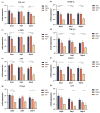
Gene expression of various proteins in hG-MSCs grown on different materials. Gene expression levels of COL1A1 (a), VEGF-A (b), ACAT2 (c), TGF-β1 (d), FAK (e), FGF-2 (f), ITG-β1 (g), and IL-8 (h) were assessed by RT-qPCR after 3, 7, and 14 days of culture, calculated using the 2−ΔΔCt method and GAPDH as a reference gene. Y-axes show the n-fold expression levels compared to the level of day 3 of TCP control (n-fold expression = 1). The data are displayed as the mean ± SD of five different donors. Differences between groups at the same time point are indicated by * and ** for p < 0.05 and p < 0.01, respectively. hG-MSCs cultured on biomaterials exhibited increased gene expression of the investigated biomarkers compared to those on TCP. Cells cultured on GPP demonstrated slightly yet significantly increased expression of VEGF-A, α-SMA, and TGF-β1 after 3 days, and COL1A1 after 14 days, compared to those grown on VSCM.
Figure 5
Figure 5
Protein production by hG-MSCs grown on different materials. The protein production of VEGF-A (a), TGF-β1 (b), and IL-8 (c) in the cells cultured on different materials for 3, 7, and 14 days was quantified by ELISA. Y-axes show the concentration of target protein in conditioned media. The data from five donors are displayed as the mean ± SD. Differences between groups at the same time point are indicated by * and ** for p < 0.05 and p < 0.01, respectively. Cells grown on VSCM presented the highest expression level of VEGF-A. VSCM showed a more significant upregulation in the production of IL-8 after 3 and 14 days. There was no significant difference in the production of TGF-β1 among the three groups.

References

    1. Wu D.T., Munguia-Lopez J.G., Cho Y.W., Ma X., Song V., Zhu Z., Tran S.D. Polymeric Scaffolds for Dental, Oral, and Craniofacial Regenerative Medicine. Molecules. 2021;26:7043. doi: 10.3390/molecules26227043. - DOI - PMC - PubMed
    1. Kassab M.M., Cohen R.E. The etiology and prevalence of gingival recession. J. Am. Dent. Assoc. 2003;134:220–225. doi: 10.14219/jada.archive.2003.0137. - DOI - PubMed
    1. Stetler K.J., Bissada N.F. Significance of the width of keratinized gingiva on the periodontal status of teeth with submarginal restorations. J. Periodontol. 1987;58:696–700. doi: 10.1902/jop.1987.58.10.696. - DOI - PubMed
    1. Tugnait A., Clerehugh V. Gingival recession-its significance and management. J. Dent. 2001;29:381–394. doi: 10.1016/S0300-5712(01)00035-5. - DOI - PubMed
    1. Cortellini P., Bissada N.F. Mucogingival conditions in the natural dentition: Narrative review, case definitions, and diagnostic considerations. J. Clin. Periodontol. 2018;45((Suppl. S20)):S190–S198. doi: 10.1111/jcpe.12948. - DOI - PubMed

LinkOut - more resources