. 2023 Nov 26;13(12):1648.
doi: 10.3390/jpm13121648.

Unraveling Extremely Damaging IRAK4 Variants and Their Potential Implications for IRAK4 Inhibitor Efficacy

Affiliations

Unraveling Extremely Damaging IRAK4 Variants and Their Potential Implications for IRAK4 Inhibitor Efficacy

Mohammed Y Behairy et al. J Pers Med. .

Abstract

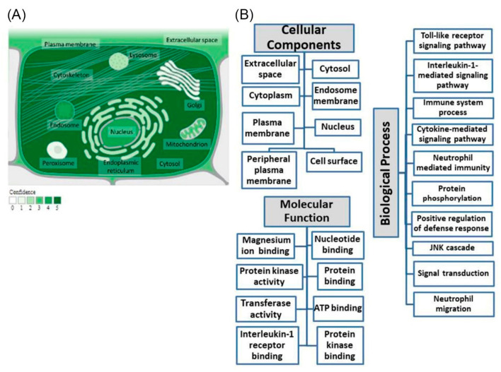
Interleukin-1-receptor-associated kinase 4 (IRAK4) possesses a crucial function in the toll-like receptor (TLR) signaling pathway, and the dysfunction of this molecule could lead to various infectious and immune-related diseases in addition to cancers. IRAK4 genetic variants have been linked to various types of diseases. Therefore, we conducted a comprehensive analysis to recognize the missense variants with the most damaging impacts on IRAK4 with the employment of diverse bioinformatics tools to study single-nucleotide polymorphisms' effects on function, stability, secondary structures, and 3D structure. The residues' location on the protein domain and their conservation status were investigated as well. Moreover, docking tools along with structural biology were engaged in analyzing the SNPs' effects on one of the developed IRAK4 inhibitors. By analyzing IRAK4 gene SNPs, the analysis distinguished ten variants as the most detrimental missense variants. All variants were situated in highly conserved positions on an important protein domain. L318S and L318F mutations were linked to changes in IRAK4 secondary structures. Eight SNPs were revealed to have a decreasing effect on the stability of IRAK4 via both I-Mutant 2.0 and Mu-Pro tools, while Mu-Pro tool identified a decreasing effect for the G198E SNP. In addition, detrimental effects on the 3D structure of IRAK4 were also discovered for the selected variants. Molecular modeling studies highlighted the detrimental impact of these identified SNP mutant residues on the druggability of the IRAK4 ATP-binding site towards the known target inhibitor, HG-12-6, as compared to the native protein. The loss of important ligand residue-wise contacts, altered protein global flexibility, increased steric clashes, and even electronic penalties at the ligand-binding site interfaces were all suggested to be associated with SNP models for hampering the HG-12-6 affinity towards IRAK4 target protein. This given model lays the foundation for the better prediction of various disorders relevant to IRAK4 malfunction and sheds light on the impact of deleterious IRAK4 variants on IRAK4 inhibitor efficacy.

Keywords: SNPs; TLR signaling; bioinformatics; innate immunity; molecular dynamics.


Conflict of interest statement

The authors declared no conflict of interest.

Figures

Figure 1
Figure 1
(A) IRAK4 subcellular localization. Color code signifies the confidence level, with a range of colors from light green, which signifies a low level, to dark green, which signifies a high level (genecards.org; accessed on 11 June 2023), with the image’s origin being compartments.jensenlab.org. (B) Investigation of IRAK4’s gene ontology. Terms of biological process, molecular function, and cellular components regarding IRAK4 are revealed (genecards.org; accessed on 20 July 2023).
Figure 2
Figure 2
Effects of the studied variants on IRAK4 3D structure produced by HOPE server: (A) with G195R, (B) with G198E, (C) with G198R, (D) with D311H, (E) with L318S, (F) with L318F, (G) with F330V, (H) with R334W, and (I) with R334Q.
Figure 3
Figure 3
A network of IRAK4 gene–gene interactions produced by GeneMANIA.
Figure 4
Figure 4
3D architecture and docking poses of HG-12-6 at IRAK4 native and SNP variants. (A) Overlaid binding modes of IRAK4/HG-12-6 models. The ligands are represented as blue sticks and orange lines at native and variant proteins, respectively. Native IRAK4 cartoon/surface is colored differently according to its structural motifs; hinge region (orange); Schellman loop (red), αDE loop (cyan); gatekeeper residue (GK; yellow), glycine-rich loop (orange); αC-helix (black); activation segment (magenta); and αG-helix/adjoining loops (blue). Right panel represents overlaid redocked and co-crystallized HG-12-6 at native IRAK4 for validating purposes of the docking protocol. (BI) Binding modes of HG-12-6 at native IRAK4 (B) and its SNP mutants: (C) F330V; (D) D311H; (E) G195R; (F) G198R; (G) L318S; (H) L318F; and (I) G198E models. Residues located within 4 Å radius of bound ligand are displayed as lines, numbered with their protein sequence, and colored based on respective motif location. Only variant residues are in bold underlined text and shown with their surrounding electron densities in 3D dot representations. Polar interactions are shown as black dashed lines.
Figure 4
Figure 4
3D architecture and docking poses of HG-12-6 at IRAK4 native and SNP variants. (A) Overlaid binding modes of IRAK4/HG-12-6 models. The ligands are represented as blue sticks and orange lines at native and variant proteins, respectively. Native IRAK4 cartoon/surface is colored differently according to its structural motifs; hinge region (orange); Schellman loop (red), αDE loop (cyan); gatekeeper residue (GK; yellow), glycine-rich loop (orange); αC-helix (black); activation segment (magenta); and αG-helix/adjoining loops (blue). Right panel represents overlaid redocked and co-crystallized HG-12-6 at native IRAK4 for validating purposes of the docking protocol. (BI) Binding modes of HG-12-6 at native IRAK4 (B) and its SNP mutants: (C) F330V; (D) D311H; (E) G195R; (F) G198R; (G) L318S; (H) L318F; and (I) G198E models. Residues located within 4 Å radius of bound ligand are displayed as lines, numbered with their protein sequence, and colored based on respective motif location. Only variant residues are in bold underlined text and shown with their surrounding electron densities in 3D dot representations. Polar interactions are shown as black dashed lines.
Figure 5
Figure 5
Analysis of molecular dynamics trajectories for HG-12-6-bound IRAK4 models. (A) Alpha-carbon RMSD of IRAK4 protein; (B) sole ligand’s RMSD; (C) buried SASA, plotted against the whole simulation timeline (200 ns).
Figure 6
Figure 6
Global stability analysis for simulated HG-12-6-bound IRAK4 complexes. (A) Monitored ΔRMSF trajectories for simulated HG-12-6/IRAK4 models along the whole molecular dynamics simulations. Alpha-carbon differences in RMSF tones are illustrated in terms of constituting residue sequence number (Ser167 to Ser460). Important structure regions are highlighted in colors; hinge region (orange); Schellman loop (red), αDE loop (cyan); gatekeeper residue (GK; yellow), glycine-rich loop (orange); αC-helix (black); activation segment (magenta); and αG-helix/adjoining loops (blue). (B) Conformation evolution of the simulated complexes across the simulation timeline. Overlaid initial, middle, and final snapshots for native and SNP variant models at 0 ns, 100 ns, and 200 ns, respectively. Complexes are shown in green, yellow, and red cartoons with respect to the initial, middle, and final extracted frames. Ligands (sticks) and SNP residues (lines) are presented in colors corresponding to their extracted frames.
Figure 6
Figure 6
Global stability analysis for simulated HG-12-6-bound IRAK4 complexes. (A) Monitored ΔRMSF trajectories for simulated HG-12-6/IRAK4 models along the whole molecular dynamics simulations. Alpha-carbon differences in RMSF tones are illustrated in terms of constituting residue sequence number (Ser167 to Ser460). Important structure regions are highlighted in colors; hinge region (orange); Schellman loop (red), αDE loop (cyan); gatekeeper residue (GK; yellow), glycine-rich loop (orange); αC-helix (black); activation segment (magenta); and αG-helix/adjoining loops (blue). (B) Conformation evolution of the simulated complexes across the simulation timeline. Overlaid initial, middle, and final snapshots for native and SNP variant models at 0 ns, 100 ns, and 200 ns, respectively. Complexes are shown in green, yellow, and red cartoons with respect to the initial, middle, and final extracted frames. Ligands (sticks) and SNP residues (lines) are presented in colors corresponding to their extracted frames.
Figure 7
Figure 7
MM_PBSA free binding energy calculations for the HG-12-6-bound IRAK4 complexes. (A) Total free binding energies and their constituting energy terms. (B) Residue-based energy contributions.

Similar articles

Cited by

References

    1. Candan G., Kahraman R., Ergen H.A. Irak-4 rs4251481 Gene Variant: As a Risk Factor on Inflammatory Bowel Disease. Turk. J. Med. Sci. 2019;49:478–482. doi: 10.3906/sag-1807-279. - DOI - PMC - PubMed
    1. Janssens S., Beyaert R. Functional Diversity and Regulation of Different Interleukin-1 Receptor-Associated Kinase (IRAK) Family Members. Mol. Cell. 2003;11:293–302. doi: 10.1016/S1097-2765(03)00053-4. - DOI - PubMed
    1. Suzuki N., Suzuki S., Duncan G.S., Millar D.G., Wada T., Mirtsos C., Takada H., Wakeham A., Itie A., Li S., et al. Severe Impairment of Interleukin-1 and Toll-like Receptor Signalling in Mice Lacking IRAK-4. Nature. 2002;416:750–754. doi: 10.1038/nature736. - DOI - PubMed
    1. Sutherland A.M., Walley K.R., Nakada T.-A., Sham A.H.P., Wurfel M.M., Russell J.A. A Nonsynonymous Polymorphism of IRAK4 Associated with Increased Prevalence of Gram-Positive Infection and Decreased Response to Toll-like Receptor Ligands. J. Innate Immun. 2011;3:447–458. doi: 10.1159/000323880. - DOI - PMC - PubMed
    1. Wang H., Zhou H., Zhang Q., Poulsen K.L., Taylor V., McMullen M.R., Czarnecki D., Dasarathy D., Yu M., Liao Y., et al. Inhibition of IRAK4 Kinase Activity Improves Ethanol-Induced Liver Injury in Mice. J. Hepatol. 2020;73:1470–1481. doi: 10.1016/j.jhep.2020.07.016. - DOI - PMC - PubMed