Chronological Changes in Sagittal Femoral Bowing after Primary Cementless Total Hip Arthroplasty: A Comparative 3D CT Study
- PMID: 38138931
- PMCID: PMC10744357
- DOI: 10.3390/jpm13121704
Chronological Changes in Sagittal Femoral Bowing after Primary Cementless Total Hip Arthroplasty: A Comparative 3D CT Study
Abstract
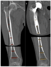
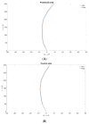
Little is known about dynamic changes of femoral anatomy after total hip arthroplasty (THA), in particular about sagittal femoral bowing (SFB). A 3D CT study was designed to evaluate the chronological changes of SFB after cementless femoral stem implantation for primary THA. Ten patients who underwent unilateral primary THA with a cementless femoral stem, with 2 consecutive CT scans (extending from the fourth lumbar vertebra to the tibial plateaus), performed before THA and at least 3 years after THA, were enrolled. The 3D models of femurs were created using image segmentation software. Using the two CT scans, SFB values of the proximal and middle thirds were calculated on the replaced and untreated sides by two different observers. Eight anatomical stems and two conical stems were involved. The post-operative CT was performed at an average follow-up of 6.5 years after THA (range: 3-12.5). The measurements performed by the two observers did not differ in the proximal and middle regions. A significant difference between the pre-operative and post-operative SFB compared to the untreated side was found in the proximal femur segment (p = 0.004). Use of a cementless stem in THA induced chronological changes in SFB of the proximal femur, after a minimum timespan of 3 years.
Keywords: anatomic stem; bowing; deformation; impingement; modification; procurvatum; revision; stem alignment; total hip arthroplasty.
Conflict of interest statement
The authors declare no conflict of interest.
Figures


References
-
- Oh Y., Fujita K., Wakabayashi Y., Kurosa Y., Okawa A. Location of Atypical Femoral Fracture Can Be Determined by Tensile Stress Distribution Influenced by Femoral Bowing and Neck-Shaft Angle: A CT-Based Nonlinear Finite Element Analysis Model for the Assessment of Femoral Shaft Loading Stress. Injury. 2017;48:2736–2743. doi: 10.1016/j.injury.2017.09.023. - DOI - PubMed
LinkOut - more resources
Full Text Sources

