Review
. 2023 Dec 7;24(24):17217.
doi: 10.3390/ijms242417217.

The Role of Neutrophils in ANCA-Associated Vasculitis: The Pathogenic Role and Diagnostic Utility of Autoantibodies

Affiliations
Review

The Role of Neutrophils in ANCA-Associated Vasculitis: The Pathogenic Role and Diagnostic Utility of Autoantibodies

Agata Walulik et al. Int J Mol Sci. .

Abstract

Recent years have brought progress in understanding the role of the neutrophil, dispelling the dogma of homogeneous cells mainly involved in the prime defence against pathogens, shedding light on their pathogenic role in inflammatory diseases and on the importance of antineutrophil-cytoplasmic antibodies' pathogenic role in ANCA-associated vasculitides vasculitis (AAV). Myeloperoxidase (MPO) and proteinase 3 (PR3) expressed in neutrophil granulocytes are the most common targets for ANCAs and contribute to the formation of MPO-ANCAs and PR3-ANCAs which, released to the bloodstream, become an excellent diagnostic tool for AAV. In this study, we focus on increasing the clinical and experimental evidence that supports the pathogenic role of ANCAs in AAV. Additionally, we discuss the diagnostic utility of ANCAs for disease activity and prognosis in AAV. Understanding the central role of ANCAs in AAV is crucial for advancing our knowledge of these complex disorders and developing targeted therapeutic strategies in the era of personalized medicine.

Keywords: ANCA-associated vasculitis (AAV); antineutrophil-cytoplasmic antibodies (ANCAs); neutrophil; proteinase 3-/ myeloperoxidase-ANCA; vasculitis.


Conflict of interest statement

The authors declare no conflict of interest.

Figures

Figure 1
Figure 1
Neutrophil development in bone marrow, detailing the localisation and characteristics of each cell among granulopoiesis.
Figure 2
Figure 2
Pathomechanism of NET formation and AAV development. Dendritic cells present antigens to naive T cells. Consequently, these T cells differentiate into T-helper-17-(Th17) cells stimulated by IL-6, TGFβ, and IL-23, and then produce interleukin-17 (IL-17). Subsequently, macrophages, under the influence of IL-17, generate cytokines like tumour necrosis factor (TNF) and interleukin-1β (IL-1β), thereby priming neutrophils for action. Simultaneously, the activation of the alternative complement pathway leads to neutrophil priming. In response to infectious stimuli, neutrophils form neutrophil extracellular traps (NETs). In individuals with compromised NET-degradation activity, persistent NETs lead to a breakdown in tolerance to specific self-antigens, notably myeloperoxidase (MPO) and proteinase 3 (PR3). Dendritic cells subsequently present these self-antigens to CD4+ T cells, triggering the production of antineutrophil-cytoplasmic antibodies (ANCAs) by B cells. Primed neutrophils express MPO and PR3 on their cell surface, binding to PR3-ANCAs and MPO-ANCAs. Concurrently, the Fc region of these ANCAs binds to the Fcγ receptor on the neutrophils, inducing excessive activation. This results in abnormal cytokine production, accompanied by the release of reactive oxygen species (ROS), lytic enzymes, and further NET formation, causing damage to vascular endothelial cells. Noteworthy angiopathic molecules found in NETs include histones dissociated from DNA and matrix metalloproteinases (MMPs). B-cell-activating factor (BAFF or TNF), produced by activated neutrophils and CD4+ T cells via IL-21 stimulate B cells, fostering continuous production of ANCAs [44].
Figure 3
Figure 3
Association between IIF staining pattern and used fixative [78].
Figure 4
Figure 4
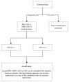
Current ANCA diagnostic recommendations in AAV [88].

References

    1. Tsukui D., Kimura Y., Kono H. Pathogenesis and pathology of anti-neutrophil cytoplasmic antibody (ANCA)-associated vasculitis. J. Transl. Autoimmun. 2021;4:100094. doi: 10.1016/j.jtauto.2021.100094. - DOI - PMC - PubMed
    1. Jennette J., Falk R., Bacon P., Basu N., Cid M., Ferrario F., Flores-Suarez L., Gross W., Guillevin L., Hagen E., et al. 2012 Revised International Chapel Hill Consensus Conference Nomenclature of Vasculitides. Arthritis Rheum. 2013;65:1–11. doi: 10.1002/art.37715. - DOI - PubMed
    1. Segelmark M., Baslund B., Wieslander J. Some patients with anti-myeloperoxidase autoantibodies have a C-ANCA pattern. Clin. Exp. Immunol. 1994;96:458–465. doi: 10.1111/j.1365-2249.1994.tb06051.x. - DOI - PMC - PubMed
    1. Radice A., Bianchi L., Sinico R.A. Anti-neutrophil cytoplasmic autoantibodies: Methodological aspects and clinical significance in systemic vasculitis. Autoimmun. Rev. 2013;12:487–495. doi: 10.1016/j.autrev.2012.08.008. - DOI - PubMed
    1. Cohen Tervaert J.W. Should proteinase-3 and myeloperoxidase anti-neutrophil cytoplasmic antibody vasculitis be treated differently: Part 2. Nephrol. Dial. Transpl. 2019;34:384–387. doi: 10.1093/ndt/gfy406. - DOI - PubMed