. 2023 Dec 7;24(24):17218.
doi: 10.3390/ijms242417218.

Sialyltransferase Mutations Alter the Expression of Calcium-Binding Interneurons in Mice Neocortex, Hippocampus and Striatum

Affiliations

Sialyltransferase Mutations Alter the Expression of Calcium-Binding Interneurons in Mice Neocortex, Hippocampus and Striatum

Senka Blažetić et al. Int J Mol Sci. .

Abstract

Gangliosides are major glycans on vertebrate nerve cells, and their metabolic disruption results in congenital disorders with marked cognitive and motor deficits. The sialyltransferase gene St3gal2 is responsible for terminal sialylation of two prominent brain gangliosides in mammals, GD1a and GT1b. In this study, we analyzed the expression of calcium-binding interneurons in primary sensory (somatic, visual, and auditory) and motor areas of the neocortex, hippocampus, and striatum of St3gal2-null mice as well as St3gal3-null and St3gal2/3-double null. Immunohistochemistry with highly specific primary antibodies for GABA, parvalbumin, calretinin, and calbindin were used for interneuron detection. St3gal2-null mice had decreased expression of all three analyzed types of calcium-binding interneurons in all analyzed regions of the neocortex. These results implicate gangliosides GD1a and GT1b in the process of interneuron migration and maturation.

Keywords: St3gal2; St3gal3; calcium-binding interneurons; cortex; hippocampus; striatum.


Conflict of interest statement

The authors declare no conflict of interest.

Figures

Figure 1
Figure 1
(A) The immunohistochemical distribution of major brain gangliosides (GM1, GD1a, GD1b, and GT1b) on sagittal and coronary brain sections of genetically modified mouse models (St3gal2-null, St3gal3-null, and St3gal2/3-double null) and wild-type mice (WT). (B) Schematic overview of the structures and biosynthetic pathways of the major brain gangliosides (GM1, GD1a, GT1b and GT1b) with responsible biosynthetic enzymes ST3GAL2 and ST3GAL3.
Figure 2
Figure 2
NeuN and GABAergic interneuron expression on coronal brain sections. (A) The immunohistochemical expression in the primary motor region (M1) and quantitative analyses (right); (B) the immunohistochemical expression in the primary sensory region (S1) and quantitative analyses (right); (C) the immunohistochemical expression in the primary visual region (V1) and quantitative analyses (right); (D) the immunohistochemical expression in the primary auditive region (Au1) and quantitative analyses (right). Figure also presents layers of the cortex that were used for layer-specific quantification of interneurons. Pictures were taken under 400 magnifications. ** p < 0.01.
Figure 3
Figure 3
(A) The immunohistochemical expression of parvalbumin (PV)-, calbindin (CB)-, and calretinin (CR)-positive interneurons on coronal brain sections in genetically modified mouse models (St3gal2-null, St3gal3-null, and St3gal2/3-double null) and wild-type (WT) mice with marked layers. Pictures were taken under 400 magnifications. (B) the number of positive interneurons in the primary motor region (M1); (C) the number of positive interneurons in the primary sensory region (S1); (D) number of positive interneurons in the primary visual region (V1); (E) the number of positive interneurons in the primary auditive region (Au1). * p < 0.05, ** p < 0.01, and *** p < 0.001.
Figure 4
Figure 4
Parvalbumin (PV) and calbindin (CB) interneuron expression through the cortical layers on coronal brain sections in genetically modified mouse models (St3gal2-null, St3gal3-null, and St3gal2/3-double null) and wild-type (WT) mice; (A) the immunohistochemical expression in the primary motor region (M1) and quantitative analyses (right); (B) the immunohistochemical expression in the primary sensory region (S1) and quantitative analyses (right); (C) the immunohistochemical expression in the primary visual region (V1) and quantitative analyses (right); (D) the immunohistochemical expression in the primary auditive region (Au1) and quantitative analyses (right). The figure also presents the layers of the cortex that were used for layer-specific quantification of interneurons. Pictures were taken under 400 magnifications. * p < 0.05, ** p < 0.01, and *** p < 0.001.
Figure 5
Figure 5
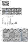
(A) Analyzed hippocampal regions, cornu ammonis, CA1, CA2 and dentate gyrus (DG); (B) the immunohistochemical NeuN and GABAergic interneuron expression in the hippocampus with quantitative analyses; (C) the expression of parvalbumin (PV)-, calbindin (CB)-, and calretinin (CR)-positive interneurons in the hippocampus of genetically modified mouse models (St3gal2-null, St3gal3-null, and St3gal2/3-double null) and wild-type (WT) mice. Pictures were taken under 100 magnifications. * p < 0.05.
Figure 6
Figure 6
(A) The immunohistochemical NeuN and GABAergic interneuron expression in the striatum with quantitative analyses. Pictures were taken under 100 (top row) and 400 (bottom row) magnifications. (B) the expression of parvalbumin (PV)-, calbindin (CB)-, and calretinin (CR)-positive interneurons in the striatum in genetically modified mouse models (St3gal2-null, St3gal3-null, and St3gal2/3-double null) and wild-type (WT) mice. Pictures were taken under 400 magnifications. Quantification is only presented for CB because PV and CR did not have any difference. * p < 0.05.
Figure 7
Figure 7
Schematic presentation of the analyzed brain regions according to Bergmann.

References

    1. Neurod6 Expression Defines New Retinal Amacrine Cell Subtypes and Regulates Their Fate|Nature Neuroscience. [(accessed on 22 June 2022)]. Available online: https://www.nature.com/articles/nn.2859. - PMC - PubMed
    1. DeFelipe J., Fariñas I. The Pyramidal Neuron of the Cerebral Cortex: Morphological and Chemical Characteristics of the Synaptic Inputs. Prog. Neurobiol. 1992;39:563–607. doi: 10.1016/0301-0082(92)90015-7. - DOI - PubMed
    1. DeFelipe J., López-Cruz P.L., Benavides-Piccione R., Bielza C., Larrañaga P., Anderson S., Burkhalter A., Cauli B., Fairén A., Feldmeyer D., et al. New Insights into the Classification and Nomenclature of Cortical GABAergic Interneurons. Nat. Rev. Neurosci. 2013;14:202–216. doi: 10.1038/nrn3444. - DOI - PMC - PubMed
    1. Gelman D.M., Marín O. Generation of Interneuron Diversity in the Mouse Cerebral Cortex. Eur. J. Neurosci. 2010;31:2136–2141. doi: 10.1111/j.1460-9568.2010.07267.x. - DOI - PubMed
    1. Parent A., Hazrati L.N. Functional Anatomy of the Basal Ganglia. I. The Cortico-Basal Ganglia-Thalamo-Cortical Loop. Brain Res. Brain Res. Rev. 1995;20:91–127. doi: 10.1016/0165-0173(94)00007-C. - DOI - PubMed

LinkOut - more resources