. 2023 Dec 8;24(24):17253.
doi: 10.3390/ijms242417253.

Rhamnogalacturonan I with β-(1,4)-Galactan Side Chains as an Ever-Present Component of Tertiary Cell Wall of Plant Fibers

Affiliations

Rhamnogalacturonan I with β-(1,4)-Galactan Side Chains as an Ever-Present Component of Tertiary Cell Wall of Plant Fibers

Tatyana Chernova et al. Int J Mol Sci. .

Abstract

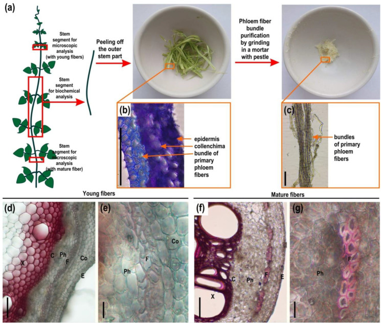
The cellulose-enriched tertiary cell walls present in many plant fibers have specific composition, architecture, machinery of formation, and function. To better understand the mechanisms underlying their mode of action and to reveal the peculiarities of fibers from different plant species, it is necessary to more deeply characterize the major components. Next to overwhelming cellulose, rhamnogalacturonan I (RG-I) is considered to be the key polymer of the tertiary cell wall; however, it has been isolated and biochemically characterized in very few plant species. Here, we add RG-I to the list from the phloem fibers of the Phaseolus vulgaris stem that was isolated and analyzed by nuclear magnetic resonance (NMR), dynamic light scattering, and immunolabeling, both within tissue and as an isolated polymer. Additionally, fibers with tertiary cell walls from nine species of dicotyledonous plants from the orders Malphigiales, Fabales, and Rosales were labeled with RG-I-related antibodies to check the presence of the polymer and compare the in situ presentation of its backbone and side chains. The obtained results confirm that RG-I is an obligatory polymer of the tertiary cell wall. However, there are differences in the structure of this polymer from various plant sources, and these peculiarities may be taxonomically related.

Keywords: NMR; cell wall; dynamic light scattering; immunochemistry; plant fibers; polysaccharides; rhamnogalacturonan I.


Conflict of interest statement

The authors declare no conflict of interest.

Figures

Figure 1
Figure 1
(a) Scheme of sample collection from common bean (Phaseolus vulgaris) stem for microscopy and biochemical analysis (red boxes); (b) cross-section of outer stem part, stained by toluidine blue; and (c) isolated phloem fiber bundles. Cross-sections of (d,e) young (with young phloem fibers) and (f,g) mature (with mature phloem fibers) part of the stem of a common bean stained by phloroglucinol-HCl. In young fibers the cell wall is not lignified. In mature fibers, only the outer layers of the cell wall undergo lignification, while the tertiary cell wall remains unlignified. C—cambium, Co—collenchyma, E—epidermis, F—fibers, Ph—phloem, X—xylem. Scale bar (bd,f) 100 µm; (e,g) 20 µm.
Figure 2
Figure 2
Immunolabeling of common bean stem cross-sections with monoclonal antibodies (a,b) INRA-RU2, specific for rhamnogalacturonan I backbone; (c,d) LM5, specific for linear β-(1,4)-d-galactan; (e,f) LM 26 specific for branched β-(1,4)-d-galactan; (g,h) LM6, specific for linear α-(1,5)-l-arabinan; (i,j) LM2; and (k,l) JIM14, specific for arabinogalactan protein. C—cambium, Co—collenchyma, E—epidermis, F—fibers, Ph—phloem, X—xylem. Scale bar (a,c,e,g,i,k) 100 µm; (b,d,f,h,j,l) 50 µm.
Figure 3
Figure 3
Immunolabeling of common bean stem cross-sections with monoclonal antibodies (a,b) LM19 and (c,d) LM20, specific for homogalacturonan with low and high levels of esterification. (e,f) LM11, specific to β-(1,4)-d-xylan; (g,h) LM21, specific for (galacto)(gluco)mannan; and (i,j) carbohydrate-binding module CBM3a that specifically recognizes the planar surface of crystalline cellulose. C—cambium, Co—collenchyma, E—epidermis, F—fibers, Ph—phloem, X—xylem. Scale bar (a,c,e,g,i) 100 µm; (b,d,f,h,j) 50 µm.
Figure 4
Figure 4
Scheme of extraction of the cell wall fractions of common bean fibers with their monosaccharide proportion (mol%).
Figure 5
Figure 5
Analysis of high-molecular-weight subfractions of (ac) buffer-extractable and (df) strongly retained by cellulose polysaccharides, isolated from the cell wall of common bean fibers. Elution profile of (a) buffer-extractable and (d) strongly retained by cellulose polysaccharides obtained after size-exclusion chromatography on a Sepharose CL-4B with indication of the high-molecular-weight subfraction (marked with a blue dotted line); (b,e) proportions of monosaccharides obtained after TFA hydrolysis; and (c,f) immunodot analysis of high-molecular-weight subfractions. Scale bar (c,f) 0.5 mm.
Figure 6
Figure 6
1H NMR spectra of rhamnogalacturonans I extracted by buffer and obtained after total destruction of the cellulose of the fiber cell wall from common bean.
Figure 7
Figure 7
Concentration dependence of the hydrodynamic radii of small and large particles of rhamnogalacturonans I extracted by buffer (black circles) and obtained after total destruction of cellulose (white circles) from the cell wall of common bean fibers.
Figure 8
Figure 8
Immunolabeling of developing fibers with tertiary cell walls on stem cross-sections of different plant species belonging to Malpighiales, Fabales, and Rosales orders with monoclonal antibodies (a,d,g,j,m,p,s,v,y) INRA-RU2; (b,e,h,k,n,q,t,w,z) LM5; and (c,f,i,l,o,r,u,x,aa) LM26. Scale bar (ax) 50 µm; (yaa) 20 µm.
Figure 9
Figure 9
β-d-galactosidase activity in the stems of flax (Linum usitatissimum), ramie (Boehmeria nivea), hops (Humulus lupulus), and common bean (Phaseolus vulgaris); tendrils of wood vetch (Vicia sylvatica); and storage root of clover (Trifolium pratense). In inserts, a larger image of the fibers are given. C—cambium, Co—collenchyma, E—epidermis, F—fibers, Ph—phloem, X—xylem. Bars are 100 µm for main figures and 50 µm for insets.
Figure 10
Figure 10
Analyzed species of Malpighiales, Fabales and Rosales, their phylogeny (following [57]) and summary of the distribution of epitopes to antibodies specific to the rhamnogalacturonan I backbone (INRA-RU2), β-(1,4)-d-galactan (LM5), and β-(1,6)-galactosyl substitution of β-(1,4)-d-galactan (LM26) in tertiary cell walls of their developing fibers. * Data for Populus spp. are given according to [7].

References

    1. Gorshkova T., Chernova T., Mokshina N., Ageeva M., Mikshina P. Plant ‘muscles’: Fibers with a tertiary cell wall. New Phytol. 2018;218:66–72. doi: 10.1111/nph.14997. - DOI - PubMed
    1. Gorshkova T., Petrova A., Mikshina P. Review: Tertiary cell wall of plant fibers as a source of inspiration in material design. Carbohydr. Polym. 2022;295:119849. doi: 10.1016/j.carbpol.2022.119849. - DOI - PubMed
    1. Chery J.G., Glos R.A.E., Anderson C.T. Do woody vines use gelatinous fibers to climb? New Phytol. 2022;233:126–131. doi: 10.1111/nph.17576. - DOI - PubMed
    1. Sousa-Baena M.S., Onyenedum J.G. Bouncing back stronger: Diversity, structure, and molecular regulation of gelatinous fiber development. Curr. Opin. Plant Biol. 2022;67:102198. doi: 10.1016/j.pbi.2022.102198. - DOI - PubMed
    1. Mellerowicz E.J., Baucher M., Sundberg B., Boerjan W. Unravelling cell wall formation in the woody dicot stem. Plant Mol. Biol. 2001;47:239–274. doi: 10.1023/A:1010699919325. - DOI - PubMed

LinkOut - more resources