. 2023 Dec 8;24(24):17263.
doi: 10.3390/ijms242417263.

Sphingomyelin Metabolism Modifies Luminal A Breast Cancer Cell Line under a High Dose of Vitamin C

Affiliations

Sphingomyelin Metabolism Modifies Luminal A Breast Cancer Cell Line under a High Dose of Vitamin C

Michela Codini et al. Int J Mol Sci. .

Abstract

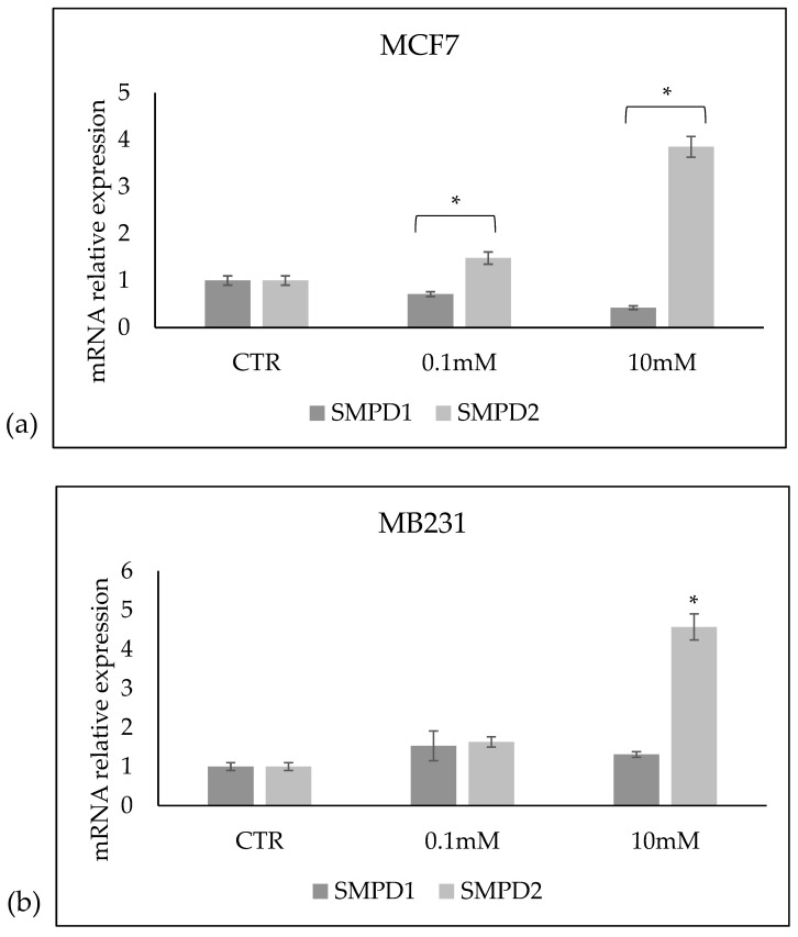
The role of sphingomyelin metabolism and vitamin C in cancer has been widely described with conflicting results ranging from a total absence of effect to possible preventive and/or protective effects. The aim of this study was to establish the possible involvement of sphingomyelin metabolism in the changes induced by vitamin C in breast cancer cells. The MCF7 cell line reproducing luminal A breast cancer and the MDA-MB-231 cell line reproducing triple-negative breast cancer were used. Cell phenotype was tested by estrogen receptor, progesterone receptor, human epidermal growth factor receptor 2 expression, and proliferation index percentage. Sphingomyelin was localized by an EGFP-NT-Lys fluorescent probe. Sphingomyelin metabolism was analyzed by RT-PCR, Western blotting and UFLC-MS/MS. The results showed that a high dose of vitamin C produced reduced cell viability, modulated cell cycle related genes, and changed the cell phenotype with estrogen receptor downregulation in MCF7 cell. In these cells, the catabolism of sphingomyelin was promoted with a large increase in ceramide content. No changes in viability and molecular expression were observed in MB231 cells. In conclusion, a high dose of vitamin C induces changes in the luminal A cell line involving sphingomyelin metabolism.

Keywords: HER-2; Ki-67; MB-231 cells; MCF7 cells; breast cancer; ceramide; estrogen receptor; progesterone receptor; sphingomyelin; vitamin C.


Conflict of interest statement

The authors declare no conflict of interest.

Figures

Figure 1
Figure 1
Effect of increasing doses of VitC on MCF7 and MB231 cell viability. Data are expressed as percentages with respect to the control sample (CTR) (100%) and represent the mean ± SD of 3 independent experiments performed in duplicate. DMSO treatment was used as a positive CTR. * p < 0.05 versus CTR.
Figure 2
Figure 2
Effect of 0.1 mM and 10 mM Vitamin C on the gene expression of GADD45A coding growth arrest and DNA-damage-inducible alpha protein, CCND1 coding cyclin D1 protein, and CDKN1A gene coding cyclin-dependent kinase inhibitor 1A protein in MCF7 and MB231 cells. GAPDH and 18S rRNA were used as housekeeping genes. mRNA relative expression levels were calculated as 2−ΔΔCt, comparing the results of the treated samples with the control sample (CTR) equal to 1, the origin of the axes. Data are expressed as the mean ± SD of 3 independent experiments performed in duplicate. * p < 0.05 versus CTR.
Figure 3
Figure 3
Effect of 0.1 mM and 10 mM Vitamin C on MCF7 cell phenotype. Immunocytochemistry analysis of ER, PR, Ki67, and HER2. Data represent the mean ± SD of ten slide readings from 2 experiments performed in duplicate. * p < 0.05 versus control sample (CTR).
Figure 4
Figure 4
Effect of 0.1 mM and 10 mM Vitamin C on MB231 cell phenotype. Immunocytochemistry analysis of ER, PR, Ki67, and HER2. ER, PR, HER2 are negative. For Ki67, the data represent the mean ± SD of ten slide readings from 2 experiments performed in duplicate.
Figure 5
Figure 5
Sphingomyelin (SM) and ceramide (CER) species in MCF7 (a,b) and MB231 (c,d) cells treated with 0.1 mM and 10 mM VitC. Data are expressed as the mean ± SD of 3 independent experiments performed in duplicate. * p < 0.05 versus control sample (CTR).
Figure 6
Figure 6
Total sphingomyelin (SM) and ceramide (CER) species in (a) MCF7 and (b) MB231 cells treated with 0.1 mM and 10 mM VitC. (c) Sum of SM and CER species in MCF7 and MB231 cells. Data are expressed as the mean ± SD of 3 independent experiments performed in duplicate. * p < 0.05 versus control sample (CTR).
Figure 7
Figure 7
Presence of sphingomyelin in control (CTR), 0.1 mM, and 10 mM treated MCF7 and Mb231 cells with the EGFP-NT-Lys fluorescent probe.
Figure 8
Figure 8
Effect of 0.1 mM and 10 mM Vitamin C on the gene expression of SMPD1 coding for acid sphingomyelinase and SMPD2 coding for neutral sphingomyelinase 1 in (a) MCF7 and (b) MB231 cells. GAPDH and 18S rRNA were used as housekeeping genes. mRNA relative expression levels were calculated as 2−ΔΔCt, comparing the results of the treated samples with the control sample (CTR) equal to 1, the origin of the axes. Data are expressed as the mean ± SD of 3 independent experiments performed in duplicate. * p < 0.05 versus CTR.
Figure 9
Figure 9
Effect of 0.1 mM and 10 mM Vitamin C on acid sphingomyelinase (aSMase) and neutral sphingomyelinase1 (nSMase) in MCF7 and MB231 cells. (a) Western blotting panel of aSMase and nSMase1 and actin as reference; (b) densitometric analysis of MCF7, the values were normalized with actin and were expressed as a percentage of the control sample (CTR). (c) densitometric analysis of MB231, the values were normalized with actin and were expressed as a percentage of the control sample (CTR). Data are expressed as the mean ± SD of 3 independent experiments performed in duplicate. * p < 0.05 versus CTR.

References

    1. Smirnoff N., Conklin P.L., Loewus F.A. Biosynthesis of Ascorbic Acid in Plants: A Renaissance. Annu. Rev. Plant Physiol. Plant Mol. Biol. 2001;52:437–467. doi: 10.1146/annurev.arplant.52.1.437. - DOI - PubMed
    1. Linster C.L., Van Schaftingen E. Vitamin C. Biosynthesis, recycling and degradation in mammals. FEBS J. 2007;274:1–22. doi: 10.1111/j.1742-4658.2006.05607.x. - DOI - PubMed
    1. Tsukaguchi H., Tokui T., Mackenzie B., Berger U.V., Chen X.Z., Wang Y., Brubaker R.F., Hediger M.A. A family of mammalian Na+-dependent L-ascorbic acid transporters. Nature. 1999;399:70–75. doi: 10.1038/19986. - DOI - PubMed
    1. Hasselholt S., Tveden-Nyborg P., Lykkesfeldt J. Distribution of vitamin C is tissue specific with early saturation of the brain and adrenal glands following differential oral dose regimens in guinea pigs. Br. J. Nutr. 2015;113:1539–1549. doi: 10.1017/S0007114515000690. - DOI - PubMed
    1. Lykkesfeldt J., Poulsen H.E. Is vitamin C supplementation beneficial? Lessons learned from randomised controlled trials. Br. J. Nutr. 2010;103:1251–1259. doi: 10.1017/S0007114509993229. - DOI - PubMed