

Blood and Brain Metabolites after Cerebral Ischemia
- PMID: 38139131
- PMCID: PMC10743907
- DOI: 10.3390/ijms242417302
Blood and Brain Metabolites after Cerebral Ischemia
Abstract
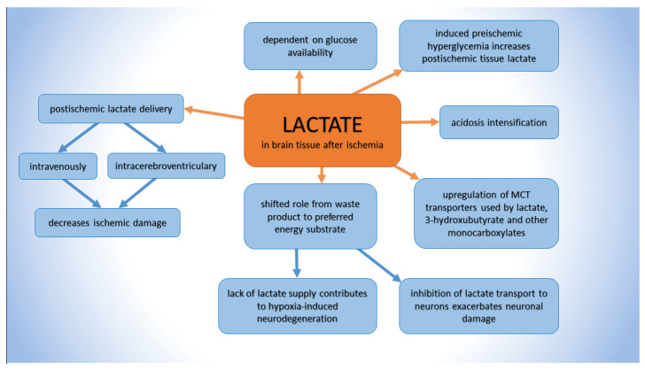
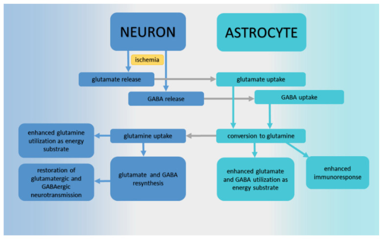
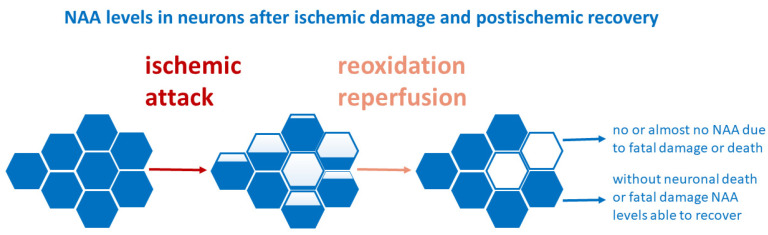
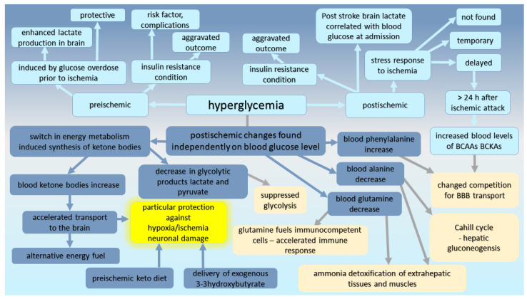
The study of an organism's response to cerebral ischemia at different levels is essential to understanding the mechanism of the injury and protection. A great interest is devoted to finding the links between quantitative metabolic changes and post-ischemic damage. This work aims to summarize the outcomes of the most studied metabolites in brain tissue-lactate, glutamine, GABA (4-aminobutyric acid), glutamate, and NAA (N-acetyl aspartate)-regarding their biological function in physiological conditions and their role after cerebral ischemia/reperfusion. We focused on ischemic damage and post-ischemic recovery in both experimental-including our results-as well as clinical studies. We discuss the role of blood glucose in view of the diverse impact of hyperglycemia, whether experimentally induced, caused by insulin resistance, or developed as a stress response to the cerebral ischemic event. Additionally, based on our and other studies, we analyze and critically discuss post-ischemic alterations in energy metabolites and the elevation of blood ketone bodies observed in the studies on rodents. To complete the schema, we discuss alterations in blood plasma circulating amino acids after cerebral ischemia. So far, no fundamental brain or blood metabolite(s) has been recognized as a relevant biological marker with the feasibility to determine the post-ischemic outcome or extent of ischemic damage. However, studies from our group on rats subjected to protective ischemic preconditioning showed that these animals did not develop post-ischemic hyperglycemia and manifested a decreased metabolic infringement and faster metabolomic recovery. The metabolomic approach is an additional tool for understanding damaging and/or restorative processes within the affected brain region reflected in the blood to uncover the response of the whole organism via interorgan metabolic communications to the stressful cerebral ischemic challenge.
Keywords: animal models; blood; cerebral ischemia; cerebral microdialysis; metabolites; stroke; tissues.
Conflict of interest statement
The authors declare no conflict of interest.
Figures
References
-
- Kocki J., Ułamek-Kozioł M., Bogucka-Kocka A., Januszewski S., Jabłoński M., Gil-Kulik P., Brzozowska J., Petniak A., Furmaga-Jabłońska W., Bogucki J., et al. Dysregulation of Amyloid-β Protein Precursor, β-Secretase, Presenilin 1 and 2 Genes in the Rat Selectively Vulnerable CA1 Subfield of Hippocampus Following Transient Global Brain Ischemia. J. Alzheimers Dis. 2015;47:1047–1056. doi: 10.3233/JAD-150299. - DOI - PMC - PubMed
-
- Schiefecker A.J., Putzer G., Braun P., Martini J., Strapazzon G., Antunes A.P., Mulino M., Pinggera D., Glodny B., Brugger H., et al. Total TauProtein as Investigated by Cerebral Microdialysis Increases in Hypothermic Cardiac Arrest: A Pig Study. Ther. Hypothermia Temp. Manag. 2021;11:28–34. doi: 10.1089/ther.2020.0016. - DOI - PubMed
Publication types
MeSH terms
Substances
Grants and funding
LinkOut - more resources
Full Text Sources
Medical
