. 2023 Dec 13;24(24):17440.
doi: 10.3390/ijms242417440.

Antarctic Krill Oil from Euphausia superba Ameliorates Carrageenan-Induced Thrombosis in a Mouse Model

Affiliations

Antarctic Krill Oil from Euphausia superba Ameliorates Carrageenan-Induced Thrombosis in a Mouse Model

Gi Ho Lee et al. Int J Mol Sci. .

Abstract

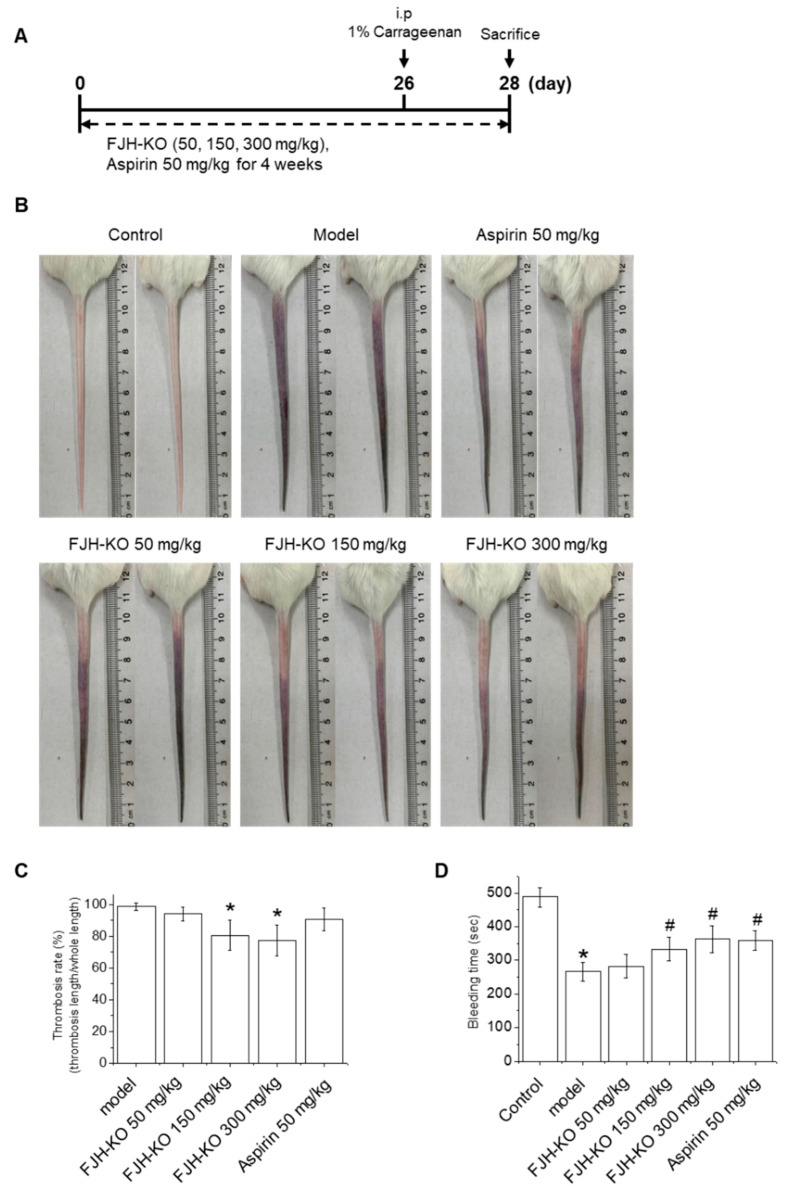
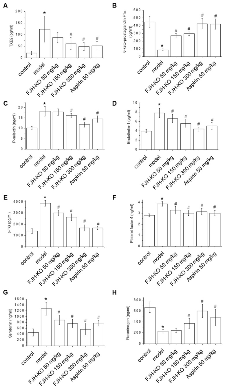
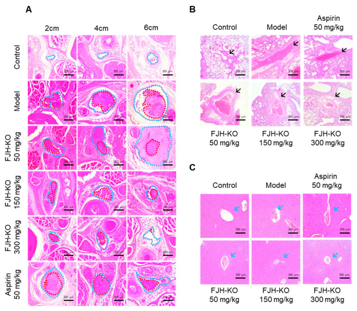
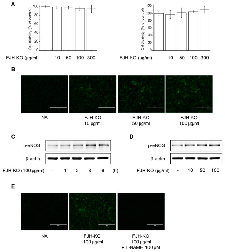
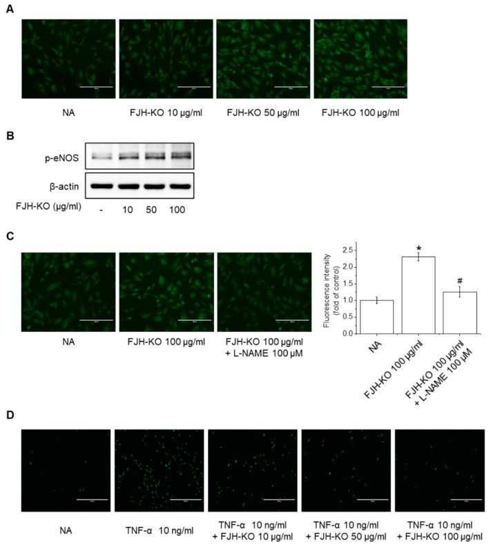
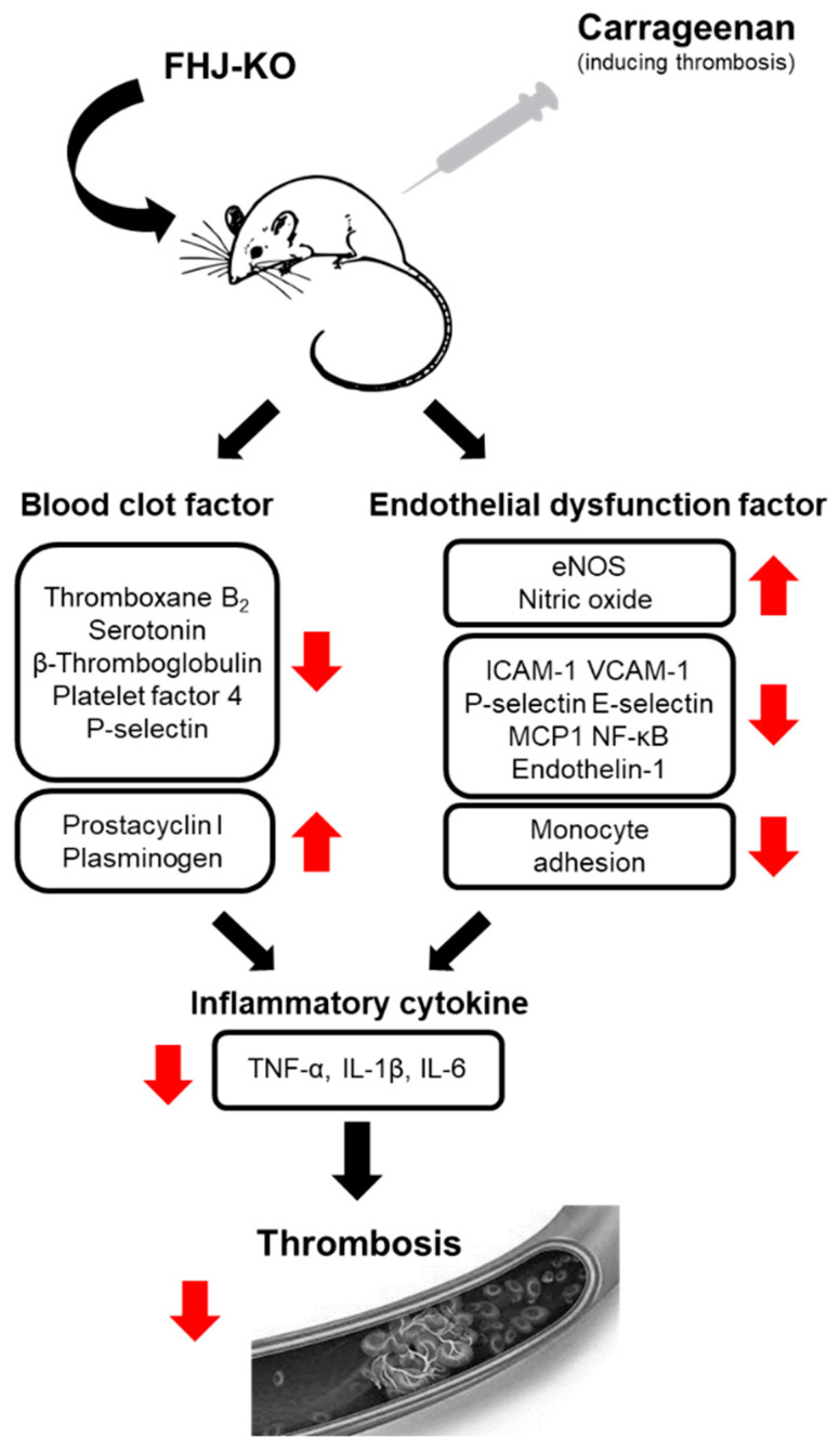
FJH-KO obtained from Antarctic krill, especially Euphausia superba, has been reported to contain high amounts of omega-3 polyunsaturated fatty acids (n-3 PUFA) and to exhibit anticancer and anti-inflammatory properties. However, its antithrombotic effects have not yet been reported. This study aimed to investigate the antithrombotic effects of FJH-KO in carrageenan-induced thrombosis mouse models and human endothelial cells. Thrombosis was induced by carrageenan injection, whereas the mice received FJH-KO pretreatment. FJH-KO attenuated carrageenan-induced thrombus formation in mouse tissue vessels and prolonged tail bleeding. The inhibitory effect of FJH-KO was associated with decreased plasma levels of thromboxane B2, P-selectin, endothelin-1, β-thromboglobulin, platelet factor 4, serotonin, TNF-α, IL-1β, and IL-6. Meanwhile, FJH-KO induced plasma levels of prostacyclin I2 and plasminogen. In vitro, FJH-KO decreased the adhesion of THP-1 monocytes to human endothelial cells stimulated by TNF-α via eNOS activation and NO production. Furthermore, FJH-KO inhibited the expression of TNF-α-induced adhesion molecules such as ICAM-1 and VCAM-1 by suppressing the NF-κB signaling pathway. Taken together, our study demonstrates that FJH-KO protects against carrageenan-induced thrombosis by regulating endothelial cell activation and has potential as an antithrombotic agent.

Keywords: carrageenan; endothelial dysfunction; inflammation; krill oil; thrombosis.


Conflict of interest statement

The authors declare no conflict of interest.

Figures

Figure 1
Figure 1
Effects of FJH-KO on carrageenan-induced thrombosis in mice. (A) Schematic diagram of carrageenan-induced thrombosis model with FJH-KO and aspirin treatment. (B) Thrombus length, (C) thrombosis rate (thrombosis length/whole tail length), and (D) bleeding time (s) in the tail of carrageenan-induced thrombosis mice. * p < 0.05 vs. the vehicle control group, # p < 0.05 vs. the carrageenan model group (n = 8).
Figure 2
Figure 2
Effect of orally administered FJH-KO on the plasma levels of (A) thromboxane B2 (TXB2), (B) 6-keto prostaglandin F1α (6-keto PGF1α), (C) P-selectin, (D) endothelin-1 (ET-1), (E) β-thromboglobulin (β-TG), (F) platelet factor 4 (PF4), (G) serotonin, and (H) plasminogen in carrageenan-induced thrombosis model mice. * p < 0.05 vs. the vehicle control group, # p < 0.05 vs. the carrageenan model group (n = 8).
Figure 3
Figure 3
Effect of orally administered FJH-KO on the plasma levels of (A) tumor necrosis factor alpha (TNF-α), (B) interleukin (IL)-1β, and (C) IL-6 in carrageenan-induced thrombosis model mice. * p < 0.05 vs. the vehicle control group, # p < 0.05 vs. the carrageenan model group (n = 8).
Figure 4
Figure 4
Effect of FJH-KO on the expression of blood clot-related mRNAs in carrageenan-induced mouse blood vessels. (A) Endothelial nitric oxide synthase (eNOS), (B) intercellular adhesion molecule 1 (ICAM-1), (C) vascular cell adhesion molecule 1 (VCAM-1), (D) P-selectin, (E) E-selectin, (F) monocyte chemoattractant protein 1 (MCP-1), (G) TNF-α, (H) IL-1β, and (I) IL-6. * p < 0.05 vs. the vehicle control group, # p < 0.05 vs. the carrageenan model group (n = 8).
Figure 5
Figure 5
Effects of FJH-KO on carrageenan-induced thrombosis in mouse tissues. (A) Hematoxylin and eosin (H&E) staining of the tail 2, 4, and 6 cm from the tail tip (Scale bar: 200 μm). Vessels and thrombi are represented by blue and red dotted lines, respectively. (B) H&E staining of lung tissue (Scale bar: 200 μm). Venous thrombosis is represented by black arrows. (C) H&E staining of liver tissue (Scale bar: 200 μm). Hepatic vein thrombosis is represented by blue arrows.
Figure 6
Figure 6
Effect of FJH-KO on eNOS phosphorylation and nitric oxide (NO) production in EA.hy926 cells. (A) Cells were treated with 10–300 μg/mL FJH-KO for 24 h. Cell viability and cytotoxicity were determined using MTT and LDH assays, respectively. (B) Cells were treated with 10–100 μg/mL FJH-KO for 3 h. NO generation was detected using DAF-2DA (Scale bar: 200 μm). (C,D) Cells were treated with 100 μg/mL FJH-KO for 1–6 h or 10–100 μg/mL FJH-KO for 3 h and assessed via Western blotting. (E) Cells were pretreated with 100 μM L-NAME for 1 h and were then treated with 100 μg/mL FJH-KO for 3 h. NO generation was detected using DAF-2DA (Scale bar: 200 μm).
Figure 7
Figure 7
Effect of FJH-KO on NO production and THP-1 monocyte adhesion in HUVEC cells. (A) Cells were treated with 10–100 μg/mL FJH-KO for 3 h. NO generation was detected using DAF-2DA (Scale bar: 200 μm). (B) Cells were treated with 10–100 μg/mL FJH-KO for 3 h and assessed via Western blotting. (C) Cells were pretreated with 100 μM L-NAME for 1 h and were then treated with 100 μg/mL FJH-KO for 3 h. NO generation was detected using DAF-2DA (Scale bar: 200 μm). (D) Cells were treated with 10–100 μg/mL of FJH-KO for 3 h, followed by incubation with 10 ng/mL TNF-α for 12 h. Endothelial cells were co-cultured with THP-1 cells for 1 h, and the adherence of endothelial cells to monocytes was assessed via fluorescence microscopy (Scale bar: 400 μm). All experiments were performed in triplicate. Data are represented as the mean ± standard deviation. * p < 0.01 vs. control cells, # p < 0.01 vs. FJH-KO-treated cells.
Figure 8
Figure 8
Effects of FJH-KO on TNF-α-induced THP-1 monocyte adhesion in EA.hy926 cells. (A) Cells were treated with 10–100 μg/mL FJH-KO for 3 h, followed by incubation with 10 ng/mL TNF-α for 12 h. Endothelial cells were co-cultured with THP-1 cells for 1 h, and the adherence of endothelial cells to monocytes was assessed via fluorescence microscopy (Scale bar: 400 μm). (B,C) Cells were pretreated with 100 μg/mL FJH-KO for 3 h, followed by 100 ng/mL TNF-α for 12 h. mRNA levels of (B) ICAM-1 and (C) VCAM-1 were measured using RT-PCR. (D,E) Cells were pretreated with 100 μM L-NAME for 1 h and then treated with 100 μg/mL FJH-KO for 3 h, followed by incubation with 10 ng/mL TNF-α for 12 h. mRNA levels of (D) ICAM-1 and (E) VCAM-1 were measured using RT-PCR. (F) Cells were pretreated with 10–100 μg/mL FJH-KO for 3 h and were then treated with 10 ng/mL TNF-α for 1 h. (G) Cells were pretreated with 100 μM L-NAME for 1 h and were then treated with 100 μg/mL FJH-KO for 3 h, followed by incubation with 10 ng/mL TNF-α for 1 h. NF-κB phosphorylation was assessed using Western blotting. (H) Cells were pretreated with 100 μM L-NAME for 1 h and were then treated with 100 μg/mL FJH-KO for 3 h, followed by incubation with 10 ng/mL TNF-α for 12 h. Endothelial cells were co-cultured with THP-1 cells for 1 h, and the adherence of endothelial cells to monocytes was assessed via fluorescence microscopy (Scale bar: 400 μm). All experiments were performed in triplicate. Data are represented as the mean ± standard deviation. * p < 0.01 vs. control cells, # p < 0.01 vs. TNF-α-treated cells, and $ p < 0.01 vs. FJH-KO-treated cells.
Figure 9
Figure 9
Schematic diagram of the protective effect of FJH-KO on thrombosis by regulating endothelial dysfunction and inflammation.

References

    1. Roth G.A., Mensah G.A., Johnson C.O., Addolorato G., Ammirati E., Baddour L.M., Barengo N.C., Beaton A.Z., Benjamin E.J., Benziger C.P., et al. Global Burden of Cardiovascular Diseases and Risk Factors, 1990–2019: Update from the GBD 2019 Study. J. Am. Coll. Cardiol. 2020;76:2982–3021. doi: 10.1016/j.jacc.2020.11.010. - DOI - PMC - PubMed
    1. Thomas H., Diamond J., Vieco A., Chaudhuri S., Shinnar E., Cromer S., Perel P., Mensah G.A., Narula J., Johnson C.O., et al. Global Atlas of Cardiovascular Disease 2000–2016: The Path to Prevention and Control. Glob. Heart. 2018;13:143–163. doi: 10.1016/j.gheart.2018.09.511. - DOI - PubMed
    1. Caron F., Anand S.S. Antithrombotic therapy in aortic diseases: A narrative review. Vasc. Med. 2017;22:57–65. doi: 10.1177/1358863X16675229. - DOI - PubMed
    1. Jorgensen L. The role of platelets in the initial stages of atherosclerosis. J. Thromb. Haemost. 2006;4:1443–1449. doi: 10.1111/j.1538-7836.2006.02006.x. - DOI - PubMed
    1. Yamashita A., Asada Y. Underlying mechanisms of thrombus formation/growth in atherothrombosis and deep vein thrombosis. Pathol. Int. 2023;73:65–80. doi: 10.1111/pin.13305. - DOI - PMC - PubMed