Review
. 2023 Dec 17;24(24):17578.
doi: 10.3390/ijms242417578.

Copper in Gynecological Diseases

Affiliations
Review

Copper in Gynecological Diseases

Rocío A Conforti et al. Int J Mol Sci. .

Abstract

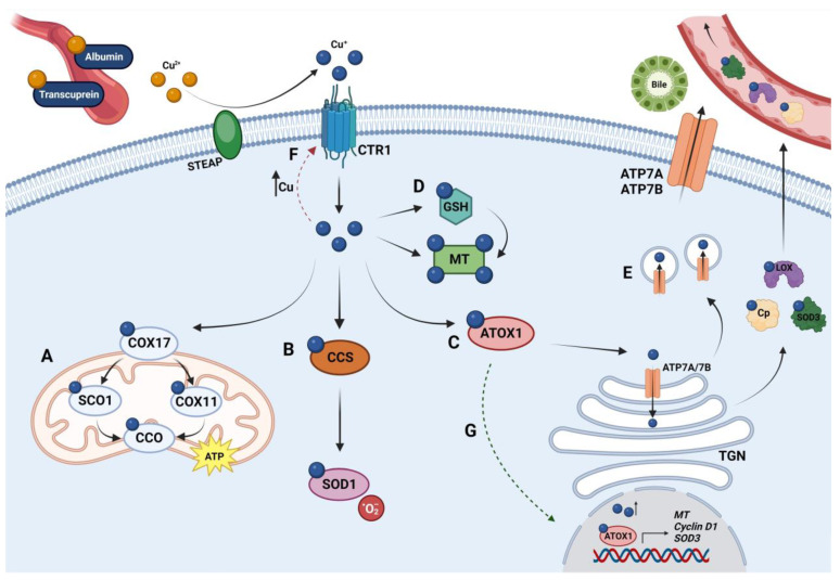
Copper (Cu) is an essential micronutrient for the correct development of eukaryotic organisms. This metal plays a key role in many cellular and physiological activities, including enzymatic activity, oxygen transport, and cell signaling. Although the redox activity of Cu is crucial for enzymatic reactions, this property also makes it potentially toxic when found at high levels. Due to this dual action of Cu, highly regulated mechanisms are necessary to prevent both the deficiency and the accumulation of this metal since its dyshomeostasis may favor the development of multiple diseases, such as Menkes' and Wilson's diseases, neurodegenerative diseases, diabetes mellitus, and cancer. As the relationship between Cu and cancer has been the most studied, we analyze how this metal can affect three fundamental processes for tumor progression: cell proliferation, angiogenesis, and metastasis. Gynecological diseases are characterized by high prevalence, morbidity, and mortality, depending on the case, and mainly include benign and malignant tumors. The cellular processes that promote their progression are affected by Cu, and the mechanisms that occur may be similar. We analyze the crosstalk between Cu deregulation and gynecological diseases, focusing on therapeutic strategies derived from this metal.

Keywords: Cu chelators; Cu ionophores; cervical cancer; copper; endometrial cancer; endometriosis; gynecological diseases; ovarian cancer; polycystic ovarian syndrome.


Conflict of interest statement

The authors declare no conflict of interest.

Figures

Figure 1
Figure 1
Schematic diagram of copper metabolism in mammals. After intestinal absorption, Cu travels through the portal vein bound to soluble proteins, such as albumin and transcuprein. On the surface of mammalian cells, metalloreductases such as STEAP 2, 3, and 4 reduce Cu2+ ions to Cu+ so that cells can absorb Cu through CTR1. (A) In the mitochondrial intermembrane space, COX17 is responsible for delivering Cu+ to either SCO1 or COX11 to contribute to the correct assembly of CCO, which utilizes Cu for energy production through oxidative phosphorylation. (B) CCS chaperone transfers Cu+ to SOD1, which is critical in the defense against oxidative stress because it catalyzes the degradation of superoxide radicals. (C) ATOX1 is responsible for providing Cu to the ATPases (ATP7A and ATP7B) that are principally located in the trans-Golgi network (TGN). ATPases pump Cu+ from the cytosol into the lumen of the TGN to promote the synthesis of cuproenzymes, such as Cp, LOX, and SOD3, which are secreted out of the cells to mediate the Cu transport through the circulatory system. (D) Since free Cu ions have the potential to generate reactive oxygen species, excess intracellular Cu+ is sequestered mainly by glutathione (GSH) and metallothioneins (MTs) that uptake Cu for storage. GSH can also deliver Cu to MTs. (E) When the cytoplasmic Cu concentration increases, ATP7A and ATP7B move within endocytic vesicles toward the plasma membrane to transfer excess Cu into the bloodstream. ATP7A is expressed in many tissues except in the liver, where it is replaced by ATP7B. In hepatocytes, ATP7B ensures the movement of Cu through the canalicular membrane for its subsequent elimination through the bile. (F) The concentration of mammalian CTR1 at the plasma membrane is negatively regulated in response to elevated Cu levels (red dotted arrow), with CTR1 being removed from the cell surface. (G) ATOX1 can carry Cu into the cell nucleus and act as a transcription factor for the expression of genes encoding cyclin D1 and SOD3 (green dotted arrow). High concentrations of cellular Cu may also stimulate the transcription of MT genes. Created with BioRender.com (accessed on 11 November 2023).

References

    1. Nevitt T., Öhrvik H., Thiele D.J. Charting the Travels of Copper in Eukaryotes from Yeast to Mammals. Biochim. Biophys. Acta (BBA)-Mol. Cell Res. 2012;1823:1580–1593. doi: 10.1016/j.bbamcr.2012.02.011. - DOI - PMC - PubMed
    1. Trumbo P., Yates A.A., Schlicker S., Poos M. Dietary Reference Intakes. J. Am. Diet. Assoc. 2001;101:294–301. doi: 10.1016/S0002-8223(01)00078-5. - DOI - PubMed
    1. Moshfegh A.J., Goldman J.D., Rhodes D.G., Friday J.E. Usual Nutrient Intake from Food and Beverages, by Gender and Age, What We Eat In America, NHANES 2017-March 2020 Prepandemic. [(accessed on 11 November 2023)];2023 Available online: www.ars.usda.gov/nea/bhnrc/fsrg.
    1. Myint Z.W., Oo T.H., Thein K.Z., Tun A.M., Saeed H. Copper Deficiency Anemia. Ann. Hematol. 2018;97:1527–1534. doi: 10.1007/s00277-018-3407-5. - DOI - PubMed
    1. Grochowski C., Blicharska E., Baj J., Mierzwińska A., Brzozowska K., Forma A., Maciejewski R. Serum Iron, Magnesium, Copper, and Manganese Levels in Alcoholism: A Systematic Review. Molecules. 2019;24:1361. doi: 10.3390/molecules24071361. - DOI - PMC - PubMed