Review
. 2023 Dec 18;24(24):17633.
doi: 10.3390/ijms242417633.

Role of Glycolytic and Glutamine Metabolism Reprogramming on the Proliferation, Invasion, and Apoptosis Resistance through Modulation of Signaling Pathways in Glioblastoma

Affiliations
Review

Role of Glycolytic and Glutamine Metabolism Reprogramming on the Proliferation, Invasion, and Apoptosis Resistance through Modulation of Signaling Pathways in Glioblastoma

Cristina Trejo-Solis et al. Int J Mol Sci. .

Abstract

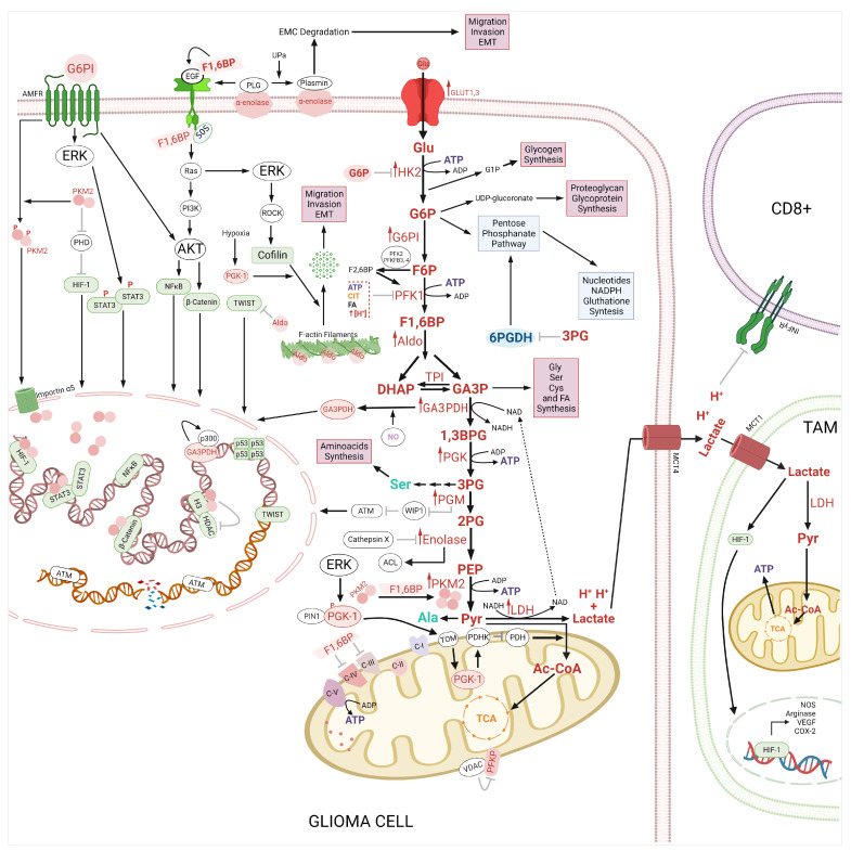
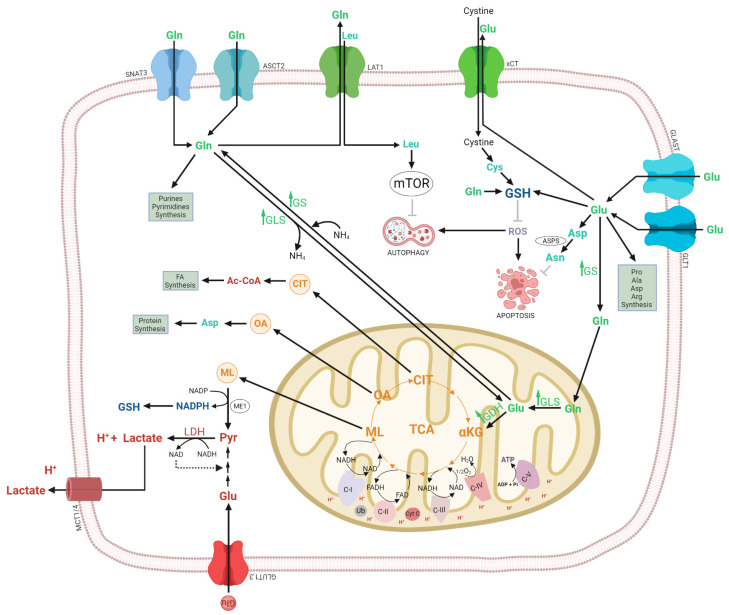
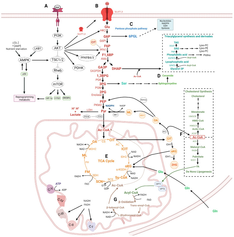
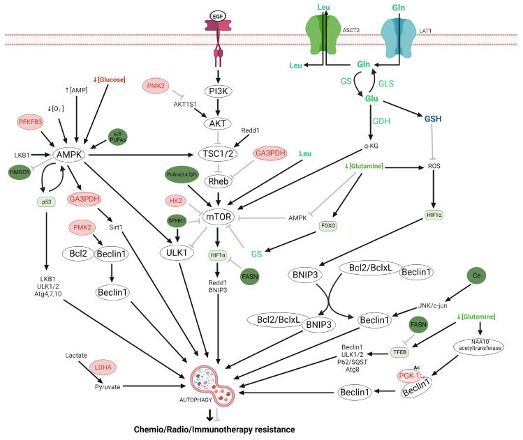
Glioma cells exhibit genetic and metabolic alterations that affect the deregulation of several cellular signal transduction pathways, including those related to glucose metabolism. Moreover, oncogenic signaling pathways induce the expression of metabolic genes, increasing the metabolic enzyme activities and thus the critical biosynthetic pathways to generate nucleotides, amino acids, and fatty acids, which provide energy and metabolic intermediates that are essential to accomplish the biosynthetic needs of glioma cells. In this review, we aim to explore how dysregulated metabolic enzymes and their metabolites from primary metabolism pathways in glioblastoma (GBM) such as glycolysis and glutaminolysis modulate anabolic and catabolic metabolic pathways as well as pro-oncogenic signaling and contribute to the formation, survival, growth, and malignancy of glioma cells. Also, we discuss promising therapeutic strategies by targeting the key players in metabolic regulation. Therefore, the knowledge of metabolic reprogramming is necessary to fully understand the biology of malignant gliomas to improve patient survival significantly.

Keywords: glioma; glucose; glutamine; metabolism; oncogenic pathways.


Conflict of interest statement

The authors declare no conflict of interest.

Figures

Figure 2
Figure 2
Role of glycolysis in glioma. Hexokinase 2 (HK) phosphorylates at glucose to obtain glucose-6 phosphate (G6P). G6P could be redirected to the pentose phosphate pathway (PPP) and at the synthesis of glycogen and UDP-glucuronate. G6PI binds to its autocrine motility factor receptor (AMFR) activating AKT and ERK kinases, which activate STAT3, NF-κB, β-Catenin and Cofilin. The VDAC2 binds to the platelet type of phosphofructokinase (PFKP) in the mitochondria inhibiting its activity. On the other hand, F1,6BP binds to EGFR and SOS blocking the EGFR/RAS/PI3K/AKT signaling pathway and inducing the formation of the tetramer of PKM2. In addition, F1,6BP inhibits the Complex-III and IV (C-III, -IV) from the electron transport chain (ETC). TPI induces the stabilization of the actin filaments and inhibits Twist-related protein (TWIST). PGK1 is phosphorylated by ERK and translocated to mitochondria via PTEN-induced kinase 1 (PIN1)/translocase of the outer membrane (TOM) complex, whereas Mitochondrial PGK1 activates the pyruvate dehydrogenase kinase 1 (PDHK1). PGM1 inactivates p53-induced phosphatase 1 (WIP1), which inhibits ATM. 2PG is metabolized to phosphoenolpyruvate (PEP) by enolase (ENO), which is inhibited by Cathepsin X. ENO induces the activation of the PI3K/AKT pathway and the expression of ATP citrate lyase (ACL). ENO is also a receptor for plasminogen (PLG), which is converted to plasmin. The nuclear PKM2 acts both as a protein kinase for histone H3 and as a transcriptional coactivator for hypoxia-inducible factor-1 α (HIF-1α), STAT3, and β-Catenin. The lactate and H+ are expelled by the monocarboxylate transporter 4 (MCT4), acidifying the tumor microenvironment, and promoting the death of cytotoxic (CD8+) T cells. Furthermore, the lactate is a metabolic fuel for tumor-associated macrophages (TAM) and induces nitric oxidase synthase (NOS), arginase, vascular endothelial grown factor (VEGF), cyclooxygenase (COX-2), and overexpression via HIF-1. Abbreviation: adenosine triphosphate (ATP), citrate (CIT), phosphofructo-2-kinase/fructose-2,6-biphosphatase 3,-4 (PFKFB-3,-4), pyruvate dehydrogenase (PDH), HIF-1α prolyl-hydroxylases (PHD) and Rho-associated protein kinase 2 (ROCK2). ↑ means overexpression, ↑ activation, ⊥ and inhibition. The figure was created with BioRender.com.
Figure 5
Figure 5
Glutamine pathway in glioma. The Glutamine (Gln) transport inside of cells by alanine/serine/cysteine transporter 2 (ASCT2) and system N transporter 3 (SNAT3) and its efflux is via LAT1 in exchange with leucine (Leu); this amino acid activates mammalian target of rapamycin (mTOR) synthesis and signaling. The Cytosolic Gln is transported to the mitochondrial and transformed to glutamate (Glu) by the glutaminase (GLS), and glutamate is converted into α-ketoglutarate (αKG) by glutamate dehydrogenase (GDH). αKG enters the TCA cycle and originates intermediates such as malate (ML), oxaloacetate (OA) and citrate (CIT), precursors of pyruvate (Pry), aspartate (Asp), acetyl CoA (Ac-CoA) and NADH and FADH, which induces the generation of glutathione (GSH), the acidification of the tumoral microenvironment, as well as protein, fatty acid (FA) and ATP synthesis. On the other hand, glutamate (Glu) is taken by cells via glutamate transporter 1 (GLT1) and glutamate/aspartate transporter (GLAST). Also, the Glu is transported outside de cell by the glutamate-cysteine antiporter (XCT) System, in exchange with cysteine. The glutamate is amidated to form glutamine by glutathione synthetase (GS). Furthermore, in the cytosol, glutamate participates in the biosynthesis of GSH and non-essential amino acids. In addition, the Asp derived from Glu is transformed by asparagine synthetase (ASPS) to asparagine (Asn), which inhibits apoptosis. Abbreviation: Proline (Pro), Alanine (Ala), Glucosa (Glu), lactate dehydrogenase (LDH), and nicotinamide adenine dinucleotide (NADPH). ↑ means activation, and inhibition. The figure was created with BioRender.com.
Figure 1
Figure 1
Interconnection of glycolysis with other metabolic pathways. (A) Regulation of cellular metabolism by PI3K/AKT/mTOR pathway and AMPK. (B) Glycolysis pathway. Hexokinase 2 (HK) phosphorylates at glucose to obtain glucose-6 phosphate (G6P), which is converted to fructose-6-phosphate (F6P) by glucose-6-phosphate isomerase (G6PI). The F6P is converted to fructose-1,6-biphosphate (F1,6BP), subsequently the F1,6BP, F1,6BP are cut by aldolase (Aldo) to produce dihydroxyacetone phosphate (DHAP), and glyceraldehyde-3-phosphate (GA3P). GA3P is transformed by glyceraldehyde-3-phosphate dehydrogenase (GA3PDH) to 1,3-bisphosphoglycerate (1,3BPG), which is converted to 3-phosphoglycerate (3PG) by a phosphoglycerate kinase (PGK). 3PG is converted to 2-phosphoglycerate (2PG) by phosphoglycerate mutase (PGM). Next, 2PG is metabolized to phosphoenolpyruvate (PEP) by enolase (ENO). PEP forms pyruvate (Pyr) by the action of pyruvate kinase (PKM2). Pyr is transformed into Lactate and H+ by Lactate dehydrogenase and acetyl-CoA by the pyruvate dehydrogenase (PDH (PDH). (C) The G6P could be redirected to the pentose phosphate pathway (PPP). (D) DHAP could be redirected to the triacylglycerol synthesis. 3PG can be a precursor by ceramide synthesis. (E) Krebs cycle. The TCA cycle starts with the combination of acetyl-CoA and oxaloacetate (OA) to produce citrate (CIT). CIT is converted into isocitrate (ICIT), which is transformed at α-ketoglutarate (αKG) with the NADH generation, Subsequently, the αKG is converted at succinyl-CoA (Su-CoA) producing NADH. Su-CoA is transformed at succinate (SCN) with the production of GTP. Next, the SCN is converted at fumarate (FM) and releases FADH2. The FM is metabolized to malate (ML), subsequently, the ML is transformed to OA and generates NADH. The OA again reacts with Ac-CoA to continue the cycle. Citrate obtained from the TCA cycle is released at cytosol and converted to Ac-CoA and OA by ATP-citrate lyase (ACLY). (F) The Ac-CoA is a substrate by de novo the fatty acids and cholesterol synthesis. (G) β-Oxidation. The fatty acids produced by De novo lipogenesis synthesis can be metabolized by the β-Oxidation pathway. Abbreviation: Phosphoinositide 3-kinase (PI3K), protein kinase B (AKT), mammalian target of rapamycin (mTOR), 5′AMP-activated protein kinase (AMPK), hypoxia inducible factor-1 (HIF-1), Sterol Regulatory Element Binding Proteins (SREBP-1), phosphofructo-2-kinase/fructose-2,6-biphosphatase (PFKFB4,-3), pyruvate dehydrogenase kinase1 (PDHK1), phosphofructokinase Type 1 (PFK1), citrate synthase (CS), aconitase (AC), isocitrate dehydrogenase 1, -2 (IDH1,-2), α-ketoglutarate dehydrogenase enzyme complex (α-KGDH), succinyl CoA synthetase (SCS), succinate dehydrogenase (SDH), fumarate hydratase (FH), Malate dehydrogenase (MDH2), Malic Enzyme (ME), ATP-citrate lyase (ACL), acetyl-CoA carboxylases (ACC), acetyl-CoA acetyltransferase (ACAT), fatty acid synthase (FASN), Stearoyl-CoA Desaturase (SCD), Hydroxymethylglutaryl-CoA Reductase (HMGCR), HMGCS: Hydroxymethylglutaryl-CoA Synthase (HMGCS), glycerol-3-phosphate acyltransferases (GPATs), generating lysophosphatidic acid, 1-acylglycerol-3-phosphate acyltransferases (AGPATs), diglyceride acyltransferase (DGAT) phosphatidate phosphatase (PAP), glutamine (Gln), glutamate (Glu). ↑ activation, ⊥ inhibition. The figure was created with BioRender.com.
Figure 3
Figure 3
Regulation of apoptotic process by glycolysis and lipid metabolism in glioma. The apoptosis is activated via death receptors (extrinsic) and mitochondrial (intrinsic). The extrinsic pathway is activated by death receptors from the tumor necrosis factor (TNF) family such as Fas (Apo/CD95) and TNF-related apoptosis-inducing ligand (TRAIL) receptors between others, both located on the cell surface, once the Fas/Fas R complex, they recruit at Fas-associated death domain (FADD) protein, which is responsible for recruiting and autoactivation of initiator procaspase-8, which in turn promotes cleaves and the catalytic activation of the effector caspase-3. Furthermore, caspase-8 hydrolyzes at Bid pro-apoptotic protein and generates tBid, which induces the Bax mitochondrial translocation inducing the release of cyt c from mitochondria to cytosol and the subsequent activation of pro-caspase9. On the other hand, an apoptotic pathway also is regulated by Hexokinase2 (HK2), pyruvate kinase (PKM2), glyceraldehyde 3-Phosphate Dehydrogenase (GAPDH), Hexadecenal (HexaD), ceramide (Ce), and sphingosine-1-phosphate (SIP). HK2, also bound voltage-dependent anion channel (VDAC) in the mitochondrial inhibiting apoptosis. The PKM2 inhibits apoptosis through binding and phosphorylation of Bcl-2, whereas Hsp90α1 mediates the binding between Bcl-2 and PKM2. Nuclear GA3PDH induces the activation of p53 and inhibition of Hsp70 at the cytoplasmic level. HexaD, Ce, CL, and SIP are inhibitors of Bax pro-apoptotic proteins. Furthermore, Cl inhibits at Bidt and fatty acid synthase (FASN), is an activator of bcl2 antiapoptotic proteína and an inhibitor of pro-apoptotic proteins including Puma, Noxa, and Bax, which are positively regulated by p53. Abbreviation: B cell lymphoma 2 (Bcl-2), B cell lymphoma-extra-large (Bcl-xL), Fas-associated via death domain (FADD), caspases (Casp)-8,-10,-9 and -3 ↑ activation, ⊥ inhibition. The figure was created with BioRender.com.
Figure 4
Figure 4
Regulation of autophagy by glycolysis, lipid metabolism, and glutamine in glioma. Autophagy could be regulated by Hexokinase2 (HK2), which directly binds and inhibits the mammalian target of rapamycin complex1 (mTORC1) leading to autophagy. Phosphofructo-2-kinase/fructose-2,6-biphosphatase3 (PFKFB3) induces autophagy by phosphorylating at 5′AMP-activated protein kinase (AMPK); glyceraldehyde 3-Phosphate dehydrogenase (GA3PDH) inhibits mTOR by binding to Rheb. PGK1 activates Beclin1 and induces autophagy; pyruvate kinase (PMK2) activates autophagy by inhibiting phosphorylation at AKT1 substrate 1 (AKT1S1) a mTORC1 inhibitor. Fatty acid synthase (FASN) inhibits hypoxia-inducible factor-1 α (HIF-1α) and transcription factor EB (TFEB), Ceramide (Ce) induces autophagy by activating at c-Jun N-terminal kinase (JNK)/c-Jun pathway. Omega-3-polyunsaturated fatty acids (ω3-PUFAS) activate at AMPK; Phosphoinositides (3,4,5)triphosphate (PtdIns (3,4,5) P3) activate at mTOR. Hydroxymethylglutaryl-coenzyme A reductase (HMGCR) activates at AMPK. Abbreviations, leucine (leu)Glutamine synthetase, glutamine (Gln), glutamate (Glu), Glutaminase (GLS), Glutamate dehydrogenase (GDH), Glutamine transport via alanine/serine/cysteine transporter 2 (ASCT2), glutathione (GSH), Forkhead box O (FOXO). ↑ activation, ⊥ inhibition. The figure was created with BioRender.com.

Similar articles

Cited by

References

    1. Crocetti E., Trama A., Stiller C., Caldarella A., Soffietti R., Jaal J., Weber D.C., Ricardi U., Slowinski J., Brandes A., et al. Epidemiology of glial and non-glial brain tumours in Europe. Eur. J. Cancer. 2012;48:1532–1542. doi: 10.1016/j.ejca.2011.12.013. - DOI - PubMed
    1. Louis D.N., Ohgaki H., Wiestler O.D., Cavenee W.K., Burger P.C., Jouvet A., Scheithauer B.W., Kleihues P. The 2007 WHO classification of tumours of the central nervous system. Acta Neuropathol. 2007;114:97–109. doi: 10.1007/s00401-007-0243-4. - DOI - PMC - PubMed
    1. Louis D.N., Perry A., Reifenberger G., von Deimling A., Figarella-Branger D., Cavenee W.K., Ohgaki H., Wiestler O.D., Kleihues P., Ellison D.W. The 2016 World Health Organization Classification of Tumors of the Central Nervous System: A summary. Acta Neuropathol. 2016;131:803–820. doi: 10.1007/s00401-016-1545-1. - DOI - PubMed
    1. Grobben B., De Deyn P.P., Slegers H. Rat C6 glioma as experimental model system for the study of glioblastoma growth and invasion. Cell Tissue Res. 2002;310:257–270. doi: 10.1007/s00441-002-0651-7. - DOI - PubMed
    1. Scott A.J., Lyssiotis C.A., Wahl D.R. Clinical Targeting of Altered Metabolism in High-Grade Glioma. Cancer J. 2021;27:386–394. doi: 10.1097/PPO.0000000000000550. - DOI - PMC - PubMed

LinkOut - more resources