. 2023 Dec 6;15(24):4630.
doi: 10.3390/polym15244630.

Evaluation of a Peptide Hydrogel as a Chondro-Instructive Three-Dimensional Microenvironment

Affiliations

Evaluation of a Peptide Hydrogel as a Chondro-Instructive Three-Dimensional Microenvironment

Rodrigo Nogoceke et al. Polymers (Basel). .

Abstract

Articular cartilage injuries are inherently irreversible, even with the advancement in current therapeutic options. Alternative approaches, such as the use of mesenchymal stem/stromal cells (MSCs) and tissue engineering techniques, have gained prominence. MSCs represent an ideal source of cells due to their low immunogenicity, paracrine activity, and ability to differentiate. Among biomaterials, self-assembling peptide hydrogels (SAPH) are interesting given their characteristics such as good biocompatibility and tunable properties. Herein we associate human adipose-derived stem cells (hASCs) with a commercial SAPH, Puramatrix™, to evaluate how this three-dimensional microenvironment affects cell behavior and its ability to undergo chondrogenic differentiation. We demonstrate that the Puramatrix™ hydrogel comprises a highly porous matrix permissible for hASC adhesion and in vitro expansion. The morphology and cell growth dynamics of hASCs were affected when cultured on the hydrogel but had minimal alteration in their immunophenotype. Interestingly, hASCs spontaneously formed cell aggregates throughout culturing. Analysis of glycosaminoglycan production and gene expression revealed a noteworthy and donor-dependent trend suggesting that Puramatrix™ hydrogel may have a natural capacity to support the chondrogenic differentiation of hASCs. Altogether, the results provide a more comprehensive understanding of the potential applications and limitations of the Puramatrix™ hydrogel in developing functional cartilage tissue constructs.

Keywords: Puramatrix; adipose-derived stem cells; chondrogenic differentiation; peptide hydrogels.


Conflict of interest statement

The authors declare no conflict of interest.

Figures

Figure 1
Figure 1
Pore size analysis using ImageJ software. The original image was used to define the scale. Then, a region of the image was selected and the threshold was defined. Finally, the particles were analyzed and information on pore size was obtained (Feret’s diameter and area).
Figure 2
Figure 2
Image analysis sequence for nuclei quantification and cell morphology analysis. To calculate the number of nuclei adhered to Puramatrix and TCPs after different periods (cell adhesion assay), the images acquired at 5× magnification were analyzed as follows: the original image (A) was processed to (1) identify nuclei using pre-existing software methods (B). Next, (2) the area of the nuclei was calculated and those that did not have specific characteristics were excluded from the analysis (C). To calculate the area of the cells in the different culture conditions, the images acquired at 20× magnification were analyzed as follows: the original image (D) was processed to (1) identify nuclei using pre-existing software methods (E). Next, (3) the cytoplasm was selected using pre-existing software methods (F), followed by calculation to verify the size of the cell area.
Figure 3
Figure 3
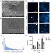
Puramatrix allows the adhesion of hASCs in a time-dependent manner. Characterization of Puramatrix™ topography using scanning electron microscopy: (A,B) view of the Puramatrix™ fibrous network. (C) Pore size (Feret’s diameter) distribution (nm). (D) Representative images of hASCs adhered to TCPs or PURA after 20 min, 40 min, and 2 h of incubation (DAPI was used to stain cell nuclei; scale bar = 1 mm). (E) Quantification of adhered cells after 20 min, 40 min, and 2 h of incubation. Each symbol (square, circle, and triangle) represents a different hASC donor. An unpaired student’s t-test was performed to compare the two treatment groups. ** p < 0.01.
Figure 4
Figure 4
Biocompatibility of the Puramatrix™ coating. A growth dynamics assay was performed via DNA extraction and quantification, while an LDH assay was performed to assess cytotoxicity (viability) and an EdU incorporation assay was performed to evaluate cell proliferation. (A) Growth dynamics of cells in TCPs and PURA after 1, 5, and 20 days in cell culture. (B) Percentage of cytotoxicity in TCPs and PURA after 5 days in cell culture. (C) Representative images of the EdU (red labeling) incorporation assay after 5 days in cell culture (scale bar = 100 μm). (D) Quantitative analysis of the percentage of EdU+ cells. Each symbol (square, circle, and triangle) represents a different hASC donor. An unpaired student’s t-test was performed to compare the two treatment groups.
Figure 5
Figure 5
Characterization of cell morphology and cell aggregate formation on Puramatrix™. Cell morphology was characterized by β-Tubulin immunostaining (green labeling). (A) Representative images of cells in TCPs and PURA (scale bar = 100 μm). (B) Quantitative analysis of the area of cell spreading in TCPs and PURA. Each symbol (square, circle, and triangle) represents a different hASC donor. (C) Representative images of cells cultured in PURA for 1, 5, 10, 15, and 20 days showing aggregate formation throughout the culture period. Red arrows indicate the cell aggregates. An unpaired t-test was used to compare the two treatment groups. ***: p < 0.005.
Figure 6
Figure 6
Ultrastructural characterization of cell morphology on Puramatrix™. (A) Cells adhered to PURA after 5 days in cell culture. (A.1) Cell aggregates interacting with the Puramatrix™ coating. (A.2) Cell cytoplasmic protrusions. (B) Cells adhered to PURA after 10 days in cell culture. (B.1) Cell aggregates stacked on top of each other. (B.2) Cell aggregates interacting with each other and with the Puramatrix™ substrate.
Figure 7
Figure 7
Chondrogenic differentiation of hASCs in Puramatrix™. (A) Safranin O staining (in Red) of hASCs induced (Chondro) and not induced (Control) to undergo chondrogenic differentiation. (B) Quantification of GAG deposition after chondrogenic induction. (C) mRNA expression levels of COL1, SOX9, COL2, ACAN, COL10, and MMP13 after chondrogenic induction. Data are represented as mean ± SD and were compared to the non-induced group (Control). Each symbol (square, circle, and triangle) represents a different hASC donor. One-way ANOVA was performed to compare the treatment groups. * p < 0.05.

References

    1. da Cunha Cavalcanti F.M.M., Doca D., Cohen M., Ferretti M. Updating on Diagnosis and Treatment of Chondral Lesion of the Knee. Rev. Bras. Ortop. Engl. Ed. 2012;47:12–20. doi: 10.1016/S2255-4971(15)30339-6. - DOI - PMC - PubMed
    1. Curl W.W., Krome J., Gordon E.S., Rushing J., Smith B.P., Poehling G.G. Cartilage Injuries: A Review of 31,516 Knee Arthroscopies. Arthroscopy. 1997;13:456–460. doi: 10.1016/S0749-8063(97)90124-9. - DOI - PubMed
    1. Liu M., Zeng X., Ma C., Yi H., Ali Z., Mou X., Li S., Deng Y., He N. Injectable Hydrogels for Cartilage and Bone Tissue Engineering. Bone Res. 2017;5:17014. doi: 10.1038/boneres.2017.14. - DOI - PMC - PubMed
    1. Marlovits S., Zeller P., Singer P., Resinger C., Vécsei V. Cartilage Repair: Generations of Autologous Chondrocyte Transplantation. Eur. J. Radiol. 2006;57:24–31. doi: 10.1016/j.ejrad.2005.08.009. - DOI - PubMed
    1. Vinatier C., Mrugala D., Jorgensen C., Guicheux J., Noël D. Cartilage Engineering: A Crucial Combination of Cells, Biomaterials and Biofactors. Trends Biotechnol. 2009;27:307–314. doi: 10.1016/j.tibtech.2009.02.005. - DOI - PubMed

LinkOut - more resources