. 2023 Dec 6;15(12):2733.
doi: 10.3390/pharmaceutics15122733.

Protocol to Induce the Temporary Opening of the Blood-Brain Barrier with Short-Time Focused Ultrasound in Rats

Affiliations

Protocol to Induce the Temporary Opening of the Blood-Brain Barrier with Short-Time Focused Ultrasound in Rats

Jorge A Rodríguez et al. Pharmaceutics. .

Abstract

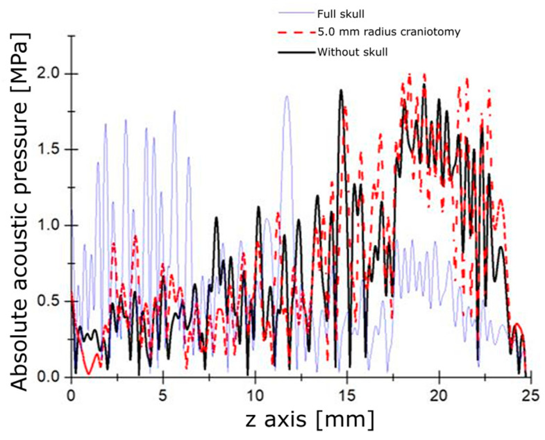
Brain neurodegenerative diseases are central nervous system (CNS) affections typically common in older adults. A new therapeutic approach for them consists of providing specific drugs to the CNS through blood circulation; however, the Blood-Brain Barrier (BBB) prevents almost 100% of neurotherapeutics from reaching the brain. There are indications that Focused Ultrasound (FUS), temporarily placed in the BBB, can achieve a controlled increase in temperature at its focus, allowing temporary, localized, and reversible opening of this barrier, which facilitates the temporary delivery of specific drugs. This work presents a FUS-based protocol for the local, temporary, and reversible opening of the BBB in Wistar rats. The proposed protocol specifies certain power, treatment times, and duty cycle to controllably increase the temperature at the region of interest, i.e., the substantia nigra. Numerical simulations using commercial software based on the finite element method were carried out to determine the optimal size of the craniotomies for nearly full-acoustic transmission. Experiments in rats were performed with the parameters used during computational simulations to determine the adequate opening of the BBB. For this, craniotomies of different sizes were made at coordinates of the substantia nigra, and FUS was applied from the exterior. The opening of the BBB was evaluated using Evans Blue (EB) as an indicator of the crossing of the dye from the blood vessels to brain tissue. Numerical simulations demonstrated a major distance reached by the ultrasound focus with a bigger diameter. Experimental results show the local, temporary, and reversible opening of the BBB through a 10 mm diameter craniotomy, which effectively allowed placing the ultrasound focus over the substantia nigra, unlike a 6 mm diameter craniotomy in which there is a deviation of the focus through that window. Moreover, from these results, it was also determined that the disruption of the BBB was reversible, with an opening duration of 6 h after FUS application. The experimental work developed in this study resulted in a minimally invasive method for the temporary opening of the BBB.

Keywords: Blood–Brain Barrier; craniotomy; focused ultrasound; reversible opening.


Conflict of interest statement

The authors declare no conflict of interest. The funders had no role in the design of the study; in the collection, analyses, or interpretation of data; in the writing of the manuscript; or in the decision to publish the results.

Figures

Figure 1
Figure 1
Experimental setup for FUS application in rats. Rats were anesthetized and placed on a stereotaxic system. The experimental setup comprises (a) the impedance coupling, (b) the FUS transducer, (c) the cone, (d) the Wistar male rat, (e) the power meter, (f) the power amplifier, (g) the digital oscilloscope, and (h) the signal generator.
Figure 2
Figure 2
Axisymmetric geometry used for obtaining the acoustic field. Boundary 1 represents the surface of the piezoelectric, and 2 is the transducer’s case. The red boundary is the symmetry axis, the blue contour represents an impedance boundary.
Figure 3
Figure 3
Modeled acoustic pressure. (Left) absolute acoustic field along the propagation axis. (Right) 2D acoustic field pattern.
Figure 4
Figure 4
Modeled heating produced by FUS. (A) temperature increase through the symmetry axis. (B) Heating pattern. (C) Complete brain and slices of rats showing the superficial opening of the BBB due to cranium heating (Scale bar, yellow line, is 5 mm).
Figure 5
Figure 5
Detailed graph of the acoustic field through 5 mm radius craniotomy.
Figure 6
Figure 6
Temperature increases for the analyzed cases.
Figure 7
Figure 7
Opening of the BBB using FUS with the transducer targeting the substantia nigra through a square craniotomy of 6 mm per side (equivalent to 3 mm radius in the models). Frame (A) corresponds to the craniotomy of 6 mm; a ruler is shown as a scale. Frame (B) is a brain extracted from the FUS-treated animal. Frames (CJ) correspond to serial cuts at the mesencephalon level in the rostral (C) to caudal (J) direction. It can be noticed that the dye location is only in the FUS-treated substantia nigra (Scale bar, yellow line, is 5 mm).
Figure 8
Figure 8
Opening of the BBB using FUS with the transducer targeting the substantia nigra through a square craniotomy of 10 mm per side (equivalent to 5 mm radius in the model). Frame (A) corresponds to the craniotomy of 10 mm; a ruler is shown as a scale. Frame (B) is a brain extracted from the FUS-treated animal. Frames (CJ) correspond to serial cuts at the mesencephalon level in the rostral (C) to caudal (J) direction. It can be noticed that the dye location is only in the FUS-treated substantia nigra (Scale bar, yellow line, is 5 mm).
Figure 9
Figure 9
Reversibility corroboration during the experiments of the opening of the BBB. Each column corresponds to different times of EB intravenous injection after FUS application. In the first row, frame (A) is the brain of an animal injected with EB at 6 h after FUS application, and the corresponding cuts are shown in frames (BD). In the second row, frame (E) of an animal injected with EB at 8 h after FUS application, and the corresponding cuts are shown in frames (FH). In the third row, frame (I) is the brain of an animal injected with EB at 12 h after FUS application, and the corresponding cuts are shown in frames (JL) (Scale bar, yellow line, is 5 mm).

References

    1. Fahn S., Sulzer D. Neurodegeneration and Neuroprotection in Parkinson Disease. NeuroRx. 2004;1:139–154. doi: 10.1602/neurorx.1.1.139. - DOI - PMC - PubMed
    1. Reeve A., Simcox E., Turnbull D. Ageing and Parkinson’s Disease: Why Is Advancing Age the Biggest Risk Factor? Ageing Res. Rev. 2014;14:19–30. doi: 10.1016/j.arr.2014.01.004. - DOI - PMC - PubMed
    1. Corti O., Hampe C., Darios F., Ibanez P., Ruberg M., Brice A. Parkinson’s Disease: From Causes to Mechanisms. Comptes Rendus-Biol. 2005;328:131–142. doi: 10.1016/j.crvi.2004.10.009. - DOI - PubMed
    1. Lee D., Dallapiazza R., De Vloo P., Lozano A. Current Surgical Treatments for Parkinson’s Disease and Potential Therapeutic Targets. Neural Regen. Res. 2018;13:1342–1345. doi: 10.4103/1673-5374.235220. - DOI - PMC - PubMed
    1. Martinez-Fong D., Bannon M.J., Trudeau L.E., Gonzalez-Barrios J.A., Arango-Rodriguez M.L., Hernandez-Chan N.G., Reyes-Corona D., Armendáriz-Borunda J., Navarro-Quiroga I. NTS-Polyplex: A Potential Nanocarrier for Neurotrophic Therapy of Parkinson’s Disease. Nanomed. Nanotechnol. Biol. Med. 2012;8:1052–1069. doi: 10.1016/j.nano.2012.02.009. - DOI - PMC - PubMed

LinkOut - more resources