. 2023 Dec 13;15(12):2772.
doi: 10.3390/pharmaceutics15122772.

IDF-11774 Induces Cell Cycle Arrest and Apoptosis by Inhibiting HIF-1α in Gastric Cancer

Affiliations

IDF-11774 Induces Cell Cycle Arrest and Apoptosis by Inhibiting HIF-1α in Gastric Cancer

Won-Ho Kim et al. Pharmaceutics. .

Abstract

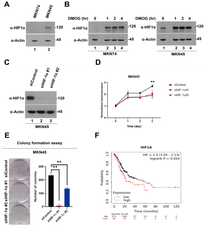
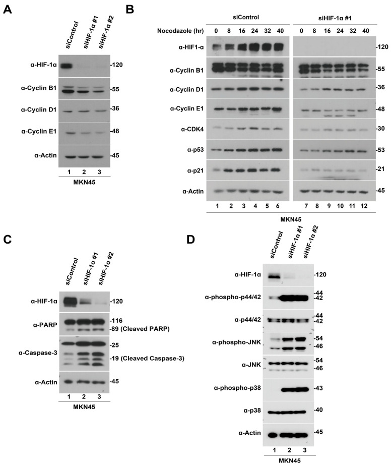
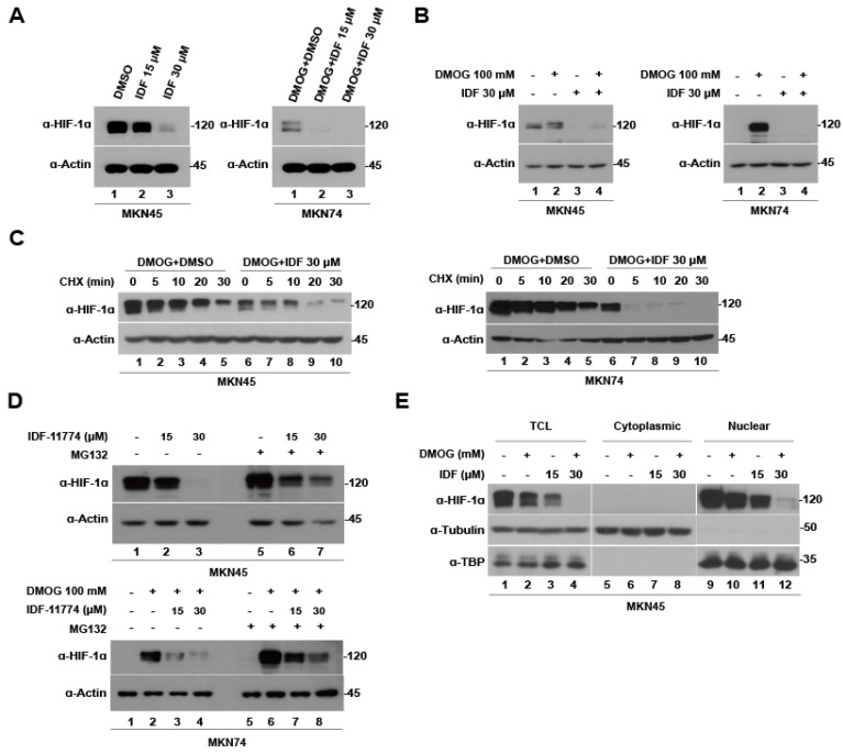
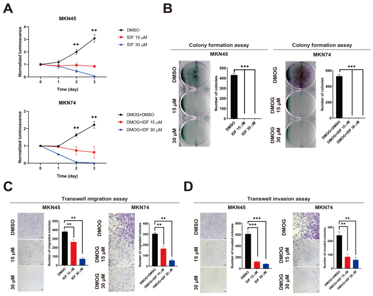
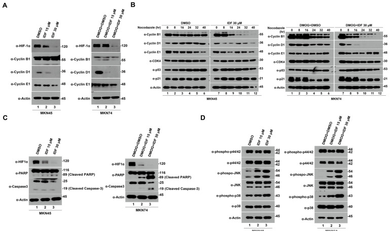
Hypoxia-inducible factor-1 alpha (HIF-1α) is a regulatory factor of intracellular oxygen supersession. The expression or increased activity of HIF-1α is closely related to various human cancers. Previously, IDF-11774 was demonstrated to inhibit HSP70 chaperone activity and suppress the accumulation of HIF-1α. In this study, we aimed to determine the effects of IDF-11774 on gastric cancer cell lines. Treatment with IDF-11774 was found to markedly decrease the proliferation, migration, and invasion of the gastric cancer cell lines. Furthermore, the phosphorylation levels of extracellular signal-regulated kinase 1/2, p38, and Jun N-terminal kinase in the mitogen-activated protein kinase signaling pathways were markedly increased in a dose-dependent manner, ultimately promoting apoptosis via the induction of cell cycle arrest. Our findings indicate that HIF-1α inhibitors are potent drugs for the treatment of gastric cancer.

Keywords: HIF-1α; HIF-1α inhibitor; IDF-11774; apoptosis; cell cycle arrest; gastric cancer.


Conflict of interest statement

The authors declare no conflict of interest.

Figures

Figure 1
Figure 1
Baseline expression of hypoxia-inducible factor-1 alpha (HIF-1α) in gastric cancer cells and HIF-1α knockdown-induced suppression of gastric cancer cell proliferation. (A) Immunoblotting analysis of HIF-1α expression under normoxia in MKN74 and MKN45 cells. (B) Immunoblotting analysis of HIF-1α expression in MKN74 and MKN45 cells after dimethyloxalylglycine treatment in the dose and time-dependent manners. (C) Immunoblotting analysis of HIF-1α expression in MKN45 cells transfected with small interfering ribonucleic acids (siRNAs) for 48 h. (D) Effects of HIF-1α knockdown using siRNAs on the viability of MKN45 cells (n = 3). Cell viability was monitored for 3 days using a CellTiter-Glo luminescent assay (normalized to control wild-type cells on day 0; ** p < 0.01). (E) Effects of HIF-1α knockdown using siRNAs on colony formation in MKN45 cells (n = 3). MKN45 cells transfected with HIF-1α siRNAs were cultured for 48 h and stained with crystal violet to visualize and determine the number of colonies (** p < 0.01). (F) Kaplan–Meier representation of the overall survival between the two groups of patients with high (red line) and low (black line) HIF-1α expression.
Figure 2
Figure 2
Hypoxia-inducible factor-1 alpha (HIF-1α) knockdown suppresses the growth of gastric cancer cells via cell cycle arrest, apoptosis, and the mitogen-activated protein kinase (MAPK) pathway. (A) MKN45 cells were transfected with HIF-1α siRNA for 48 h and then lysed and immunoblotted with HIF-1α, cell cycle arrest-associated proteins (Cyclin B1, D1, and E1), and β-actin. (B) MKN45 cells were transfected with HIF-1α siRNA for 48 h and nocodazole for 16 h, and then released into G1 for up to 40 h; nocodazole was replaced with fresh media. Samples were lysed and immunoblotted with HIF-1α, cell cycle arrest-associated proteins (Cyclin B1, D1, E1, Cyclin-dependent kinase 4, p53, and p21), and β-actin. (C) MKN45 cells were transfected with HIF-1α siRNA for 48 h and then lysed and immunoblotted with HIF-1α, apoptosis-associated proteins (poly(ADP ribose) polymerase, cleaved caspase-3), and β-actin. (D) MKN45 cells were transfected with HIF-1α siRNAs for 48 h and then lysed and immunoblotted with HIF-1α, MAPK pathway proteins (phospho-extracellular signal-regulated protein kinases (ERK), ERK, phosphor-Jun N-terminal kinase (JNK), JNK, phosphor-p38, and p38), and β-actin.
Figure 3
Figure 3
IDF-11774 promotes hypoxia-inducible factor-1 alpha (HIF-1α) degradation in gastric cancer cells. (A) MKN74 and MKN45 cells were treated with 15 μM and 30 μM IDF-11774 for 4 h and then lysed and immunoblotted with HIF-1α and β-actin. (B) After induction of hypoxia with dimethyloxalylglycine for 4 h, MKN74 and MKN45 cells were treated with 15 μM and 30 μM IDF-11774 for 4 h, and then lysed and immunoblotted with HIF-1α and β-actin. (C) MKN74 and MKN45 cells were treated with DMSO or 30 μM IDF-11774 for 4 h. Thereafter, de novo protein synthesis was inhibited using the cycloheximide treatment. Lysis and immunoblotting with HIF-1α and β-actin were performed 0, 5, 10, 20, and 30 min after cycloheximide treatment. (D) MKN74 and MKN45 cells were treated with DMSO, 15 μM or 30 μM IDF-11774 for 4 h, followed by inhibition of the proteasome using MG132. Lysis and immunoblotting with HIF-1α were subsequently performed. (E) Fractionation assay with total cell lysate, nuclear and cytoplasmic HIF-1α in MKN74 and MKN45 cells after treatment with 30 μM IDF-11774 for 4 h.
Figure 4
Figure 4
IDF-11774 inhibits the proliferation, migration, and invasion of gastric cancer cells. (A) Effect of IDF-11774 (15 and 30 μM) on the viability of MKN45 and MKN74 cells (n = 3). Cell viability was monitored for 3 days using a CellTiter-Glo luminescent assay (normalized to control wild-type cells at day 0; ** p < 0.01). (B) Effects of IDF-11774 (15 and 30 μM) on colony formation in MKN45 and MKN74 cells (n = 3). MKN45 and MKN74 cells transfected with hypoxia-inducible factor-1 alpha (HIF-1α) short interfering ribonucleic acids were cultured for 48 h and stained with crystal violet to visualize and determine the number of colonies (*** p < 0.001). (C) Effects of IDF-11774 (15 and 30 μM) on the migration of MKN45 and MKN74 cells. Cell migration was estimated using a Transwell migration assay, and data are presented as the mean ± standard deviation of the number of migrated cells (n = 5, (** p < 0.01). (D) Effects of IDF-11774 (15 and 30 μM) on the invasion of MKN45 and MKN74 cells. Cell invasion was estimated using a Transwell migration assay, and the data are presented as the mean ± standard deviation of the number of migrated cells (n = 5, (** p < 0.01).
Figure 5
Figure 5
IDF-11774 induces cell cycle arrest and apoptosis by downregulating hypoxia-inducible factor-1 alpha (HIF-1α) in gastric cancer cells. (A) MKN45 and MKN74 cells were treated with 30 μM IDF-11774 for 4 h and then lysed and immunoblotted with HIF-1α, cell cycle arrest-associated proteins (Cyclin B1, D1, and E1), and β-actin. (B) MKN45 and MKN74 cells were treated with 30 μM IDF-11774 for 4 h and nocodazole for 16 h. Thereafter, the cells were released into G1 for up to 40 h; nocodazole was replaced with fresh media. Samples were lysed and immunoblotted with HIF-1α, cell cycle arrest-associated proteins (Cyclin B1, D1, E1, cyclin-dependent kinase 4, p53, and p21), and β-actin. (C) MKN45 and MKN74 cells were treated with 30 μM IDF-11774 for 4 h and then lysed and immunoblotted with HIF-1α, apoptosis-associated proteins (poly(ADP ribose) polymerase, cleaved caspase-3), and β-actin. (D) MKN45 and MKN74 cells were treated with 30 μM IDF-11774 for 4 h and then lysed and immunoblotted with HIF-1α, mitogen-activated protein kinase pathway proteins (phospho-extracellular signal-regulated protein kinases (ERK), ERK, phosphor-Jun N-terminal kinase (JNK), JNK, phosphor-p38, and p38), and β-actin.
Figure 6
Figure 6
Graphical overview of the effect of IDF-11774 on the gastric cancer cell lines, MKN45 and MKN74. IDF-11774 mediates the degradation of hypoxia-inducible factor-1 alpha, arresting cell cycle progression and increasing mitogen-activated protein kinase activation, ultimately causing apoptosis.

Similar articles

Cited by

References

    1. Sung H., Ferlay J., Siegel R.L., Laversanne M., Soerjomataram I., Jemal A., Bray F. Global Cancer Statistics 2020: GLOBOCAN Estimates of Incidence and Mortality Worldwide for 36 Cancers in 185 Countries. CA Cancer J. Clin. 2021;71:209–249. doi: 10.3322/caac.21660. - DOI - PubMed
    1. Dulak A.M., Schumacher S.E., van Lieshout J., Imamura Y., Fox C., Shim B., Ramos A.H., Saksena G., Baca S.C., Baselga J., et al. Gastrointestinal adenocarcinomas of the esophagus, stomach, and colon exhibit distinct patterns of genome instability and oncogenesis. Cancer Res. 2012;72:4383–4393. doi: 10.1158/0008-5472.CAN-11-3893. - DOI - PMC - PubMed
    1. Deng N., Goh L.K., Wang H., Das K., Tao J., Tan I.B., Zhang S., Lee M., Wu J., Lim K.H., et al. A comprehensive survey of genomic alterations in gastric cancer reveals systematic patterns of molecular exclusivity and co-occurrence among distinct therapeutic targets. Gut. 2012;61:673–684. doi: 10.1136/gutjnl-2011-301839. - DOI - PMC - PubMed
    1. Viale P.H. The American Cancer Society’s Facts & Figures: 2020 Edition. J. Adv. Pract. Oncol. 2020;11:135–136. doi: 10.6004/jadpro.2020.11.2.1. - DOI - PMC - PubMed
    1. Li T., He Y., Zhong Q., Yu J., Chen X. Advances in Treatment Models of Advanced Gastric Cancer. Technol. Cancer Res. Treat. 2022;21:15330338221090353. doi: 10.1177/15330338221090353. - DOI - PMC - PubMed