. 2023 Dec 2;11(12):1805.
doi: 10.3390/vaccines11121805.

Prophylactic Vaccination and Intratumoral Boost with HER2-Expressing Oncolytic Herpes Simplex Virus Induces Robust and Persistent Immune Response against HER2-Positive Tumor Cells

Affiliations

Prophylactic Vaccination and Intratumoral Boost with HER2-Expressing Oncolytic Herpes Simplex Virus Induces Robust and Persistent Immune Response against HER2-Positive Tumor Cells

Zahid Delwar et al. Vaccines (Basel). .

Abstract

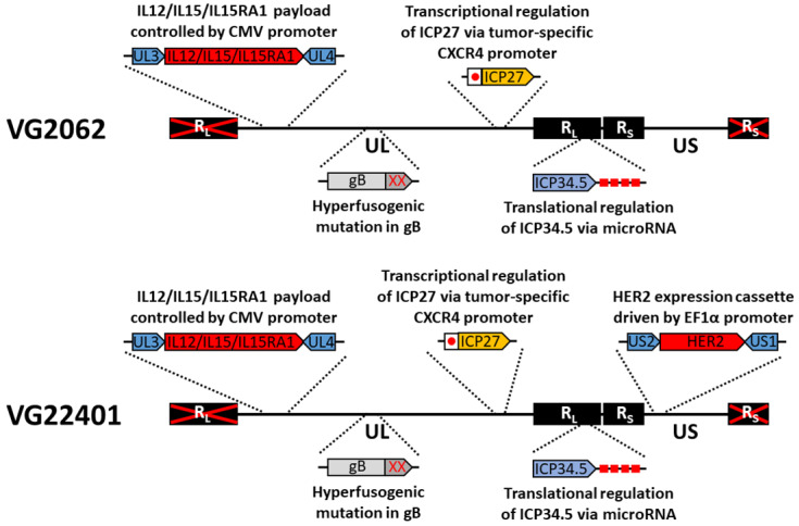
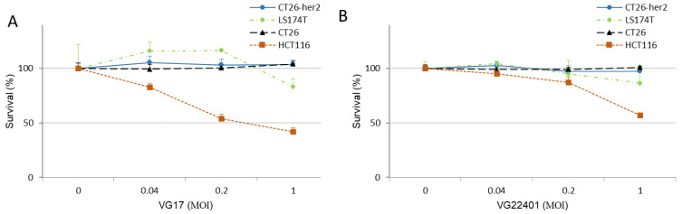
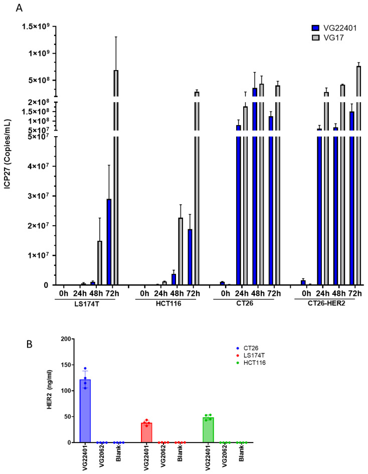
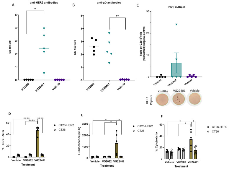
The development of effective cancer vaccines remains a significant challenge due to immune tolerance and limited clinical benefits. Oncolytic herpes simplex virus type 1 (oHSV-1) has shown promise as a cancer therapy, but efficacy is often limited in advanced cancers. In this study, we constructed and characterized a novel oHSV-1 virus (VG22401) expressing the human epidermal growth factor receptor 2 (HER2), a transmembrane glycoprotein overexpressed in many carcinomas. VG22401 exhibited efficient replication and HER2 payload expression in both human and mouse colorectal cancer cells. Mice immunized with VG22401 showed significant binding of serum anti-HER2 antibodies to HER2-expressing tumor cells, inducing antibody-dependent cell-mediated cytotoxicity (ADCC) and complement-dependent cytotoxicity (CDC). Furthermore, mice primed with VG22401 and intratumorally boosted with the same virus showed enhanced antitumor efficacy in a bilateral syngeneic HER2(+) tumor model, compared to HER2-null backbone virus. This effect was accompanied by the induction of anti-HER2 T cell responses. Our findings suggest that peripheral priming with HER2-expressing oHSV-1 followed by an intratumoral boost with the same virus can significantly enhance antitumor immunity and efficacy, presenting a promising strategy for cancer immunotherapy.

Keywords: HER2; cancer vaccine; herpes simplex virus; oncolytic virus.


Conflict of interest statement

All authors are current employees of Virogin Biotech Canada Ltd., have an ownership interest (including stock, patents, etc.) in Virogin Biotech Canada Ltd. This study was wholly funded by Virogin Biotech Canada Ltd., and patent applications have been filed to cover the technologies detailed herein.

Figures

Figure 1
Figure 1
Schematic diagram of HER2-expressing virus.
Figure 2
Figure 2
Cytotoxic effect of VG22401 and VG17 in human and mouse cancer cells in vitro. Cell survival percentages were determined using a Cell Counting KIT 8 assay to evaluate the cytotoxic effects of VG17 (A), and VG22401 (B) viruses in human cancer cell monolayers (HCT116 and LS174T) and mouse cancer cell monolayers (CT26 and CT26-HER2) at 72 h after infection with MOIs of 0.04, 0.2, and 1.
Figure 3
Figure 3
Virus replication and payload expression in human and mouse colorectal cancer cells in vitro. (A) Cells were infected with VG22401 or VG17 viruses. LS174T and HCT116 human colon cancer cells were infected at MOI = 0.001; CT26 and CT26-HER2 murine colon cancer cells were infected at MOI = 3. All infections were carried out in duplicate. Total DNA was extracted from the samples at the indicated times post-infection. To determine viral yield, ICP27 gene copy numbers were quantified by qPCR. (B) To determine HER2 payload expression, CT26, LS174T, and HCT116 cells were infected with HER2-expressing VG22401, or with the backbone virus VG2062. LS174T and HCT116 human colon cancer cells were infected at MOI = 3; CT26 murine colon cancer cells were infected at MOI = 6. Supernatants were collected 48 h post-infection. All infections were carried out in duplicate. Concentration of HER2 in the supernatant of the infected cells was analyzed by ELISA.
Figure 4
Figure 4
Induction of humoral and cellular immune response by an HSV-1 expressing HER2. Humoral immune response to HER2 (A) or oHSV-1 (B) after vaccination with VG22401 (black dots) and VG2062 (green triangles) viruses was evaluated by ELISA. Serum isolated from mice 35 days post-vaccination was diluted in series and incubated on plates pretreated with recombinant HER2 protein or with HSV-1 envelope glycoprotein D (gD). Anti-HER2 and anti-gD IgG antibody levels were measured with HRP-conjugated goat anti-mouse IgG and TMB, and absorbance at 450 nm was quantified (optical density). Plots show median values. An unpaired t-test was performed, and two-tailed p-values < 0.05 (*) and p < 0.002 (**) are shown. (C) Cellular immune response was measured by IFNγ ELISPOT assay with splenocytes isolated from mice. ELISPOT bar graph shows mean frequencies of IFNγ spots induced by HER2 peptide mix stimulation per 2.5 × 10⁵ splenocytes. Error bars indicate SEM. The bottom panel shows representative wells with IFNγ spots after stimulation with HER2 peptide mix. (DF) Binding of serum anti-HER2 antibodies to HER2-expressing tumor cells. CT26-HER2 and HER2-negative parental CT26 cells were co-incubated with 1:100 dilution of serum followed by APC-conjugated, anti-mouse IgG antibody. The antibody binding to cell-surface-expressed HER2 was measured by flow cytometry (*, p < 0.05; ****, p < 0.0001) (D). Antibody-dependent cell-mediated cytotoxicity (ADCC) was measured against CT26-HER2 and CT26 cells using mFcgRIV ADCC Reporter Bioassay. Target cells were seeded, incubated at 37 °C overnight, and pre-incubated with 1:500 dilution of serum for 15 min at room temperature. Effector cells were subsequently applied per the manufacturer’s instructions. Effector to target ratio = 10:1 was tested. After 6 h of co-incubation, Bio-Glo™ Reagent was added, and luminescence was measured (*, p < 0.05) (E). Complement-dependent cytotoxicity (CDC) was assessed against CT26-HER2 and CT26 cells. Target cells were pre-incubated with 1:100 dilution of serum for 1 h at room temperature, and rabbit complement (1:100 dilution) was subsequently added for 2.5 h. Lactate dehydrogenase (LDH) released in the culture medium was measured and the percentage cytolysis was reported (*, p < 0.05) (F).
Figure 5
Figure 5
HER2 payload improves the antitumor effect of oHSV-1 and induces a T cell response in a HER2-vaccinated bilateral, syngeneic murine tumor model. Immunocompetent BALB/c mice were randomized into three groups and primed/boosted with VG2062 virus, VG22401 virus, or vehicle control. The mice were subsequently inoculated with CT26-HER2 tumors on both the left and right flanks. Once a tumor bump was visible, the tumors located on the right flank were injected intratumorally with a single dose of the same viruses used for immunization (1 × 10⁷ PFU/mouse), or with vehicle control. Tumor volumes were measured using a caliper, and the tumor growth curve illustrates the regression of the injected and abscopal tumors. The upper panel illustrates the tumor volume of individual animals, while the lower panel displays the average tumor volume (n = 10) for each group (A). The Kaplan–Meier survival curve was constructed using the time it took for mice tumors to grow to 1500 mm3 after intratumoral treatment (B), while the body weight change curve shows the post-treatment-changes in body weight (C). Cellular immune response was measured by IFNγ ELISPOT assay with splenocytes isolated from mice prime/boosted with VG2062 virus (black dots) and VG22401 virus (green triangles) when tumor size reached 1500 mm3. ELISPOT bar graph shows mean frequencies of IFNγ spots induced by HER2 peptide mix stimulation per 2.5 × 10⁵ splenocytes. Error bars indicate SEM. Ordinary two-way ANOVA, with Dunn’s multiple comparisons test, was performed to analyze the ELISPOT results. Two-tailed p-values are shown. The bottom panel shows representative wells with IFNγ spots after HER2 peptide mix stimulation (D). Asterisks (*) represent p values below 0.05.

Similar articles

Cited by

References

    1. Hemminki O., dos Santos J.M., Hemminki A. Oncolytic viruses for cancer immunotherapy. J. Hematol. Oncol. 2020;13:84. doi: 10.1186/s13045-020-00922-1. - DOI - PMC - PubMed
    1. Varghese S., Rabkin S.D. Oncolytic herpes simplex virus vectors for cancer virotherapy. Cancer Gene Ther. 2002;9:967–978. doi: 10.1038/sj.cgt.7700537. - DOI - PubMed
    1. Ning J., Wakimoto H. Oncolytic herpes simplex virus-based strategies: Toward a breakthrough in glioblastoma therapy. Front. Microbiol. 2014;5:303. doi: 10.3389/fmicb.2014.00303. - DOI - PMC - PubMed
    1. Ferrucci P.F., Pala L., Conforti F., Cocorocchio E. Talimogene Laherparepvec (T-VEC): An Intralesional Cancer Immunotherapy for Advanced Melanoma. Cancers. 2021;13:1383. doi: 10.3390/cancers13061383. - DOI - PMC - PubMed
    1. Martuza R.L., Malick A., Markert J.M., Ruffner K.L., Coen D.M. Experimental therapy of human glioma by means of a genetically engineered virus mutant. Science. 1991;252:854–856. doi: 10.1126/science.1851332. - DOI - PubMed