. 2023 Nov 30;15(12):2356.
doi: 10.3390/v15122356.

Identification of Adenovirus E1B-55K Interaction Partners through a Common Binding Motif

Affiliations

Identification of Adenovirus E1B-55K Interaction Partners through a Common Binding Motif

Nafiseh Chalabi Hagkarim et al. Viruses. .

Abstract

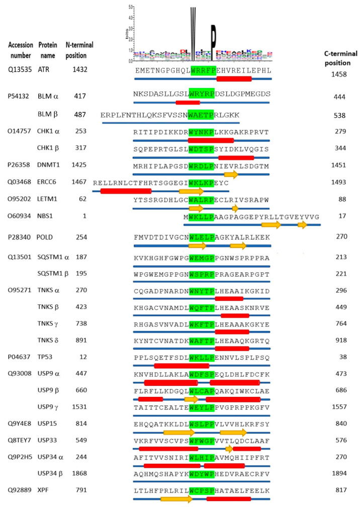
The adenovirus C5 E1B-55K protein is crucial for viral replication and is expressed early during infection. It can interact with E4orf6 to form a complex that functions as a ubiquitin E3 ligase. This complex targets specific cellular proteins and marks them for ubiquitination and, predominantly, subsequent proteasomal degradation. E1B-55K interacts with various proteins, with p53 being the most extensively studied, although identifying binding sites has been challenging. To explain the diverse range of proteins associated with E1B-55K, we hypothesized that other binding partners might recognize the simple p53 binding motif (xWxxxPx). In silico analyses showed that many known E1B-55K binding proteins possess this amino acid sequence; therefore, we investigated whether other xWxxxPx-containing proteins also bind to E1B-55K. Our findings revealed that many cellular proteins, including ATR, CHK1, USP9, and USP34, co-immunoprecipitate with E1B-55K. During adenovirus infection, several well-characterized E1B-55K binding proteins and newly identified interactors, including CSB, CHK1, and USP9, are degraded in a cullin-dependent manner. Notably, certain binding proteins, such as ATR and USP34, remain undegraded during infection. Structural predictions indicate no conservation of structure around the proposed binding motif, suggesting that the interaction relies on the correct arrangement of tryptophan and proline residues.

Keywords: MRE11; PR619; USP; adenovirus; cullin; early region 1B; p53; p53 binding motif.


Conflict of interest statement

The authors declare no conflict of interest.

Figures

Figure 1
Figure 1
The interaction of HAdV-C5 E1B-55K with novel cellular binding partners. Lysates from HEK293 cells were immunoprecipitated with antibodies (either mouse or rabbit as appropriate) against the HAdV-C5 E1B-55K protein. After immunoblotting, interacting proteins were detected with the antibodies shown. Known binding proteins such as NBS1, MRE11, and hnRNPUL1 are included for comparison (left column). “Control” is an irrelevant antibody included as a negative control, raised against either collagen type IV (rabbit) or vimentin (mouse). Images represent the results of three repeated experiments. WCL, whole cell lysate.
Figure 2
Figure 2
The interaction of adenovirus E1B-55K proteins with the xWxxxPx motif. (A) HEK293 cell lysates were mock incubated or incubated with either biotin-linked ETFSDLWKLLPENNVLS peptide (WP peptide) or ETFSDLAKLLAENNVLS peptide (AA peptide), (i) 1 µg/mL, (ii) 5 µg/mL and (iii) 20 µg/mL for 2 h. Streptavidin (SA) beads were added for a further 90 min. After washing bound proteins were released with SDS sample buffer and fractionated by PAGE. Bound HAdV-C5 E1B-55K was detected by immunoblotting. (B) HER2 cell lysate was incubated with WP peptide or AA peptide (10 µg/mL) as in (A). Bound HAdV-A12 E1B-55K was detected by immunoblotting. (C) HeLa cells were transfected with pcDNA3 constructs expressing HA-tagged E1B-55K proteins from Ads 4, 34, 40, and 9. After 48 h, cell lysates were incubated or mock incubated with WP peptide (10 µg/mL) for 2 h. After incubation with streptavidin beads samples were processed as in (A) and then immunoblotted for the HA tag. Images represent the results of three repeated experiments. WCL, whole cell lysate.
Figure 3
Figure 3
Degradation of novel binding partners during HAdV-C5 infection is dependent on cullin function. The left-hand blots display novel E1B-55K binding partners, while the right-hand blots serve as controls. HeLa cells were infected with HAdV-C5 at an infectivity of 4 pfu/cell, either in the presence or absence of 5 mM MLN4924. Cells were harvested at the times shown, post-infection, fractionated by PAGE, and immunoblotted with the antibodies as indicated. For the cullin 5 blots, * indicates the NEDDylated form. Images represent the results of three repeated experiments.
Figure 4
Figure 4
HAdV-C5 E1B-55K binds to MRE11 in the absence of NBS1. (A,B) U2OS or NBS1-negative cells were infected with H5pm4155 (E4orf3, E4orf6) at 10 pfu/cell for 48 h. Cells were harvested and immunoprecipitated with antibodies against either HAdV-C5 E1B-55K (A) or MRE11 (B). After PAGE and immunoblotting, co-immunoprecipitated MRE11 (A) or HAdV-C5 E1B-55K (B) was detected. Control, immunoprecipitated with an irrelevant antibody as in Figure 1. (C) Immunoblot showing no expression of NBS1 in NBS1 cells. (D) NBS1 cells were infected with HAdV-C5 (5 pfu/cell) and harvested at the times shown. Lysates were immunoblotted for MRE11, HAdV-C5 E1B-55K, and GAPDH as a loading control. Images represent the results of three repeated experiments. WCL, whole cell lysate.
Figure 5
Figure 5
The effect of the DUB inhibitor PR619 on adenovirus infection. HeLa cells were treated for 1 h with 20 µM PR619 and infected with (A) HAdV-C5 (5 pfu/cell) or (B) dl1520 (10 pfu/cell). Cultures were then incubated in the presence of 20 µM PR619 and harvested at the times shown. Lysates were subjected to immunoblotting as shown. Images represent the results of three repeated experiments. L4, L4-100K.
Figure 6
Figure 6
Structural prediction of xWxxxPx-containing motifs. The accession numbers of candidate proteins were obtained from the UniProtKB databank. Proteins containing xWxxxPx motifs (green background highlighted typeset) are listed alphabetically. The N- and C-terminal positions of each protein primary sequence are shown. Where a protein contains more than one xWxxxPx motif they are designated by Greek letters (α, β, γ, etc.). A weblogo cartoon of amino acids in the sequence of the proteins listed is shown at the top of the figure. Average predicted structural propensities for each amino acid in each protein are illustrated as either α-helix (red cylinder), β-strand (yellow arrow), or random coil (thin blue cylinder) below the primary sequence.

References

    1. Benko M., Aoki K., Arnberg N., Davison A.J., Echavarria M., Hess M., Jones M.S., Kajan G.L., Kajon A.E., Mittal S.K., et al. ICTV Virus Taxonomy Profile: Adenoviridae 2022. J. Gen. Virol. 2022;103:001721. doi: 10.1099/jgv.0.001721. - DOI - PMC - PubMed
    1. Khanal S., Ghimire P., Dhamoon A.S. The Repertoire of Adenovirus in Human Disease: The Innocuous to the Deadly. Biomedicines. 2018;6:30. doi: 10.3390/biomedicines6010030. - DOI - PMC - PubMed
    1. Lynch J.P., 3rd, Kajon A.E. Adenovirus: Epidemiology, Global Spread of Novel Serotypes, and Advances in Treatment and Prevention. Semin. Respir. Crit. Care Med. 2016;37:586–602. doi: 10.1055/s-0036-1584923. - DOI - PMC - PubMed
    1. Berk A.J. Recent lessons in gene expression, cell cycle control, and cell biology from adenovirus. Oncogene. 2005;24:7673–7685. doi: 10.1038/sj.onc.1209040. - DOI - PubMed
    1. Ip W.H., Dobner T. Cell transformation by the adenovirus oncogenes E1 and E4. FEBS Lett. 2020;594:1848–1860. doi: 10.1002/1873-3468.13717. - DOI - PubMed

MeSH terms

Substances