Review
. 2023 Dec 12;15(12):2413.
doi: 10.3390/v15122413.

Picornavirus 3C Proteins Intervene in Host Cell Processes through Proteolysis and Interactions with RNA

Affiliations
Review

Picornavirus 3C Proteins Intervene in Host Cell Processes through Proteolysis and Interactions with RNA

Somnath Mondal et al. Viruses. .

Abstract

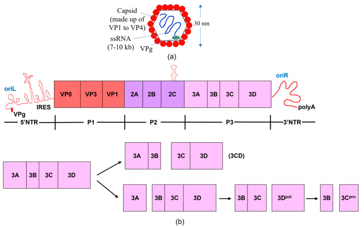
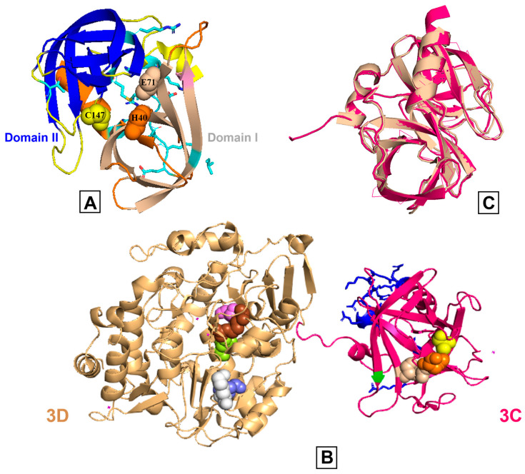
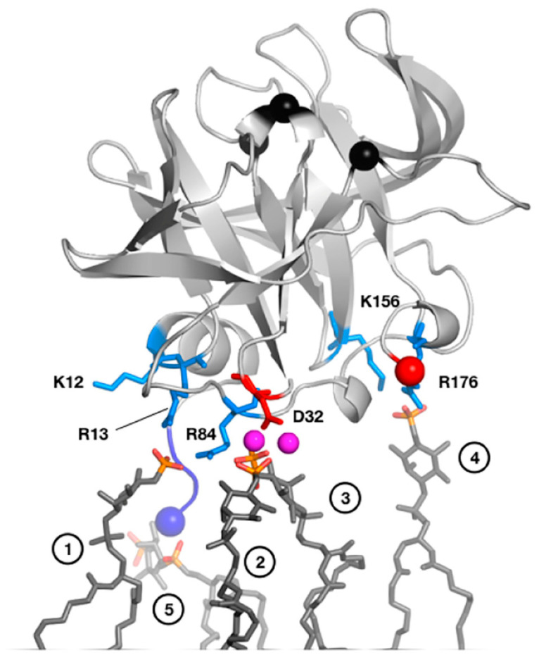
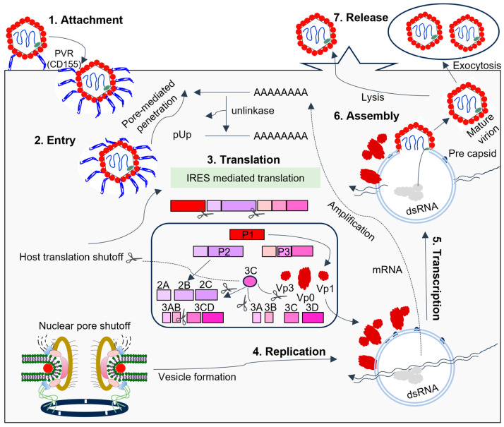
The Picornaviridae family comprises a large group of non-enveloped viruses with enormous impact on human and animal health. The picornaviral genome contains one open reading frame encoding a single polyprotein that can be processed by viral proteases. The picornaviral 3C proteases share similar three-dimensional structures and play a significant role in the viral life cycle and virus-host interactions. Picornaviral 3C proteins also have conserved RNA-binding activities that contribute to the assembly of the viral RNA replication complex. The 3C protease is important for regulating the host cell response through the cleavage of critical host cell proteins, acting to selectively 'hijack' host factors involved in gene expression, promoting picornavirus replication, and inactivating key factors in innate immunity signaling pathways. The protease and RNA-binding activities of 3C are involved in viral polyprotein processing and the initiation of viral RNA synthesis. Most importantly, 3C modifies critical molecules in host organelles and maintains virus infection by subtly subverting host cell death through the blocking of transcription, translation, and nucleocytoplasmic trafficking to modulate cell physiology for viral replication. Here, we discuss the molecular mechanisms through which 3C mediates physiological processes involved in promoting virus infection, replication, and release.

Keywords: 3C protease; host cell defense; picornavirus; protein dynamics; protein structure; virus replication.


Conflict of interest statement

The authors declare no conflict of interest.

Figures

Figure 1
Figure 1
The genome and proteome of poliovirus. (a) Structure of poliovirus. (b) The processing of polyproteins and the poliovirus genome are shown schematically. The poly (A) tail, broad open reading frame, and 3′ and 5′ non-translated regions (NTR) comprise the poliovirus genome. The type II internal ribosome entry site (IRES) and a cloverleaf structure comprise the 5′NTR. The virus-encoded 3B (VPg) protein is attached to the 5′ end of the RNA. The viral genome encodes a single polyprotein; the P1 region encodes the structural proteins of the virus, while the P2 and P3 portions encode the non-structural proteins. The P3 region is cleaved by viral proteases via two main pathways, resulting in the production of the 3C and 3D proteins, either separately or collectively as 3CD. The RNA-dependent RNA polymerase 3D replicates the viral RNA, while 3C, a protease, cleaves at specific, conserved motifs located within flexible linkers that separate discrete proteins within the viral polyprotein. Because 3CD lacks polymerase activity and has a unique protease specificity that enables it to cleave the P1 capsid region differently than 3C, the functional activity changes when the 3C and 3D proteins are combined to produce 3CD [27,28,29,30].
Figure 2
Figure 2
Comparison of poliovirus 3C and 3CD proteins. (A) The poliovirus 3C protease X-ray crystal structure (PDB: 1L1N) is shown, and the β-barrels of domains I and II are represented in wheat and blue colors, respectively. The catalytic triad (His40 (orange), Glu71 (light wheat), Cys147 (yellow)) is shown in spheres and the substrate pockets are in different colors. Side chains are colored by heteroatoms and the RNA binding region (E81-H89) is highlighted. (B) The X-ray crystal structure of poliovirus 3CD (PDB: 2IJD); the wheat and pink colors represent 3D and 3C, respectively. The active site residues are shown on both subdomains using spheres (3C: His40 (orange), Glu71 (light wheat), Cys147 (yellow); 3D, starting at the first residue of the 3D domain: Arg174 (brown), Asp233 (slate), Ser288 (violet), Asn297 (lemon), Lys359 (white)). The RNA binding region of 3C is colored blue. (C) The 3C region of 3C (pink) and 3CD (wheat) are structurally comparable as evidenced by an overlay with an RMSD value of 0.46 angstroms.
Figure 3
Figure 3
MD-derived model of PV 3C interactions with a PI4P-containing lipid membrane. Residues involved in interactions include Arg13 in the N-terminal alpha helix and Arg84 in the RNA-binding region. NMR studies with short-chain, soluble PI4P lipids largely confirm this model. In this model, PV 3C interacts with five clustered PI4P lipid molecules as shown in dark grey sticks with phosphates colored orange (phosphorus) and red (oxygen). The following specific interactions are listed, with numbers in parenthesis denoting the panel’s head group: (1) R13 and R84; (2) D32, mediated by sodium ions; (3) D32 mediated by sodium; (4) K156 and R176; and (5) α-amino group of G1. Reprinted from Structure, 25(12), D. Shengjuler, Y.M. Chan, S. Sun, I. Moustafa, Z.-L. Li, D.W. Gohara, M. Buck, P.S. Cremer, D.D. Boehr, C.E. Cameron. The RNA-binding site of poliovirus 3C protein doubles as a phosphoinositide-binding domain, 1875–1886, Copyright (2017) with permission from Elsevier.
Figure 4
Figure 4
The picornavirus life cycle involves 3C interfering with host components to facilitate RNA replication. Endocytosis, promoted by the attachment of the virus to host receptors, allows the virus to enter the host cell. In PV, the receptor is CD155. Viral proteases cleave translation initiation factors, halting cellular cap-dependent translation. Replication occurs in endoplasmic reticulum-derived membrane vesicles in viral factories, using genomic single-stranded positive-strand RNA (ssRNA(+)) to create a double-stranded RNA intermediate. Transcription and replication produce viral mRNAs and new ssRNA(+) genomes. The packaging of genomic RNA into prepared procapsids leads to cell death and virus release. During SVV infection, the virion first attaches itself to the host cell ANXTR1 receptor before internalizing itself through an endocytic pathway [92,93]. Unlike other picornaviruses, SVV has a distinct uncoating mechanism and binding mode with its receptor.
Figure 5
Figure 5
Translation initiation can be (a) cap-dependent and/or (b) IRES-dependent. Using the color code, only the primary factors mentioned in the text have been depicted for simplicity. Cellular mRNAs functionally pseudo-circularize as a result of the interaction between eIF4G and PABP. Regarding the FMDV genome, the 5′-3′ long-range communication among the 3′-UTR with the S fragment (a large stem-loop formed by the folding of the terminal structure at the 5′ end), in addition to 3′ UTR-IRES interaction, permits a comparable circumstance that is probably stabilized by binding factors for RNA. IRES-driven translational initiation requires a few eIFs and IRES-transacting factors (ITAFs), i.e., PCBP2, PTB, and Germin 5 which modulate IRES activity. 3C is crucial because it guarantees the internal initiation of translation by cleaving certain eIFs and ITAFs.
Figure 6
Figure 6
Chemical structure of various 3C protease inhibitors.

Similar articles

Cited by

References

    1. Yi J., Peng J., Yang W., Zhu G., Ren J., Li D., Zheng H. Picornavirus 3C—A Protease Ensuring Virus Replication and Subverting Host Responses. J. Cell Sci. 2021;134:jcs253237. doi: 10.1242/jcs.253237. - DOI - PubMed
    1. Ng C.S., Stobart C.C., Luo H. Innate Immune Evasion Mediated by Picornaviral 3C Protease: Possible Lessons for Coronaviral 3C-like Protease? Rev. Med. Virol. 2021;31:1–22. doi: 10.1002/rmv.2206. - DOI - PMC - PubMed
    1. Sun D., Chen S., Cheng A., Wang M. Roles of the Picornaviral 3C Proteinase in the Viral Life Cycle and Host Cells. Viruses. 2016;8:82. doi: 10.3390/v8030082. - DOI - PMC - PubMed
    1. Minor P.D. Poliovirus Biology. Structure. 1996;4:775–778. doi: 10.1016/S0969-2126(96)00084-6. - DOI - PubMed
    1. Weidman M.K., Yalamanchili P., Ng B., Tsai W., Dasgupta A. Poliovirus 3C Protease-Mediated Degradation of Transcriptional Activator P53 Requires a Cellular Activity. Virology. 2001;291:260–271. doi: 10.1006/viro.2001.1215. - DOI - PubMed