Review
. 2024 Apr;271(4):1947-1958.
doi: 10.1007/s00415-023-12134-x. Epub 2023 Dec 23.

Immune checkpoint inhibitor-related myositis and myocarditis: diagnostic pitfalls and imaging contribution in a real-world, institutional case series

Affiliations
Review

Immune checkpoint inhibitor-related myositis and myocarditis: diagnostic pitfalls and imaging contribution in a real-world, institutional case series

Alex Vicino et al. J Neurol. 2024 Apr.

Abstract

Background: Immune checkpoint inhibitors (ICIs) are reshaping the prognosis of many cancers, but often cause immune-related adverse events (irAEs). Among neurological irAEs, myositis is the most frequently reported. Our aim is to describe clinical and non-clinical characteristics, treatment and outcome of all irMyositis (skeletal limb-girdle and/or ocular myositis) and irMyocarditis cases in our reference center.

Methods: We retrospectively enrolled all irMyositis/irMyocarditis patients seen between 2018 and 2022. We reviewed demographics, clinical characteristics, biological, neurophysiological, imaging workup, treatment and outcome.

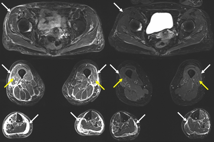
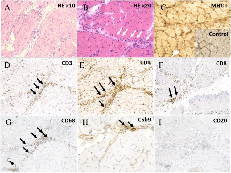
Results: We included 14 consecutive patients. The most frequent treatments were pembrolizumab (35%) or ipilimumab-nivolumab combination (35%). Limb-girdle, ocular (non-fluctuating palpebral ptosis and/or diplopia with or without ophthalmoparesis) and cardiac phenotypes were equally distributed, overlapping in 40% of cases. Ocular involvement was frequently misdiagnosed; review of brain MRIs disclosed initially missed signs of skeletal myositis in one patient and ocular myositis in 3. Seven patients had other co-existing irAEs. When performed, myography showed a myogenic pattern. CK was elevated in 8/15 patients, troponin-T in 12/12 and troponin-I in 7/9 tested patients. ICI were discontinued in all cases, with further immunosuppressive treatment in nine patients. In most cases, neurological and cardiological outcome was good at last follow-up.

Conclusion: Myositis is a potentially severe irAE. Despite its heterogeneous presentation, some highly suggestive clinical symptoms, such as ocular involvement, or radiological signs should raise physicians' attention to avoid misdiagnosis. We thus recommend a multidisciplinary assessment (including complete neuromuscular evaluation) even in case of isolated myocarditis. Our series underlines the importance of an early diagnosis, since suspension of ICI and adequate treatment are usually associated with good functional outcome.

Keywords: Imaging; Immune-checkpoint-inhibitor; Immune-related-adverse-event; Myocarditis; Myositis.


Conflict of interest statement

The authors declare that they have no conflict of interest.

Figures

Fig. 1
Fig. 1
Muscle MRI, patient 2, at time of symptoms (left) and control after 5 months (right). Dixon “water” T2 weighted images, showing symmetrical (with slight right predominance) signs of myositis in vastus lateralis muscles (yellow arrows), and fasciitis (white arrows), predominant in the legs (yellow arrows)
Fig. 2
Fig. 2
Brain MRI, patients 3 (axial view) and 1 (coronal view). Signs of orbital inflammation. In T2 fat-suppression weighted sequence (A, C), T2-hypersignal in oculomotor muscle (orange arrowheads) and intraorbital fat (orange arrow). In T1 SPACE 3D fat-saturation weighted sequence (B, D), presence of muscle (green arrowhead) and intraorbital fat (green arrow) enhancement after gadolinium injection
Fig. 3
Fig. 3
Signs of skeletal myositis on [68Ga]-DOTATOC PET/CT scan. Pathological uptake of the trapezius muscle (red arrows) (SUV scale 0–2); A Maximum intensity projection imaging. B Sagittal PET image, C Sagittal CT image and D Sagittal fused PET/CT image
Fig. 4
Fig. 4
Cardiac MRI, patient 4. Late gadolinium enhancement images obtained with cardiac magnetic resonance in basal and mid-ventricular short axis orientation, and long-axis four-chamber view in a patient with ICI-induced myocarditis. Myocardial enhancement is detected in basal septal, basal inferolateral, mid anterolateral and apical segments with a sub-epicardial distribution (arrows)
Fig. 5
Fig. 5
Muscle biopsy, vastus lateralis muscle, patient 2. A, B hematoxylin and eosin (HE) stain of vastus lateralis muscle, showing abundant, diffuse inflammatory infiltrate predominant in the perimysial area (white arrows). C Overexpression of MHC1 in comparison to healthy control (box). Immunohistochemistry showing (D) T lymphocytic infiltrate (CD3) of both CD4 and CD8 subtypes (E, F), macrophage infiltrate (G) (CD68) (black arrows). H Presence of complement deposition (C5b9, black arrows) in perimysial capillaries. I Absence of B lymphocytes (CD20)

References

    1. Ribas A, Wolchok JD. Cancer immunotherapy using checkpoint blockade. Science. 1979;2018(359):1350–1355. doi: 10.1126/science.aar4060. - DOI - PMC - PubMed
    1. Fife BT, Pauken KE. The role of the PD-1 pathway in autoimmunity and peripheral tolerance. Ann N Y Acad Sci. 2011;1217:45–59. doi: 10.1111/j.1749-6632.2010.05919.x. - DOI - PubMed
    1. Hodi FS, O’Day SJ, McDermott DF, Weber RW, Sosman JA, Haanen JB, et al. Improved survival with ipilimumab in patients with metastatic melanoma. N Engl J Med. 2010;363:711–723. doi: 10.1056/NEJMoa1003466. - DOI - PMC - PubMed
    1. Hargadon KM, Johnson CE, Williams CJ. Immune checkpoint blockade therapy for cancer: an overview of FDA-approved immune checkpoint inhibitors. Int Immunopharmacol. 2018;62:29–39. doi: 10.1016/j.intimp.2018.06.001. - DOI - PubMed
    1. Postow MA, Sidlow R, Hellmann MD. Immune-related adverse events associated with immune checkpoint blockade. N Engl J Med. 2018;378:158–168. doi: 10.1056/NEJMra1703481. - DOI - PubMed

MeSH terms

Substances