. 2024 Jul 8;52(12):6964-6976.
doi: 10.1093/nar/gkad1222.

The BRCA2 R2645G variant increases DNA binding and induces hyper-recombination

Affiliations

The BRCA2 R2645G variant increases DNA binding and induces hyper-recombination

Lucia Alvaro-Aranda et al. Nucleic Acids Res. .

Erratum in

Abstract

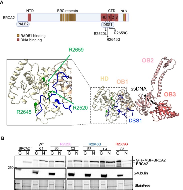
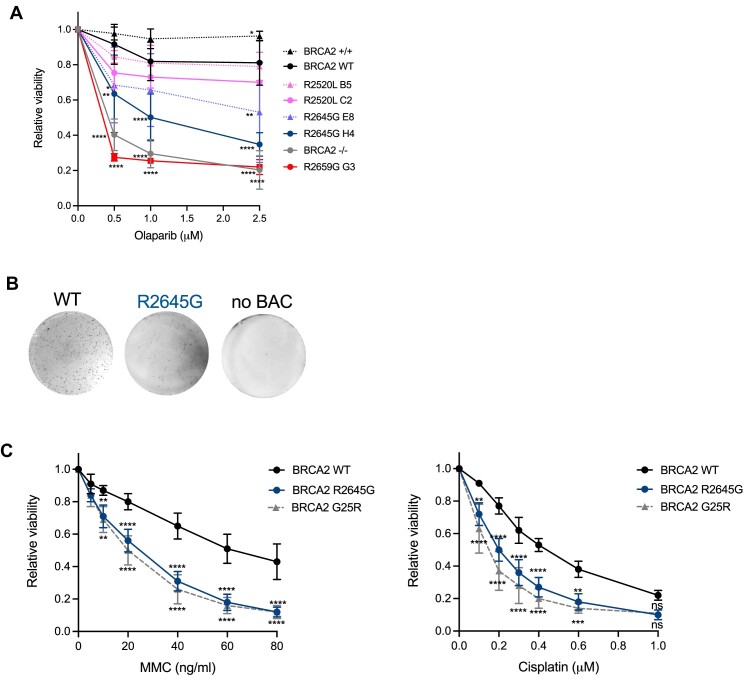
BRCA2 tumor suppressor protein ensures genome integrity by mediating DNA repair via homologous recombination (HR). This function is executed in part by its canonical DNA binding domain located at the C-terminus (BRCA2CTD), the only folded domain of the protein. Most germline pathogenic missense variants are located in this highly conserved region which binds to single-stranded DNA (ssDNA) and to the acidic protein DSS1. These interactions are essential for the HR function of BRCA2. Here, we report that the variant R2645G, identified in breast cancer and located at the DSS1 interface, unexpectedly increases the ssDNA binding activity of BRCA2CTDin vitro. Human cells expressing this variant display a hyper-recombination phenotype, chromosomal instability in the form of chromatid gaps when exposed to DNA damage, and increased PARP inhibitor sensitivity. In mouse embryonic stem cells (mES), this variant alters viability and confers sensitivity to cisplatin and Mitomycin C. These results suggest that BRCA2 interaction with ssDNA needs to be tightly regulated to limit HR and prevent chromosomal instability and we propose that this control mechanism involves DSS1. Given that several missense variants located within this region have been identified in breast cancer patients, these findings might have clinical implications for carriers.


Figures

Graphical Abstract
Graphical Abstract
Figure 1.
Figure 1.
BRCA2 variants R2520L and R2645G at the DSS1 interface do not alter BRCA2 nuclear localization. (A) (Top) Scheme of BRCA2 showing its main functional domains and the selected VUS. Figure created with BioRender.com. (Bottom) Human model of BRCA2CTD based on mouse Brca2CTD (1MJE) showing the localization of the residues affected by the selected VUS (in green). In this model, BRCA2CTD (in white to dark pink) binds to DSS1 (in blue) and a ssDNA (in black). (B) Nuclear/cytoplasmic fractionation of stable clones bearing BRCA2 VUS presented in (A).
Figure 2.
Figure 2.
The BRCA2 R2645G variant confers sensitivity to genotoxic agents. (A) Quantification of the relative cell viability monitored by MTT assay in human DLD1 cells upon treatment with increasing doses of the PARP inhibitor Olaparib, as indicated. The data represent the mean ± SD of 2–6 independent experiments. A two-way ANOVA test with Dunnett's multiple comparisons test was used to calculate the statistical significance of differences (P-values show significant differences compared to the BRCA2 WT clone). Only significant values are shown. (B) Representative images of methylene blue staining of HAT-resistant colonies of mES cells expressing no BAC, BRCA2 WT or the BRCA2 R2645G variant. (C) Quantification of the relative cell viability monitored by an MTT assay upon treatment with increasing doses of the MMC (left) or cisplatin (right), as indicated. The data represent the mean ± SD of three independent experiments. A two-way ANOVA test with Dunnett's multiple comparisons test was used to calculate the statistical significance of differences (the P-values show significant differences compared to the BRCA2 WT clone). Only significant values are shown.
Figure 3.
Figure 3.
The BRCA2 R2645G variant enhances BRCA2CTD binding to ssDNA in vitro. (A) (Top) Representative EMSA and (bottom) quantification comparing the ssDNA (167 nt) binding activity of BRCA2CTD WT, BRCA2CTD R2520L and BRCA2CTD R2645G at increasing concentrations, as indicated. The data represent the mean from three independent experiments and it was fitted to a single-site binding curve. Error bars, SD. (B) (Left) Representative EMSA showing BRCA2CTD WT binding to ssDNA (167nt) at the specified concentrations after incubation with 5 μM DSS1, as indicated, for 15 min at 37ºC. (Right) Quantification of (B). The data represent the mean from three independent experiments and it was fitted to a single-site binding curve. Error bars, SD.
Figure 4.
Figure 4.
Human DLD1 expressing BRCA2 R2645G exhibit hyper-recombination. (A) (left) Scheme of the cell-based HR assay. (Right) Frequency of mCherry positive cells transfected with the promoter-less donor plasmid (AAVS1-2A-mCherry) without (−TALEN) (filled circles) or with (+TALEN) nucleases (open circles). The error bars represent the mean ± SEM of two to four independent experiments. Two-way ANOVA test with Dunnett's multiple comparisons test. The p-values show significant differences compared to the BRCA2 WT clone. (B) (top) Schematic representation of the experiment timing and the dose of MMC used to detect chromosomal aberrations and representative images of metaphase spreads of DLD1 BRCA2-deficient cells (BRCA2−/−) or BRCA2−/− cells stably expressing BRCA2 WT or BRCA2-R2645G (c. E8), as indicated, treated with 3 μM MMC for 1h. The type of chromosomal aberrations observed is indicated with numbers and magnified below. (Bottom). Quantification of chromosomal gaps/small breaks from the same cells either left untreated or upon treatment with MMC, as indicated. Data represent the median and 25% and 75% quartiles of three independent experiments where 50 metaphase spreads were analyzed in each experimental data set. Statistical difference was determined by the Kruskal-Wallis test followed by Dunn's multiple comparison test. ns, not significant, *P< 0.05, **P< 0.01, ***P< 0.001, ****P< 0.0001.
Figure 5.
Figure 5.
R2645G segregates with breast cancer in high-risk families. (A) Individual pedigrees (A–C) containing the R2645G variant. A number inside an icon indicates the number of individuals. Black filed portions in icons denote report-confirmed cancers, and yellow portions denote unconfirmed cancers. The patient's current age or decease age is indicated under individuals’ icons. Below is the cancer location, histology (where available), and age at diagnosis: BR – breast cancer, Brain – brain tumor, CRC – colorectal cancer, ES – esophageal cancer, HN – head and neck cancer, OV – ovarian cancer, PR – Prostate cancer, Skin – skin cancer; IDC – invasive ductal, MED – medullary, MEL – melanoma, OTHER – other histology (non-serous), SER – serous. Variant carriers are indicated (only index cases were tested). (B) Working model: Based on previous literature, the active form of BRCA2 would be a monomer that is favored by its interaction with DSS1(10). Upon a DSB, BRCA2 WT bound to DSS1 locates and loads RAD51 at the ssDNA/dsDNA junction. This allows the formation of a nucleoprotein filament of RAD51 on ssDNA, the limiting step to promote HR. The variant BRCA2-R2645G makes more stable or higher affinity complexes with ssDNA than the BRCA2 WT protein (which might eventually be released from the DNA); this results in hyper-recombination activity in cells expressing this variant. As a consequence, cells expressing BRCA2-R2645G display a higher frequency of chromosomal instability in the form of chromatid gaps when challenged with MMC and higher sensitivity to different genotoxic agents including PARPi, MMC and cisplatin. Figure created with BioRender.com.

References

    1. Carreira A., Hilario J., Amitani I., Baskin R.J., Shivji M.K.K., Venkitaraman A.R., Kowalczykowski S.C.. The BRC repeats of BRCA2 modulate the DNA-binding selectivity of RAD51. Cell. 2009; 136:1032–1043. - PMC - PubMed
    1. Jensen R.B., Carreira A., Kowalczykowski S.C.. Purified human BRCA2 stimulates RAD51-mediated recombination. Nature. 2010; 467:678–683. - PMC - PubMed
    1. Patel K.J., Yu V.P., Lee H., Corcoran A., Thistlethwaite F.C., Evans M.J., Colledge W.H., Friedman L.S., Ponder B.A., Venkitaraman A.R.. Involvement of Brca2 in DNA repair. Mol. Cell. 1998; 1:347–357. - PubMed
    1. Julien M., Ghouil R., Petitalot A., Caputo S.M., Carreira A., Zinn-Justin S.. Intrinsic disorder and phosphorylation in BRCA2 facilitate tight regulation of multiple conserved binding events. Biomol. 2021; 11:1060. - PMC - PubMed
    1. Yang H., Jeffrey P.D., Miller J., Kinnucan E., Sun Y., Thoma N.H., Zheng N., Chen P.-L., Lee W.-H., Pavletich N.P.. BRCA2 function in DNA binding and recombination from a BRCA2-DSS1-ssDNA structure. Science. 2002; 297:1837–1848. - PubMed

MeSH terms

LinkOut - more resources