Potent immunogenicity and neutralization of recombinant adeno-associated virus expressing the glycoprotein of severe fever with thrombocytopenia virus
- PMID: 38143087
- PMCID: PMC10898983
- DOI: 10.1292/jvms.23-0375
Potent immunogenicity and neutralization of recombinant adeno-associated virus expressing the glycoprotein of severe fever with thrombocytopenia virus
Abstract
Severe fever with thrombocytopenia syndrome (SFTS) is an infectious disease caused by a tick-borne virus called severe fever with thrombocytopenia syndrome virus (SFTSV). In recent years, human infections through contact with ticks and through contact with the bodily fluids of infected dogs and cats have been reported; however, no vaccine is currently available. SFTSV has two glycoproteins (Gn and Gc) on its envelope, which are vaccine-target antigens involved in immunogenicity. In the present study, we constructed novel SFTS vaccine candidates using an adeno-associated virus (AAV) vector to transport the SFTSV glycoprotein genome. AAV vectors are widely used in gene therapy and their safety has been confirmed in clinical trials. Recently, AAV vectors have been used to develop influenza and SARS-CoV-2 vaccines. Two types of vaccines (AAV9-SFTSV Gn and AAV9-SFTSV Gc) carrying SFTSV Gn and Gc genes were produced. The expression of Gn and Gc proteins in HEK293T cells was confirmed by infection with vaccines. These vaccines were inoculated into mice, and the collected sera produced anti-SFTS antibodies. Furthermore, sera from AAV9-SFTSV Gn infected mice showed a potent neutralizing ability, similar to previously reported SFTS vaccine candidates that protected animals from SFTSV infection. These findings suggest that this vaccine is a promising candidate for a new SFTS vaccine.
Keywords: adeno-associated virus; glycoprotein; severe fever with thrombocytopenia syndrome virus; vaccine.
Conflict of interest statement
AAV9-SFTSV vaccine design and preparation
AAV9-SFTSV vaccines (AAV9-SFTSV Gn and AAV9-SFTSV Gc) carrying the Gn and Gc genomic regions of SFTSV were prepared by reverse genetics using HEK293T cells and the three plasmids. The whole genome of YG1 strain [24] was used as Polymerase Chain Reaction (PCR) template of the SFTSV M segment (GenBank accession number is AB817987.1). Three plasmids: pAAV2/9n (pRC9), pAdDeltaF6 (pHelper), and pAAV-CAG-GFP (pAAV) were purchased from Addgene (Watertown, MA, USA). pRC9 and pHelper were used without further editing. pAAV was edited to obtain two plasmids: pAAV-CAG-Gn and pAAV-CAG-Gc. The eGFP genome of pAAV was cut out by restriction enzyme (5′ side: BamHI, 3′ side: EcoRV) and replaced with Gn and Gc sequences (including signal sequence and transmembrane region, Fig. 1A) amplified by PCR from the whole genome of SFTSV YG1 strain. Adherent HEK293T cells cultured in Dulbecco’s modified Eagle’s medium (DMEM) supplemented with 5% fetal bovine serum (FBS) were seeded at 1.0 × 10⁵ cells/cm2 one-day before transfection. Three plasmids (pAAV-CAG-Gn or pAAV-CAG-Gc, pRC9, pHelper at a weight ratio of 1:1:1, 0.5 µg/105 cells) mixed with the same weight of PEI-PRO (Polyplus-transfection, Illkirch, France) in phosphate-buffered saline (PBS) solution was added to the medium for transfection and incubated in 5% CO2 incubator at 37°C for 96 hr. One-tenth volume of Lysis Buffer (1% Triton-X, 200 mM Tris, 20 mM MgCl2, pH 7.2) and 50 µ/mL of Benzonase (Merck, Burlington, MA, USA) were added to the culture medium. The mixture was incubated at 37°C with shaking for 1 hr. After removing the debris by centrifugation (10,000 ×
Quantification of AAV9 by real-time PCR
The copy number of the AAV9-SFTSV vaccine vector genome were determined by real-time PCR targeting the inverted terminal repeats (ITRs) of AAV. The residual genome in the samples was removed by DNase I (Fujifilm Wako Pure Chemical Corp., Osaka, Japan) treatment (37°C for 15 min and 95°C for 10 min in the attached DNase I Buffer). The AAV9 capsid protein was disassembled using Protease K (Fujifilm Wako Pure Chemical Corporation) treatment (50°C for 1 hr and 95°C for 10 min in Proteinase K Buffer; 100 mM Tris pH 8.0, 50 mM EDTA, 1% SDS). The primer set (Table 1) was used under the same conditions as in a previous study [1]. AAV9 (37825-AAV9; Addgene) was used as a standard with a known vector genome concentration, and the concentration in the samples was measured by comparing it to a calibration curve (linearity of 2.4 × 10⁴–108 vg/mL was confirmed beforehand).
Western blot
Western blotting was used to confirm the expression of target proteins in cells infected with AAV9-SFTSV vaccines. Adherent HEK293T cells cultured in 5% FBS/DMEM were seeded at 1.25 × 10⁵ cells/cm2 one-day before infection and infected with AAV9-SFTSV Gn and AAV9-SFTSV Gc, respectively, at MOI 1 × 10⁶. After 7 days of infection, the culture medium was removed and cells were lysed in Sample Buffer (Fujifilm Wako Pure Chemical Corp.) diluted 1 × with water and subjected to SDS-PAGE (c-PAGEL HR, ATTO, Tokyo, Japan) and membrane transfer (Trans-Blot Turbo; BIO-RAD, Hercules CA, USA) according to the manufacturer’s recommended protocol. Primary antibodies (Rabbit Anti-SFTS Gn poly, Rabbit Anti-SFTS Gc poly; NOVUS Biologicals, Centennial, CO, USA) were diluted in Solution 1 (Toyobo, Osaka, Japan) and incubated for 2 hr at room temperature. After washing three times with TBS-T, the secondary antibody (Anti Rabbit IgG HRP Conjugate; Promega Corp., Madison, WI, USA) was diluted with Solution 2 (Toyobo, Osaka, Japan) and reacted for 1 hr at room temperature. After washing three times with TBS-T, the samples were luminesced with Super Signal West Femto Maximum Sensitive Substrate (Thermo Fischer Scientific, Waltham MA, USA) and detected using ImageQuant 800 (Cytiva, Marlborough, MA, USA).
Immunofluorescence staining (IF)
IF was used to detect target protein expression in AAV9-SFTSV vaccine-infected cells. Adherent HEK293T cells were infected in the same manner as for western blotting. After 7 days of infection, the culture medium was removed, and the cells were fixed with methanol, blocked with blocking solution (1% NDS, 1% BSA, 0.1% Triton/PBS), and then the same antibody as Western Blot was used as the primary blotting. Goat anti-Rabbit IgG (H+L) Cross-Adsorbed Secondary Antibody Alexa Fluor 594 (Thermo Fischer Scientific) was used as the secondary antibody. The antibodies were diluted in a staining solution (1% NDS/PBS). Nuclei were stained with Hoechst stain (Dojin Chemical Laboratory, Kumamoto, Japan) and observed under a fluorescence microscope (ZOE Fluorescence Cell Imager; BIO-RAD). PBS was used as the washing solution for each step.
Immunity testing of mice with the AAV9-SFTSV vaccines
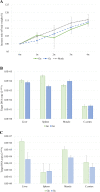
Mice were purchased from the Japan SLC Corporation and bred at the animal facility of the Tokyo University of Agriculture and Technology (Approval No. R03-4). Six to eight-week-old male C57BL/6NCrSlc mice were divided into three groups of six mice each. Each group was inoculated intramuscularly with AAV9-SFTSV Gn, AAV9-SFTSV Gc, and Formulation Buffer (mock group). A dose of 2 × 10¹¹ vg/animal (1 × 10¹³ vg/mL, 50 µL/animal) was used. Weighing was performed before and every week after inoculation. Four weeks after inoculation, the mice were euthanized by whole blood collection under anesthesia, and the serum was separated. The liver, spleen, muscles (inoculation site), and cerebral cortex were collected.
Nucleic acid extraction from mouse organs and quantification of specific mRNA or DNA using real-time PCR
Real-time PCR was used to confirm the biodistribution and mRNA expression of glycoproteins after inoculation with AAV9-SFTSV vaccines. To extract nucleic acids from mouse organs, magLEAD6gC (Precision System Science Co., Ltd., Chiba, Japan) for DNA extraction and the RNeasy Mini Kit (Qiagen, Venlo, Netherlands) for RNA extraction were used according to the recommended protocols of the manufacturer. To quantify the amounts of Gn and Gc genomes in the extracted DNA and RNA, real-time PCR with the One Step PrimeScript RT-PCR Kit (Takara Bio Inc., Kusatsu, Japan) was performed following the recommended protocol of the manufacturer. The reaction temperature and time were set according to the same protocol. However, reverse transcription was not performed because a reverse transcription reagent was not added when measuring the DNA. Additionally, the RNA assay was performed without reverse transcriptase to avoid the influence of residual DNA. The primer set used for quantifying Gn and Gc genomes is shown in Table 1, and primers targeting beta-actin (Mm02619580_g1; Thermo Fischer Scientific) were used to standardize nucleic acid amounts. We used the ΔΔCq method [13] to compare the expression level of nucleic acids. The amplification efficiencies of all primers were confirmed beforehand (data not shown). To calculate the amount of DNA and RNA, the following calculations were performed: These calculations pertained specifically to the Gn genome; however, the same equation was used for Gc.
ΔCq Gn=(Cq of Gn-injected mouse organs, using Gn primer set)−(Cq of Gn-injected mouse organs, using beta-actin primer set)
ΔCq mock=(Cq of mock mouse organ using Gn primer set)−(Cq of mock mouse organ using beta-actin primer set)
ΔCq mock average=Average of ΔCq mock for each organ.
ΔΔCq of each mouse=ΔCq Gn−ΔCq mock average
DNA amount=2 ^ (−ΔΔCq)
ΔCq Gn=[(Cq of Gn-injected mouse organ using Gn primer set, reverse transcriptase+)−(Cq of Gn-injected mouse organ using Gn primer set, reverse transcriptase−)]−[(Cq of Gn-injected mouse organ using beta-actin primer set, reverse transcriptase+)−(Cq of Gn-injected mouse organ using beta-actin primer set, reverse transcriptase−)]
ΔCq mock=[(Cq of mock mouse organ, using Gn primer set, reverse transcriptase+)−(Cq of mock mouse organ, using Gn primer set, reverse transcriptase−)]−[(Cq of mock mouse organ, using beta-actin primer set, reverse transcriptase+)−(Cq of mock mouse organ, using beta-actin primer set, reverse transcriptase−)]
ΔCq mock average=Average of ΔCq mock for each organ.
ΔΔCq of each mouse=ΔCq Gn−ΔCq mock average
RNA amount=2 ^ (−ΔΔCq)
Enzyme-linked immuno sorbent assay (ELISA)
The titer of SFTSV-specific antibodies in AAV9-infected mouse serum was determined using ELISA. Coat the wells of 96-well plates with the SFTSV-infected Huh7 cell lysate (provided by the National Institute of Infectious Diseases) as antigen by incubating at 4°C overnight. As a negative control, lysates of SFTSV-uninfected Huh7 cells were fixed in the same way. After washing once with TBS-T, cells were blocked with 20% Blocking One (nacalai tesque, Kyoto, Japan) at room temperature for 1 hr. After washing once with TBS-T, infected mouse serum diluted 100–409,600 times in 4-fold increments with 20% Blocking One was added and left at 37°C for 2 hr. After washing three times with TBS-T, a secondary antibody (Purified Recomb Protein A/G Peroxidase Conjugated; Thermo Fischer Scientific) diluted 20,000 times with 20% Blocking One/TBS-T was added and left at 37° C for 1 hr. After washing three times with TBS-T, the ELISA POD Substrate TMB Kit (nacalai tesque) was added and allowed to develop color (room temperature for 30 min). The reaction was stopped by adding dilute sulfuric acid. For each dilution rate, the OD405 value of the lysate of infected cells minus the OD405 value of the lysate of uninfected cells was used as the measurement value, and the cut-off value was set by adding twice the standard deviation to the mean value measured in the 100-fold diluted sera of the mock group. The maximum dilution rate that exceeded the cut-off value in the sera from each AAV9-inoculated individual was defined as the limiting dilution rate. Sera from each individual were assayed in triplicates.
Neutralizing assay
Recombinant VSV SFTS pseudovirus (rVSV-SFTSV) expressing the SFTSV glycoprotein on its envelope was used to measure neutralizing ability, as described in a previous paper [25, 26]. Sera from mice inoculated with the AAV9-SFTSV vaccine were diluted in PBS in 2-fold steps from 5–320 times. The diluted sera were mixed with equal volumes of the rVSV-SFTSV solution and incubated at 37°C for 1 hr. A mixture of serum and rVSV-SFTSV (20 µL/well) was added to the culture medium (Eagle’s minimal essential medium, E-MEM/5% FBS, 100 µL/well) of Vero9013 cells prepared at 70–80% semi-confluency in 96-well plates (three wells per condition). After 2 hr at 37°C, the culture medium was removed, washed once with fresh culture medium, and 100 µL/well of fresh culture medium was added again and incubated at 37°C for 24 hr. The luciferase reaction was performed using the recommended protocol of the luciferase reaction reagent (Bright-Glo; Promega), and images were captured using ImageQuant 800. Relative reaction intensities were calculated using ImageQuant TL and the average reaction intensities of mock sera (six mice, three wells/cell) at the same dilution rate were set to 100. The mean of the response intensities at each dilution rate in the mock group minus two standard deviations was set as the cut-off value, and the maximum dilution rate below the cut-off value in the serum obtained from each AAV9 inoculated individual was set as the limiting dilution rate.
Statistical analysis
Dunnett’s test was performed for each measurement point to determine the change in mouse body weight gain rate after vaccination with AAV9-SFTSV. The Wilcoxon test was used to compare genomic levels in each organ between the SFTSV Gn- and Gc-inoculated groups. The Kruskal-Walls test followed by a Steel-Dwass test was used to compare genomic levels between organs within each vaccination group. The Wilcoxon test was used to compare antibody levels in each organ.
Figures


References
-
- Aurnhammer C, Haase M, Muether N, Hausl M, Rauschhuber C, Huber I, Nitschko H, Busch U, Sing A, Ehrhardt A, Baiker A. 2012. Universal real-time PCR for the detection and quantification of adeno-associated virus serotype 2-derived inverted terminal repeat sequences. Hum Gene Ther Methods 23: 18–28. doi: 10.1089/hgtb.2011.034 - DOI - PubMed
-
- Dong F, Li D, Wen D, Li S, Zhao C, Qi Y, Jangra RK, Wu C, Xia D, Zhang X, Deng F, Chandran K, Zou Z, Yuan F, Zheng A. 2019. Single dose of a rVSV-based vaccine elicits complete protection against severe fever with thrombocytopenia syndrome virus. NPJ Vaccines 4: 5. doi: 10.1038/s41541-018-0096-y - DOI - PMC - PubMed
MeSH terms
Substances
LinkOut - more resources
Full Text Sources
Miscellaneous

