

Intussusception in Incisional Hernia: A Case Report and Literature Review
- PMID: 38143605
- PMCID: PMC10748933
- DOI: 10.7759/cureus.49346
Intussusception in Incisional Hernia: A Case Report and Literature Review
Abstract
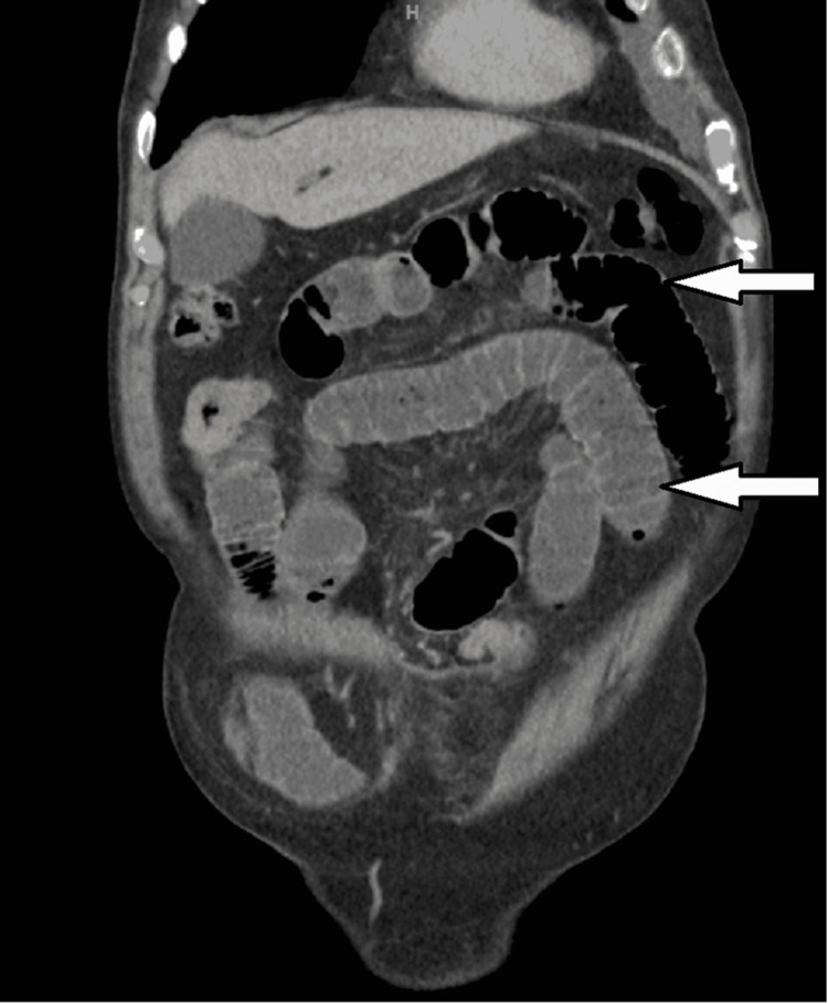
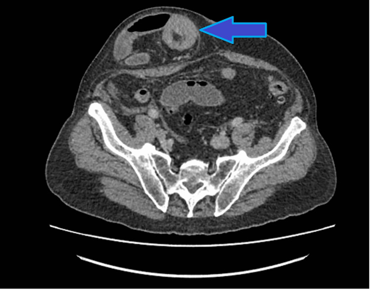
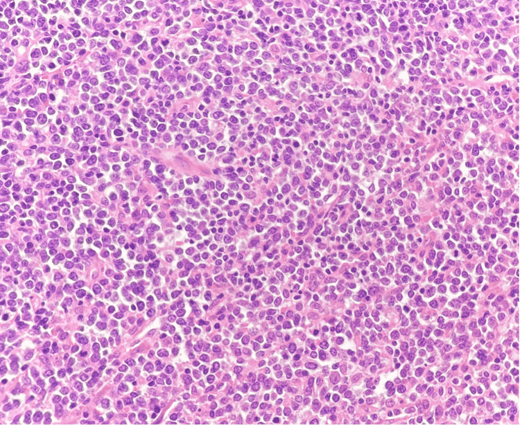
Intussusception in adults is a rare condition. Most frequently, intussusception involves the small intestine and, very rarely, the large intestine. In this report, we present the case of a 79-year-old male who was admitted with symptoms and signs of bowel obstruction due to an incarcerated incisional hernia (a tender irreducible incisional hernia associated with nausea and vomiting). His CT scan confirmed intussusception in his incisional hernia, showing the target sign. An emergency laparotomy, small bowel resection, and anastomosis were done. The histopathology report revealed the cause of intussusception to be a polypoid small bowel B cell lymphoma. It is necessary to excise the affected bowel segment in order to treat adult intussusception because it is commonly associated with malignant organic lesions. Computed tomography is the most sensitive imaging modality for intussusception; thus, we must consider a low threshold for a scan for patients presenting with abdominal pain.
Keywords: bowel obstruction; incarcerated incisional hernia; incisional hernia; intussusception; surgical management of intussusception.
Copyright © 2023, Hassan et al.
Conflict of interest statement
The authors have declared that no competing interests exist.
Figures

References
Publication types
LinkOut - more resources
Full Text Sources