Case Reports
. 2023 Nov 23;15(11):e49274.
doi: 10.7759/cureus.49274. eCollection 2023 Nov.

Dual Lumen Microcatheter in Percutaneous Biliary Drainage for Postoperative Bile Leakage: A Case Report

Affiliations
Case Reports

Dual Lumen Microcatheter in Percutaneous Biliary Drainage for Postoperative Bile Leakage: A Case Report

Yasuyuki Onishi et al. Cureus. .

Abstract

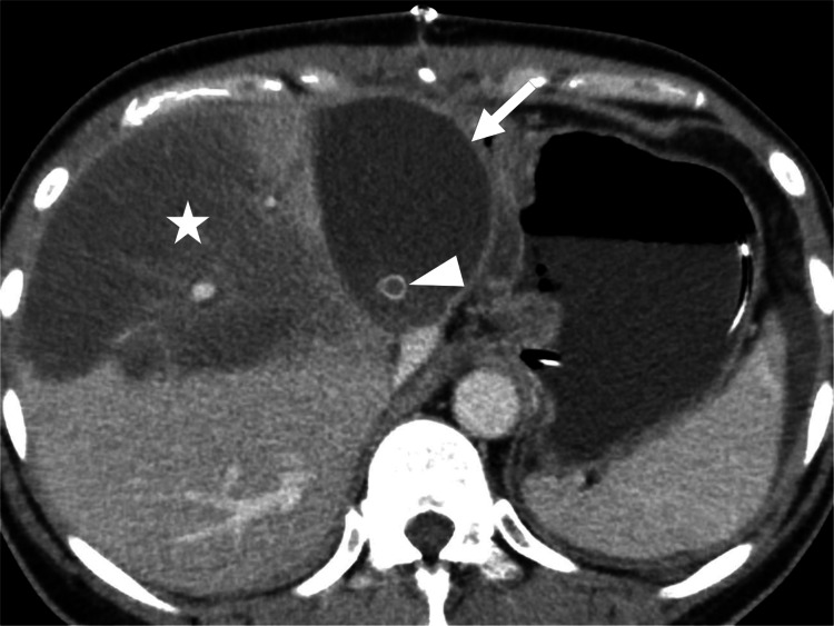
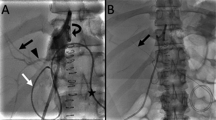
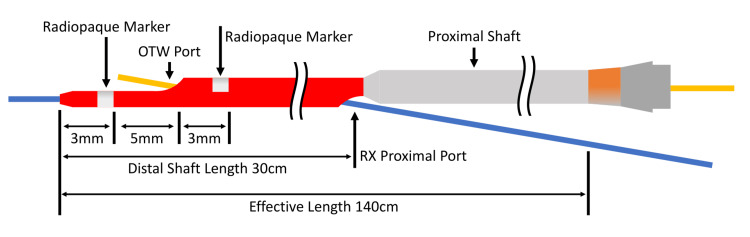
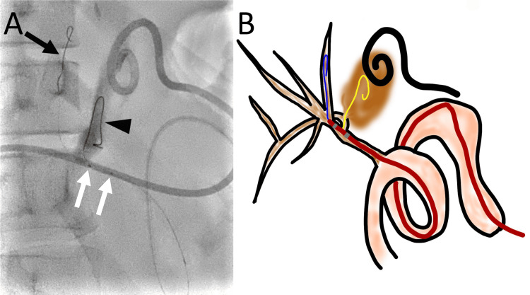
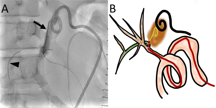
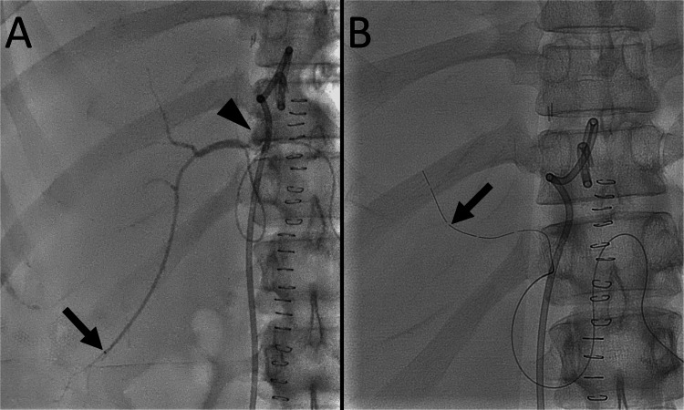
Percutaneous biliary intervention is widely accepted as an effective and safe treatment for various types of bile duct diseases. We present the case of a 44-year-old woman who developed bile leakage after a living-donor liver transplantation for locally advanced cholangiocarcinoma. A percutaneous drainage tube was placed in the segment 8 bile duct via the blind end of the jejunum. However, the bile leakage was unchanged. Bile leakage from the right posterior hepatic duct was suspected. Using a dual lumen microcatheter, a percutaneous drainage tube was placed in the segment 7 bile duct via the blind end of the jejunum, which reduced the bile leakage. These results suggest that a dual lumen microcatheter is a valuable tool for navigating the biliary tree during difficult percutaneous biliary interventions.

Keywords: bile leak; dual lumen microcatheter; living donor liver transplantation; percutaneous biliary drainage; post-operative complication.


Conflict of interest statement

The authors have declared that no competing interests exist.

Figures

Figure 1
Figure 1. Contrast-enhanced CT of the upper abdomen showing a biloma (arrow) adjacent to the right lobe liver graft; the anterior portion of the liver shows hypodensity (star) due to occlusion of a segmental hepatic vein (arrowhead) that was reconstructed using an artificial graft.
Figure 2
Figure 2. Percutaneous biliary drainage tube placement in the right anterior segmental bile duct
(A) A frontal radiograph with a contrast injected through the drainage tube (curved arrow) in the biloma shows the segment 8 bile duct (black arrow) and the right posterior hepatic duct (arrowhead). A seeking catheter (white arrow) is advanced from the blind end of the jejunum. The catheter is inserted through the blind end of the jejunum (star). (B) A frontal radiograph shows a drainage tube (arrow) placed in the segment 8 bile duct.
Figure 3
Figure 3. Crusade catheter
The catheter has an over-the-wire (OTW) lumen and a rapid-exchange (RX) lumen. There are two radiopaque markers at the distal end of the catheter, and the OTW port is located between the two markers. The distal and proximal shaft diameters are 2.9-F and 3.2F. Image credit: Authors
Figure 4
Figure 4. Advancement of a guidewire to the biloma through the defect at the right posterior hepatic duct using a Crusade catheter
(A) A right oblique radiograph shows the advancement of one guidewire (arrow) to the segment 8 bile duct and another guidewire (arrowhead) to the biloma. The two markers of the Crusade catheter are observed (white arrows). (B) A schematic drawing shows a guidewire (blue) that was advanced to the segment 8 bile duct, which was used as a safety wire. The Crusade catheter is advanced on the first guidewire along the rapid-exchange lumen. The position of the Crusade catheter is adjusted to advance the second guidewire (yellow) to the right posterior hepatic duct through the over-the-wire lumen. The second guidewire (yellow) is then advanced to the biloma (brown) through the defect at the right posterior hepatic duct. Image credit: Authors Manufacturer details: Crusade catheter; Kaneka Corporation, Minato City, Tokyo, Japan
Figure 5
Figure 5. Advancement of a guidewire to the right posterior segmental bile duct using a Crusade catheter
(A) A right oblique radiograph shows one guidewire (arrow) that is advanced to the biloma. The other guidewire (arrowhead) is advanced to the segment 6 bile duct. (B) A schematic drawing shows the guidewire (yellow) that was advanced to the biloma and used as a safety wire. The Crusade catheter is advanced using the guidewire along the rapid-exchange lumen. The position of the Crusade catheter is adjusted to advance another guidewire (green) to the right posterior segmental bile duct. Subsequently, the guidewire is advanced to the segment 6 bile duct. Image credit: Authors Manufacturer details: Crusade catheter; Kaneka Corporation, Minato City, Tokyo, Japan
Figure 6
Figure 6. Percutaneous biliary drainage tube placement in the right posterior segmental bile duct
(A) A frontal radiograph with contrast injected using a microcatheter (arrow) advanced to the segment 6 bile duct shows leakage of the contrast medium (arrowhead) into the biloma. (B) A frontal radiograph shows a drainage tube (arrow) placed in the segment 7 bile duct.

References

    1. Society of Interventional Radiology Quality improvement standards for percutaneous cholecystostomy and percutaneous transhepatic biliary interventions. Devane AM, Annam A, Brody L, et al. J Vasc Interv Radiol. 2020;31:1849–1856. - PubMed
    1. CIRSE standards of practice on percutaneous transhepatic cholangiography, biliary drainage and stenting. Das M, van der Leij C, Katoh M, Benten D, Hendriks BM, Hatzidakis A. Cardiovasc Intervent Radiol. 2021;44:1499–1509. - PubMed
    1. Percutaneous transhepatic biliary drainage. Covey AM, Brown KT. Tech Vasc Interv Radiol. 2008;11:14–20. - PubMed
    1. Dual lumen microcatheters for complex percutaneous coronary interventions. Oreglia JA, Garbo R, Gagnor A, Gasparini GL. Cardiovasc Revasc Med. 2018;19:298–305. - PubMed
    1. Dual lumen microcatheters for recanalisation of chronic total occlusions: a EuroCTO Club expert panel report. Pyxaras SA, Galassi AR, Werner GS, et al. EuroIntervention. 2021;17:0–70. - PMC - PubMed

Publication types

LinkOut - more resources