. 2023 Dec 1;5(4):290-306.
doi: 10.1089/bioe.2022.0036. Epub 2023 Dec 15.

Endoplasmic Reticulum Calcium Mediates Drosophila Wing Development

Affiliations

Endoplasmic Reticulum Calcium Mediates Drosophila Wing Development

Laura Faith George et al. Bioelectricity. .

Abstract

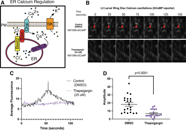
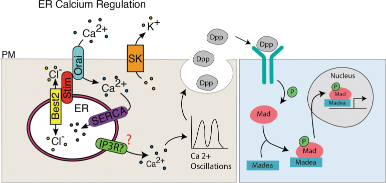
Background: The temporal dynamics of morphogen presentation impacts transcriptional responses and tissue patterning. However, the mechanisms controlling morphogen release are far from clear. We found that inwardly rectifying potassium (Irk) channels regulate endogenous transient increases in intracellular calcium and bone morphogenetic protein (BMP/Dpp) release for Drosophila wing development. Inhibition of Irk channels reduces BMP/Dpp signaling, and ultimately disrupts wing morphology. Ion channels impact development of several tissues and organisms in which BMP signaling is essential. In neurons and pancreatic beta cells, Irk channels modulate membrane potential to affect intracellular Ca++ to control secretion of neurotransmitters and insulin. Based on Irk activity in neurons, we hypothesized that electrical activity controls endoplasmic reticulum (ER) Ca++ release into the cytoplasm to regulate the release of BMP.

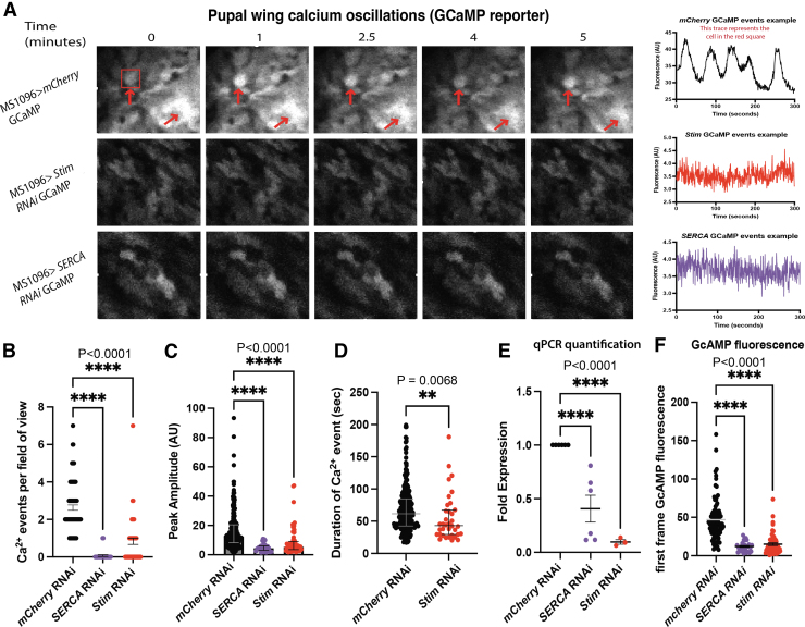
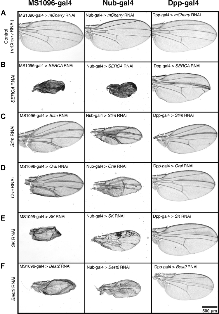
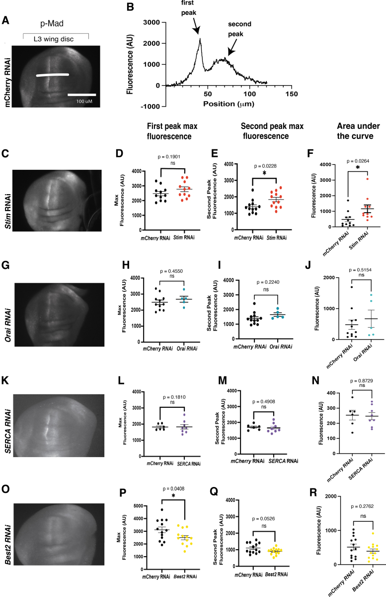
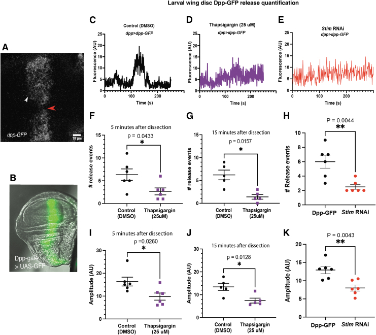
Materials and methods: To test this hypothesis, we reduced expression of four proteins that control ER calcium, Stromal interaction molecule 1 (Stim), Calcium release-activated calcium channel protein 1 (Orai), SarcoEndoplasmic Reticulum Calcium ATPase (SERCA), small conductance calcium-activated potassium channel (SK), and Bestrophin 2 (Best2) using RNAi and documented wing phenotypes. We use live imaging to study calcium and Dpp release within pupal wings and larval wing discs. Additionally, we employed immunohistochemistry to characterize Small Mothers Against Decapentaplegic (SMAD) phosphorylation downstream of the BMP/Dpp pathway following RNAi knockdown.

Results: We found that reduced Stim and SERCA function decreases amplitude and frequency of endogenous calcium transients in the wing disc and reduced BMP/Dpp release.

Conclusion: Our results suggest control of ER calcium homeostasis is required for BMP/Dpp release, and Drosophila wing development.

Keywords: BMP; Drosophila wing development; Orai; SERCA; Stim; bestrophin; bone morphogenic protein; calcium; endoplasmic reticulum; transients; wing venation.


Conflict of interest statement

No competing financial interests exist.

Figures

FIG. 1.
FIG. 1.
Endogenous transient changes in cytoplasmic calcium are mediated by ER calcium stores. (A) A schematic shows how five proteins work together to mediate ER calcium stores. SERCA pumps cytoplasmic calcium into the ER. Ion channels like bestrophins act as counterbalances to maintain membrane potential by passively allowing chloride to move into and out of the ER. Stim senses calcium levels in the ER and works with Orai to open to allow extracellular calcium to pass into the cytoplasm. SK is a potassium channel that works as a counterbalance to Orai, letting potassium out of the cytoplasm. (B) Sequential images of GCaMP fluorescence show transient changes in cytoplasmic calcium in an MS1096>GCaMP wing control (top) and with SERCA inhibition with thapsigargin (bottom row). (C) Quantification of GCaMP fluorescence changes over time in MS1096>GCaMP control (DMSO, black) and thapsigargin (purple) show reduced amplitude of changes in fluorescence. (D) Amplitudes of individual changes in GCaMP fluorescence are compared in DMSO control (black) and thapsigargin (purple) treated larval wing discs. DMSO, dimethyl sulfoxide; ER, endoplasmic reticulum; PM, plasma membrane; SERCA, sarcoEndoplasmic reticulum calcium ATPase; SK, small conductance calcium-activated potassium channel.
FIG. 2.
FIG. 2.
Pupal wings have endogenous transient changes in cytoplasmic calcium that are mediated by ER calcium. (A) Representative sequential images (left) and fluorescence profiles (right) of GCaMP fluorescence in pupal wings in MS1096>GCaMP (top panels), MS1096>GCaMP; Stim RNAi (middle panels), MS1096>GCaMP; SERCA RNAi (bottom panels) show that reducing Stim and SERCA expression reduce calcium transients. (B) Quantification of calcium events per field of view out of seven possible shows reduced number of calcium events with SERCA and stim RNAi. (C) Quantification of GCaMP fluorescence peak amplitude shows that SERCA and stim RNAi reduces GCaMP fluorescence peak amplitude. (D) Duration of calcium events is reduced with stim RNAi. (E) Quantitative RT-PCR shows that RNAi knockdown of SERCA and stim reduced expression of each gene. (F) GCaMP fluorescence values of the first frame in each video used for analysis. Shown is every ROI taken from each genotype, to compare relative GCaMP (calcium) expression after knockdown. ROI, region of interest; RT-PCR, reverse transcription polymerase chain reaction.
FIG. 3.
FIG. 3.
Wing-specific knockdown of SERCA, Stim, Orai, SK, and Best2 have severe wing phenotypes. (A) Representative images of female adult wings from MS1096>mcherryRNAi (left), nub>mcherryRNAi (middle); dpp>mcherryRNAi (right) are controls for effects of driver lines. (B) Representative images show female adult wings from MS1096>SERCA RNAi (left) and nub>SERCA RNAi (middle) dpp>SERCA RNAi (right) are reduced in size, have bristle patterning defects, and disrupted venation. (C) Representative images show female adult wings from MS1096>stim RNAi (left) and nub>stim RNAi (middle) dpp>stim RNAi (right) have thickened veins and are reduced in size. (D) Representative images show female adult wings from MS1096>orai RNAi (left) and nub>orai RNAi (middle) dpp>orai RNAi (right) have thickened veins and are reduced in size. (E) Representative images show female adult wings from MS1096>SK RNAi (left) completely lack venation and are reduced in size. nub>SK RNAi (middle) have disrupted venation pattern and are reduced in size. dpp>SK RNAi (right) are slightly reduced in size and lack the anterior cross vein. (F) Representative images show female adult wings from MS1096>best2 RNAi (left) and nub>best2 RNAi (middle) completely lack venation and are reduced in size. dpp>best2 RNAi (right) are slightly reduced in size and lack the anterior cross vein.
FIG. 4.
FIG. 4.
Phosphorylation of Mad is altered with the loss of Stim and Best2. (A) A representative image of a MS1096>mcherryRNAi shows a wild-type pattern of p-Mad staining for control. (B) A representative trace of fluorescence across the wing disc from anterior to posterior shows anterior (first peak) and posterior peak fluorescence (second peak) and area under the curve. A representative image of MS1096>Stim RNAi shows an increase in p-Mad fluorescence (C), a trend toward an increase in peak fluorescence for anterior peaks and a significant increase in fluorescence of posterior peaks (D, E), and an increase in area under the curve (F). A representative image of a wing disc expressing orai RNAi (G) shows a trend toward an increase in average fluorescence anterior and posterior peak fluorescence and area under the curve that is not significant (H–J). A representative image of a p-Mad stained MS1096>SERCA RNAi (K) wing disc is not significantly different from controls (L–N). A representative image of MS1096>Best2 RNAi shows a significant decrease in p-Mad staining fluorescence area under the curve (P) and a trend toward a decrease in peak fluorescence (Q, R).
FIG. 5.
FIG. 5.
Representative images of p-Mad stained pupal wings show that knockdown of ER calcium regulatory proteins disrupts BMP signaling in the pupal wing. (A) A schematic shows where Dpp signaling specifies wing veins (B) MS1096>mCherry RNAi pupal wings serve as controls. (C) MS1096>SERCA RNAi pupal wings have increased thickness of p-Mad staining along longitudinal veins but reduced p-Mad staining in cross veins. (D) MS1096>stim RNAi pupal wings have increased p-Mad staining. (E) A representative image shows MS1096>Orai RNAi increased the domain of p-Mad staining in pupal wings. (F) A representative image shows disruption of p-Mad staining in MS1096>best2 RNAi adult wings. BMP, bone morphogenetic protein.
FIG. 6.
FIG. 6.
Reduction of SERCA and Stim function reduces number and amplitude of Dpp-GFP release events. (A) A representative image of a dpp>dpp-gfp wing disc shows where Dpp-GFP release events were measured. (B) A representative image of dpp>gfp shows the expression zone. (C) A representative trace of Dpp-GFP fluorescence over time from a dpp>dpp-gfp wing disc shows a dramatic change in fluorescence associated with Dpp-GFP release. (D) A representative trace of Dpp-GFP fluorescence over time in a dpp>dpp-gfp incubated thapsigargin shows that SERCA inhibition reduces amplitude of Dpp-GFP fluorescence changes. (E) A representative trace of Dpp-GFP fluorescence over time from a dpp>dpp-gfp;stim RNAi wing disc shows that knockdown of Stim reduces Dpp-GFP release events. (F, G) Graphs show that the number of Dpp-GFP release events is significantly reduced by incubation in thapsigargin for 5 min (F) and 15 min (G). (H) A graph shows that knockdown of Stim reduces the number of Dpp-GFP release events. (I, J) A graph shows that the amplitude of Dpp-GFP release events is reduced by incubation in thapsigargin for 5 min (I) and 15 min (J). (K) A graph shows that Stim knockdown reduces the amplitude of changes in Dpp-GFP fluorescence. GFP, green fluorescent protein.
FIG. 7.
FIG. 7.
Potential model for ER calcium-mediated regulation of BMP/Dpp signaling. Together SERCA, Orai, Stim, SK, and Best2 help regulate the flux of ER calcium. Calcium movement out of the ER potentially through IP3R regulates calcium oscillations that in turn regulate the fusion of BMP/Dpp containing vesicles impacting downstream signaling.

References

    1. Basson MA. Signaling in cell differentiation and morphogenesis. Cold Spring Harb Perspect Biol 2012;4(6):a008151. - PMC - PubMed
    1. Simin K, Bates EA, Horner MA, et al. . Genetic analysis of punt, a type II Dpp receptor that functions throughout the Drosophila melanogaster life cycle. Genetics 1998;148(2):801–813. - PMC - PubMed
    1. Letsou A, Arora K, Wrana JL, et al. . Drosophila Dpp signaling is mediated by the punt gene product: A dual ligand-binding type II receptor of the TGF beta receptor family. Cell 1995;80(6):899–908. - PubMed
    1. Newfeld SJ, Mehra A, Singer MA, et al. . Mothers against Dpp participates in a DDP/TGF-beta responsive serine-threonine kinase signal transduction cascade. Development 1997;124(16):3167–3176. - PubMed
    1. Chen Y, Riese MJ, Killinger MA, et al. . A genetic screen for modifiers of Drosophila decapentaplegic signaling identifies mutations in punt, Mothers against Dpp and the BMP-7 homologue, 60A. Development 1998;125(9):1759–1768. - PubMed

LinkOut - more resources