. 2023 Oct 13;3(6):516-527.
doi: 10.1021/acsbiomedchemau.3c00031. eCollection 2023 Dec 20.

The C-Terminal of NaV1.7 Is Ubiquitinated by NEDD4L

Affiliations

The C-Terminal of NaV1.7 Is Ubiquitinated by NEDD4L

Katharine M Wright et al. ACS Bio Med Chem Au. .

Abstract

NaV1.7, the neuronal voltage-gated sodium channel isoform, plays an important role in the human body's ability to feel pain. Mutations within NaV1.7 have been linked to pain-related syndromes, such as insensitivity to pain. To date, the regulation and internalization mechanisms of the NaV1.7 channel are not well known at a biochemical level. In this study, we perform biochemical and biophysical analyses that establish that the HECT-type E3 ligase, NEDD4L, ubiquitinates the cytoplasmic C-terminal (CT) region of NaV1.7. Through in vitro ubiquitination and mass spectrometry experiments, we identify, for the first time, the lysine residues of NaV1.7 within the CT region that get ubiquitinated. Furthermore, binding studies with an NEDD4L E3 ligase modulator (ubiquitin variant) highlight the dynamic partnership between NEDD4L and NaV1.7. These investigations provide a framework for understanding how NEDD4L-dependent regulation of the channel can influence the NaV1.7 function.


Conflict of interest statement

The authors declare no competing financial interest.

Figures
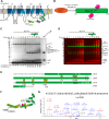
Figure 1
Figure 1
NaV1.7 gets ubiquitinated in its C-terminal domain. (A) Scheme representation of the NaV1.7 topology. Each domain and transmembrane helix are labeled accordingly. The cytoplasmic regions are shown as loops. The C-terminal of NaV1.7 is shown as green cylinders representing alpha-helices as displayed in the crystallographic structures of other isoforms NaV1.4 and NaV1.5. The location of CaM is also highlighted in magenta. The canonical NEDD4L PY binding motif is indicated at residues 1955–1958. The two auxiliary beta subunits (β1 and β2) are shown in yellow. (B) Scheme of the GST-fusion with a precision protease cleavage sequence construct of NaV1.7CTspanning residues 1761–1988 (green). Coexpression was performed with full-length CaM (magenta). (C) In vitro ubiquitination time course of FLNEDD4L in the absence and presence of the NaV1.7CT-CaM substrate. Equal amounts of sample were taken at the indicated time points and quenched with reducing loading buffer. The purified NaV1.7CT-CaM protein is in the last lane. Unmodified FLNEDD4L and NaV1.7CT substrate protein bands were quantified by a densitometry analysis as a function of time. All assays were repeated at least twice with good reproducibility (n = 2). Gel is stained with colloidal Coomassie Blue. (D) Fluorescent Western blot analysis of the in vitro ubiquitination assays as carried out in C using anti-ubiquitin (red) and anti-NEDD4L (green) antibodies. (n = 2.) (E) NaV1.7CT sequence coverage after band excision from the in vitro assay. Identified peptides are highlighted in shaded green regions. Bold, red lysine residue with a yellow square above them indicate Ub-modification and the numbers in parentheses indicate total number of PSMs for each lysine identified from MS. Lysine residues detected by MS but not modified with ubiquitin are highlighted in bold, white. The IQ region is highlighted in bold and a purple box; the PY motif is bold and underlined. (F) Scheme of NaV1.7CT-CaM with the ubiquitinated lysine sites labeled with yellow squares based on location. (G) Representative MS/MS spectrum and sequence coverage of the peptide containing a Ub-modification on NaV1.7CT-CaM Lys1930. Lower case k indicates Ub-modification and lower case m indicates an oxidized methionine.
Figure 2
Figure 2
Lys63-linked ubiquitin chains are assembled on NaV1.7CT. (A) LC/MS/MS sequence coverage of ubiquitin after di-Ub-NaV1.7CT band excision from the in vitro assay. Identified peptides are highlighted in shaded, boxed gray regions. Bold, red letters with a yellow square above them are the identified Lysine residues with ubiquitin chain formation. (B) Ubiquitin lysine chain linkage of NaV1.7CT modified by FLNEDD4L in vitro ubiquitination assay was analyzed by LC/MS/MS. The peptide spectrum matches of each ubiquitin Lysine residue seen with a Ub-modification is represented as a pie graph. (C) Representative MS/MS spectrum and sequence coverage of the peptide containing a Ub-modified Lys63. Lower case k indicates Ub-modification. (D) In vitro ubiquitination assays of NaV1.7CT-CaM in the presence of UbWT, UbK48R, UbK63R, or UbK48/63R. Samples were quenched with reducing 2× SDS-PAGE loading buffer at 0, 10, and 30 min, and the gel was stained with a colloidal Coomassie Blue stain. Unmodified NaV1.7CT bands were quantified by a densitometry analysis as a function of time. All assays were repeated at least twice with good reproducibility (n = 2).
Figure 3
Figure 3
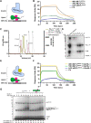
Binding of NaV1.7CT-CaM to FLNEDD4L is enhanced by a ubiquitin variant. (A) Scheme of the SPR binding kinetics between NaV1.7CT-CaM and FLNEDD4L. The ligand NaV1.7CT-CaM was covalently attached to a CM5 chip. The analyte, FLNEDD4L, recognizes the PY motif of the NaV1.7CT unstructured tail (black line). (B) FLNEDD4L binding to NaV1.7CT-CaM was evaluated by parallel kinetics. Each binding sensorgram at 6.25, 100, and 400 nM of FLNEDD4L were fit with a one-to-one binding model. N = 2. (C) Size exclusion chromatogram profile for NaV1.7CT-CaM + FLNEDD4L–UbvNL.1 (solid gold line) showing a ∼ 0.5 mL shift to the left of the FLNEDD4L–UbvNL.1 peak (dashed maroon line) indicating interaction and larger molecular weight. Molecular weight standard (Biorad) is shown in gray. Bold numbers indicated each protein within the molecular weight standard: 1. thyroglobulin, 2. γ-globulin, 3. ovalbumin, 4. myoglobin, 5. vitamin B12. (D) SDS-PAGE gel showing the elution fractions from (B). The FLNEDD4L–UbvNL.1-NaV1.7CT-CaM complex elutes are ∼12 mL and excess NaV1.7CT-CaM elutes at 14 mL. (E) Scheme of the SPR binding kinetics as carried out in (A) with the analyte being FLNEDD4L + 100 nM of ubiquitin variant, UbvNL.1. (F) FLNEDD4L + UbvNL.1 binding to NaV1.7CT-CaM was evaluated by parallel kinetics SPR. Each binding sensorgram at 6.25, 100, and 400 nM of FLNEDD4L + UbvNL.1 was fit with a one-to-one binding model. N = 2. (G) In vitro ubiquitination time course of NaV1.7CT-CaM in the absence and presence of UbvNL.1. Equal amounts of samples were taken at the indicated time points and quenched with reducing loading buffer. Unmodified NaV1.7CT substrate band was quantified by a densitometry analysis as a function of time. N = 2.
Figure 4
Figure 4
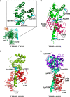
Mapping the locations of the ubiquitinated lysine residues of NaV1.7CT–CaM by sequence and structural analysis. (A) CryoEM structure of full-length human NaV1.7 (PDB ID 7W9K) with a zoomed in view of the C-terminal domain (CTD). NaV1.7 is shown in forest green, the β1 subunit in cyan, and the β2 subunit in orange. The position of the identified lysine residues of NaV1.7CT-CaM are shown in cyan sticks. (B) Structure of NaV1.5CT with apo-CaM (PDB ID 4OVN). NaV1.5CT is shown in green and apo-CaM in hot pink. The corresponding position of the identified lysine residues of NaV1.7CT-CaM are shown in cyan sticks. (C) Structure of NaV1.5CT with calcium-bound CaM (PDB ID 6MUD) structurally aligned to (A). Similarly to (A), NaV1.5CT is shown in split pea and calcium-CaM in red. The corresponding positions of the identified lysine residues of NaV1.7CT-CaM are shown in cyan sticks. (D) Structure of NaV1.5CT with apo-CaM and an FGF13 (PDB ID 4DCK) structurally aligned to (A). Similarly to (A), NaV1.5CT is shown in lime green, apo-CaM in dark red, and FGF13 in purple. The corresponding positions of the identified lysine residues of NaV1.7CT-CaM are shown in cyan sticks.

References

    1. Hodgkin A. L.; Huxley A. F. A quantitative description of membrane current and its application to conduction and excitation in nerve. J. Physiol. 1952, 117, 500–544. 10.1113/jphysiol.1952.sp004764. - DOI - PMC - PubMed
    1. Fozzard H. A.; Hanck D. A. Structure and function of voltage-dependent sodium channels: comparison of brain II and cardiac isoforms. Physiol. Rev. 1996, 76, 887–926. 10.1152/physrev.1996.76.3.887. - DOI - PubMed
    1. Catterall W. A. From ionic currents to molecular mechanisms: the structure and function of voltage-gated sodium channels. Neuron 2000, 26, 13–25. 10.1016/S0896-6273(00)81133-2. - DOI - PubMed
    1. Hille B. Ionic channels in excitable membranes. Current problems and biophysical approaches. Biophys. J. 1978, 22, 283–294. 10.1016/S0006-3495(78)85489-7. - DOI - PMC - PubMed
    1. Goldin A. L.; Barchi R. L.; Caldwell J. H.; Hofmann F.; Howe J. R.; Hunter J. C.; Kallen R. G.; Mandel G.; Meisler M. H.; Netter Y. B.; Noda M.; Tamkun M. M.; Waxman S. G.; Wood J. N.; Catterall W. A. Nomenclature of voltage-gated sodium channels. Neuron 2000, 28, 365–368. 10.1016/S0896-6273(00)00116-1. - DOI - PubMed