. 2023 Dec 8:13:1280977.
doi: 10.3389/fonc.2023.1280977. eCollection 2023.

GIPC1 regulates MACC1-driven metastasis

Affiliations

GIPC1 regulates MACC1-driven metastasis

Franziska Siegel et al. Front Oncol. .

Abstract

Background: Identification of cancer metastasis-relevant molecular networks is desired to provide the basis for understanding and developing intervention strategies. Here we address the role of GIPC1 in the process of MACC1-driven metastasis. MACC1 is a prognostic indicator for patient metastasis formation and metastasis-free survival. MACC1 controls gene transcription, promotes motility, invasion and proliferation of colon cancer cells in vitro, and causes tumor growth and metastasis in mice.

Methods: By using yeast-two-hybrid assay, mass spectrometry, co-immunoprecipitation and peptide array we analyzed GIPC1 protein binding partners, by using the MACC1 gene promoter and chromatin immunoprecipitation and electrophoretic mobility shift assay we probed for GIPC1 as transcription factor. We employed GIPC1/MACC1-manipulated cell lines for in vitro and in vivo analyses, and we probed the GIPC1/MACC1 impact using human primary colorectal cancer (CRC) tissue.

Results: We identified MACC1 and its paralogue SH3BP4 as protein binding partners of the protein GIPC1, and we also demonstrated the binding of GIPC1 as transcription factor to the MACC1 promoter (TSS to -60 bp). GIPC1 knockdown reduced endogenous, but not CMV promoter-driven MACC1 expression, and diminished MACC1-induced cell migration and invasion. GIPC1 suppression reduced tumor growth and metastasis in mice intrasplenically transplanted with MACC1-overexpressing CRC cells. In human primary CRC specimens, GIPC1 correlates with MACC1 expression and is of prognostic value for metastasis formation and metastasis-free survival. Combination of MACC1 and GIPC1 expression improved patient survival prognosis, whereas SH3BP4 expression did not show any prognostic value.

Conclusions: We identified an important, dual function of GIPC1 - as protein interaction partner and as transcription factor of MACC1 - for tumor progression and cancer metastasis.

Keywords: GIPC1; MACC1; patient survival prognosis; protein-protein interaction; transcription factor.


Conflict of interest statement

The authors declare that the research was conducted in the absence of any commercial or financial relationships that could be construed as a potential conflict of interest. The author(s) declared that they were an editorial board member of Frontiers, at the time of submission. This had no impact on the peer review process and the final decision.

Figures
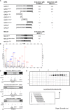
Figure 1
Figure 1
GIPC1 is a newly identified protein binding partner of the metastasis inducer protein MACC1. (A) Physical interaction of GIPC1 and different mutant polypeptides thereof were analyzed for binding to MACC1 and to SH3BP4 by Y2H in Y187 yeast cells. The C-terminal region of GIPC1 (aa 222-333) was identified to be essential for these interactions. MACC1 and mutants thereof were analyzed for binding to GIPC1 by Y2H. The SH3-domain of MACC1 (aa 552-618) was identified to be essential for this interaction. The original results of the b-galactosidase staining of transformed yeast colonies are shown in Supplementary Figure 1 . (B) Tandem MS spectrum for GIPC peptide obtained from cLC-ESI-MS analysis. The sequence of this peptide was verified by the presence of complementary B ions (NH2-terminus–derived fragment ions) and Y ions (COOH-terminus–derived fragment ions). We performed two independent IPs of MACC1 from SW620 cells with high endogenous MACC1 expression employing two different MACC1 antibodies (four experiments), which were subjected to shot-gun MS. In total, 1203 proteins were identified, (C) Validation of the physical interaction of GIPC1 to MACC1 and of GIPC1 to SH3BP4 by co-immunoprecipitation in SW480 cells. No interaction with GIPC1 was found when using MACC1 mutants lacking the SH3 domain confirming the co-immunoprecipitation data (input: 2% of total protein). (D) Pepspot analysis. The peptide array displays 98 peptides in 5 rows and 20 columns. The peptides represent overlapping 15mer-sequences of the GIPC1 C-terminus (222-333). The array was probed for binding MACC1. Spots 13-19 show sufficient spot signal intensity and were selectively recognized by MACC1. Spot-sequence correlation and deducing the GIPC1 binding site for MACC1.
Figure 2
Figure 2
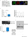
GIPC1 is a newly identified transcription factor and binds to the MACC1 gene promoter. (A) Left: Subcellular distribution of GIPC1 and MACC1 in the cytoplasm (C), the membranous fraction (M) and the nucleus (N) of SW620 cells. GAPDH (cytoplasm), CLTC (cytoplasm and membranous fraction) and HIST1H3A (nucleus) served as controls. Interestingly, GIPC1 and MACC1 were both found also in the nucleus. Right: By immunofluorescence, the subcellular localization of GIPC1 and MACC1 was validated, predominantly the presence of both proteins in the nuclei. These data are in harmony with those from in silico predictions (https://www.genecards.org/cgi-bin/carddisp.pl?gene=GIPC1). The intended co-staining for GIPC1 and MACC1 in SW620 cells is shown with GIPC1 in green, MACC1 in red and DAPI in blue. Scale bar 10 mm. (B) Left: Impact of GIPC1 on the MACC1 promoter activity. MACC1 promoter luciferase reporter construct was co-transfected along with pGL4.74 Renilla plasmid in SW620-shcontrol and SW620-shGIPC1 cells. After 24 h of transfection, luciferase activity was measured and was normalized to Renilla luciferase activity to correct for the variation in transfection efficiencies. Luciferase activity from the SW620-shcontrol cells was set to 100%. shGIPC1-transfected cells showed a significantly reduced MACC1 promoter activity. Right: Fragments of the MACC1 promoter with deletions at the 5’ end and possessing a common 3’-end were inserted into pGL4.17 and were transfected into SW620-shGIPC1 and SW620-shControl cells. Luciferase activity was normalized to Renilla values and expressed as percentage reduction of luciferase activity in SW620-shGIPC1 as compared with SW620-shcontrol cells. The MACC1 promoter fragment -18 to -133 bp is sufficient to demonstrate MACC1 promoter activity loss, which is not further enhanced by larger deletions. (C) ChIP assay. SW620 chromatin was immunoprecipitated with a GIPC1 antibody and quantified by agarose gel electrophoresis, using a primer set specific for the MACC1 promoter and GAPDH. Non-immune IgG and input DNA without any immunoprecipitation with antibody served as negative and positive controls respectively. The binding of GIPC1 to the MACC1 promoter is demonstrated. (D) EMSA. 5’-end biotin labeled oligonucleotide corresponding to human MACC1 promoter were incubated alone as well as with nuclear extracts from SW620 cells along with 100x molar excess of unlabeled competitor sequence or with the antibody specific for GIPC1 to indicate specificity<city/> of the protein-DNA complexes. The reactions were analyzed by polyacrylamide gel electrophoresis. GIPC1 binds to the first 60 bp of the MACC1 promoter.
Figure 3
Figure 3
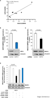
GIPC1 regulates MACC1 expression and MACC1-induced cell motility in vitro. (A) Correlation of GIPC1 and MACC1 expression levels in different CRC cell lines. GIPC1 and MACC1 expression levels were determined in the same sample of a variety of several CRC cell lines by qRT-PCR, normalized using G6PDH and sorted by ascending MACC1/G6PDH mRNA levels of CRC cell lines (HT29, WIDR, HCT116, HCT15, HCA7, DLD1, Caco-2, SW48). We found a positive statistically significant correlation with a Pearson r of 0.9188 and a P value of 0.0013. (B, C) Impact of GIPC1 knock-down in SW620 cells on (B) GIPC1 expression and (C) on MACC1 expression on mRNA and protein levels (both P=0.002). shRNA transfection was done to knock down GIPC1. Stable clones were isolated and validated. Total RNA was isolated from these cells, reverse transcribed and quantified using real time PCR. The data is normalized for G6PDH. Results are shown as means ± SEM of three independent experiments performed in duplicate. (D) Expression of MACC1 in dependence of MACC1, SH3BP4 and GIPC1 knock down in ectopically MACC1-overexpressing cells SW480/MACC1 cells (CMV-promoter-driven). Cells were treated with siRNAs either acting on MACC1, on SH3BP4 or on GIPC1, compared to control siRNA-treated cells. Treatment with MACC1 siRNA resulted in decreased MACC1 expression (P<0.001), whereas siRNA acting on SH3BP4 or GIPC1 did not affect the CMV promoter-driven forced MACC1 expression, neither at RNA nor at protein level.
Figure 4
Figure 4
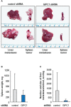
GIPC1 silencing reduces CRC metastasis in mice (A) Tumor growth at the spleen and liver metastasis formation of SW620 cells is reduced after GIPC1 knock-down. SW620/shControl cells and SW620/shGIPC1 cells were intraspenically transplanted, Left: Isolated organs, spleens and livers, originating from intrasplenically transplanted primary SW620-tumors and metastasis after treatment with control shRNA, exemplified for two animals. Right: Isolated organs, spleens and livers, with primary SW620-tumors and metastasis after treatment with GIPC1 shRNA, exemplified for two animals. A clear reduction of tumor growth and metastasis formation is observed after GIPC1 shRNA treatment. (B) Spleen weight. This is illustrated by clearly reduced weight of spleens inclusive primary tumors of shGIPC1-mice vs. control mice. (C) Most importantly, GIPC1 knock-down resulted in reduced number and size of metastases/animal of shGIPC1-mice vs. control mice.
Figure 5
Figure 5
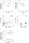
GIPC1 is prognostic for CRC metastasis and improves CRC patient prognosis when combined with MACC1. (A, B) Expression of GIPC1 mRNA (A) and SH3BP4 (B) was determined by quantitative real time RT-PCR with RNA isolated from microdissected tumor cell population of primary, not (yet) metastasized tumors in stages I, II and III (n=59). GIPC1 expression serves as prognostic factor for the formation of distant metastases in CRC patients (P=0.034), with significantly worse metastasis-free survival in the GIPC1 high-expressing patients. SH3BP4, the only relative of MACC1 with a 43.7% of aa sequence identity, was determined simultaneously in the same tumors from the same RNA sample, but did not show any prognostic value. (C) For expression of MACC1 in the same patient cohort ((P<0.0001; even same RNA sample) and for patient characteristics: see (3). Analyzing an expression correlation of MACC1 and GIPC1 in the CRC tumors, we found a positive statistically significant correlation with a Pearson r of 0.505 and a P value of <0.0001. (D) For correlation analysis of MACC1 expression and GIPC1 expression, tumors were classified due to low and high expressing groups, we found a statistically significant expression for GIPC1 and MACC1 expression (P=0.014). (E) Combining high MACC1 and high GIPC1 (qRT-PCR values) vs. low MACC1 and low GIPC1 patients showed improved prognostic values (P<0.0001) when compared to MACC1 expression alone (3) or GIPC1 expression alone (A).

References

    1. Siegel RL, Miller KD, Wagle NS, Jemal A. Cancer statistics, 2023. CA Cancer J Clin (2023) 73:17–48. doi: 10.3322/caac.21763 - DOI - PubMed
    1. Siegel RL, Wagle NS, Cercek A, Smith RA, Jemal A. Colorectal cancer statistics, 2023. CA Cancer J Clin (2023) 73:233–54. doi: 10.3322/caac.21772 - DOI - PubMed
    1. Stein U, Walther W, Arlt F, Schwabe H, Smith J, Fichtner I, et al. . MACC1, a newly identified key regulator of HGF-MET signaling, predicts colon cancer metastasis. Nat Med (2009) 15:59–67. doi: 10.1038/nm.1889 - DOI - PubMed
    1. Radhakrishnan H, Walther W, Zincke F, Kobelt D, Imbastari F, Erdem M, et al. . MACC1-the first decade of a key metastasis molecule from gene discovery to clinical translation. Cancer Metastasis Rev (2018) 37:805–20. doi: 10.1007/s10555-018-9771-8 - DOI - PubMed
    1. Stein U, Burock S, Herrmann P, Wendler I, Niederstrasser M, Wernecke KD, et al. . Circulating MACC1 transcripts in colorectal cancer patient plasma predict metastasis and prognosis. PloS One (2012) 7:e49249. doi: 10.1371/journal.pone.0049249 - DOI - PMC - PubMed

LinkOut - more resources