. 2023 Nov 23:31:101165.
doi: 10.1016/j.omtm.2023.101165. eCollection 2023 Dec 14.

Hepatocyte-targeted siTAZ therapy lowers liver fibrosis in NASH diet-fed chimeric mice with hepatocyte-humanized livers

Affiliations

Hepatocyte-targeted siTAZ therapy lowers liver fibrosis in NASH diet-fed chimeric mice with hepatocyte-humanized livers

Xiaobo Wang et al. Mol Ther Methods Clin Dev. .

Abstract

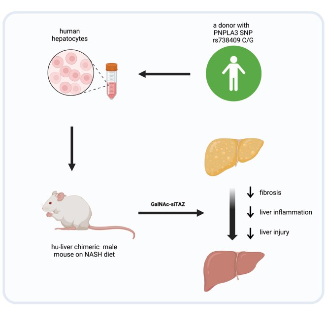
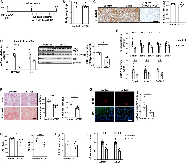
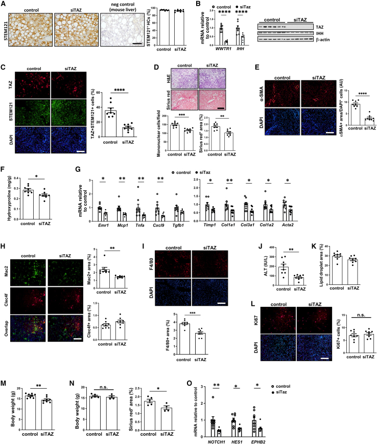
Nonalcoholic steatohepatitis (NASH) is emerging as the most common cause of liver disease. Although many studies in mouse NASH models have suggested therapies, translation to humans is poor, with no approved drugs for NASH. One explanation may lie in differences between mouse and human hepatocytes. We used NASH diet-fed chimeric mice reconstituted with human hepatocytes (hu-liver mice) to test a mechanism-based hepatocyte-targeted small interfering RNA (siRNA), GalNAc-siTaz, shown previously to block the progression to fibrotic NASH in mice. Following ablation of endogenous hepatocytes, male mice were reconstituted with human hepatocytes from a single donor with the rs738409-C/G PNPLA3 risk variant, resulting in ∼95% human hepatocyte reconstitution. The mice were then fed a high-fat choline-deficient l-amino acid-defined diet for 6 weeks to induce NASH, followed by six weekly injections of GalNAc-siTAZ to silence hepatocyte-TAZ or control GalNAc-siRNA (GalNAc-control) while still on the NASH diet. GalNAc-siTAZ lowered human hepatic TAZ and IHH, a TAZ target that promotes NASH fibrosis. Most important, GalNAc-siTAZ decreased liver inflammation, hepatocellular injury, hepatic fibrosis, and profibrogenic mediator expression versus GalNAc-control, indicating that GalNAc-siTAZ decreased the progression of NASH in mice reconstituted with human hepatocytes. In conclusion, silencing TAZ in human hepatocytes suppresses liver fibrosis in a hu-liver model of NASH.

Keywords: GalNAc-siRNA; IHH; MASH; NASH/MASH therapy, NASH, TAZ, Indian hedgehog; WWTR1; hepatocyte-humanized mice; liver fibrosis.


Conflict of interest statement

The laboratory of I.T. received research funding, material support, and technical input from the Takeda Pharmaceutical Company to study siTAZ therapeutics, for which Columbia University holds a patent.

Figures

None
Graphical abstract
Figure 1
Figure 1
Treatment of hu-liver NASH mice with GalNAc-siTAZ lowers liver inflammation and fibrosis (A) Experimental scheme. Humanized mice were fed the HF-CDAA diet for 6 weeks and then injected once weekly for 6 additional weeks with 5 mg/kg GalNAc-siTAZ (siTAZ) or control GalNAc construct (control) while still on the diet. At 12 weeks, the 2 cohorts were analyzed for systemic and liver endpoints. (B) Body weight. (C) Livers were immunostained with anti-STEM121, and the percentages of hepatocytes that were STEM121+ were quantified; liver from a mouse fed the HF-CDAA diet for 12 weeks was used as a negative control for anti-STEM121 staining. Scale bar, 100 μm. (D) Liver WWTR1 and IHH mRNA and immunoblots of YAP, TAZ, IHH, and β-actin; a longer exposure (long) of the TAZ immunoblot is included. The densitometric data for IHH is shown. (E) Livers were assayed for the inflammatory mRNAs Cxcl9, Tnfa, Emr1, Tgfb1, and Mcp1 and the fibrosis-related mRNAs Spp1, Acat2, and Col1a1 mRNAs. (F) Livers were stained with H&E (upper row) and Sirius red (lower row) and then quantified for inflammatory cells per field and percentage of Sirius red area. Scale bar, 200 μm. (G) Livers were immunostained with anti-αSMA and quantified for the αSMA+ area, which was normalized for cellularity by dividing by the number of DAPI+ cells. Scale bar, 200 μm. (H) Plasma ALT and AST. (I) Percentage of lipid droplet area in the livers. (J) Liver NOTCH1 and HES1 mRNA. Values shown for all of the graphs are means ± SEM; n = 6 control mice and 7 siTAZ mice. ∗p < 0.05; ∗∗p < 0.01; ∗∗∗p < 0.001; ∗∗∗∗p < 0.0001. The data were analyzed using the Student’s t test.
Figure 2
Figure 2
A repeat experiment showing that GalNAc-siTAZ lowers NASH progression in hu-liver mice A second cohort of hu-liver mice were treated with GalNAc-siTAZ or control GalNAc construct exactly as depicted in Figure 1A. (A) Livers were immunostained with anti-STEM121, and the percentages of hepatocytes that were STEM121+ were quantified; liver from a mouse fed the HF-CDAA diet for 12 weeks was used as a negative control for anti-STEM121 staining. Scale bar, 100 μm. (B) Liver WWTR1 and IHH mRNA and immunoblots of TAZ, IHH, and β-actin. (C) Livers were immunostained with anti-TAZ and anti-STEM121 and then quantified for the percentage of TAZ+cells among STEM121+cells. Scale bar, 200 μm. (D) Livers were stained with H&E (upper row) and Sirius red (lower row) and then quantified for inflammatory cells per field and percentage of Sirius red area. Scale bar, 200 μm. (E) Livers were immunostained with anti-αSMA and quantified as in Figure 1G. Scale bar, 200 μm. (F) Hydroxyproline content. (G) Livers were assayed for the inflammatory mRNAs Emr1, Mcp1, Tnfa, Cxcl9, and Tgfb1 and the fibrosis-related mRNAs Timp1, Col1a1, Col3a1, Col1a2, and Acta2. (H) Livers were immunostained with anti-Mac2 and anti-Clec4f and quantified for the percentage positive area of each. Scale bar, 200 μm. (I) Livers were immunostained with anti-F4/80 and quantified for the percentage of positive area. Scale bar, 200 μm. (J) Plasma ALT. (K) Percentage of lipid droplet area in the livers. (L) Livers were immunostained with anti-Ki67 and quantified for the percentage of positive area. Scale bar, 200 μm. (M) Body weight. (N) The livers of a subgroup mice from each cohort (5 control, 4 siTAZ) with similar body weights were assayed for percentage of Sirius red+. (O) Liver NOTCH1, HES1, and EPHB2 mRNA. Values shown for all of the graphs are means ± SEM; n = 8 mice per group. ∗p < 0.05; ∗∗p < 0.01; ∗∗∗p < 0.001; ∗∗∗∗p < 0.0001. The data were analyzed using the Student’s t test.

References

    1. Younossi Z.M., Golabi P., Paik J.M., Henry A., Van Dongen C., Henry L. The global epidemiology of nonalcoholic fatty liver disease (NAFLD) and nonalcoholic steatohepatitis (NASH): a systematic review. Hepatology. 2023;77:1335–1347. - PMC - PubMed
    1. Sanyal A.J., Van Natta M.L., Clark J., Neuschwander-Tetri B.A., Diehl A., Dasarathy S., Loomba R., Chalasani N., Kowdley K., Hameed B., et al. Prospective study of outcomes in adults with nonalcoholic fatty liver disease. N. Engl. J. Med. 2021;385:1559–1569. - PMC - PubMed
    1. Harrison S.A., Allen A.M., Dubourg J., Noureddin M., Alkhouri N. Challenges and opportunities in NASH drug development. Nat. Med. 2023;29:562–573. - PubMed
    1. Santhekadur P.K., Kumar D.P., Sanyal A.J. Preclinical models of non-alcoholic fatty liver disease. J. Hepatol. 2018;68:230–237. - PMC - PubMed
    1. Wang X., Zheng Z., Caviglia J.M., Corey K.E., Herfel T.M., Cai B., Masia R., Chung R.T., Lefkowitch J.H., Schwabe R.F., Tabas I. Hepatocyte TAZ/WWTR1 promotes inflammation and fibrosis in nonalcoholic steatohepatitis. Cell Metab. 2016;24:848–862. - PMC - PubMed

LinkOut - more resources