Observational Study
. 2023 Sep;2(9):10.1056/evidoa2300046.
doi: 10.1056/evidoa2300046. Epub 2023 Jul 20.

Long-Term Dysfunction of Taste Papillae in SARS-CoV-2

Affiliations
Observational Study

Long-Term Dysfunction of Taste Papillae in SARS-CoV-2

Qin Yao et al. NEJM Evid. 2023 Sep.

Abstract

Background: We sought to determine whether ongoing taste disturbance in the postacute sequelae of coronavirus disease 2019 period is associated with persistent virus in primary taste tissue.

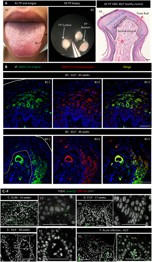
Methods: We performed fungiform papillae biopsies on 16 patients who reported taste disturbance lasting more than 6 weeks after molecularly determined severe acute respiratory syndrome coronavirus 2 (SARS-CoV-2) infection. Then, on multiple occasions, we rebiopsied 10 of those patients who still had taste complaints for at least 6 months postinfection. Fungiform papillae obtained from other patients before March 2020 served as negative controls. We performed hematoxylin and eosin staining to examine fungiform papillae morphology and immunofluorescence and fluorescence in situ hybridization to look for evidence of persistent viral infection and immune response.

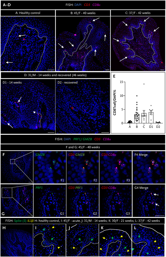
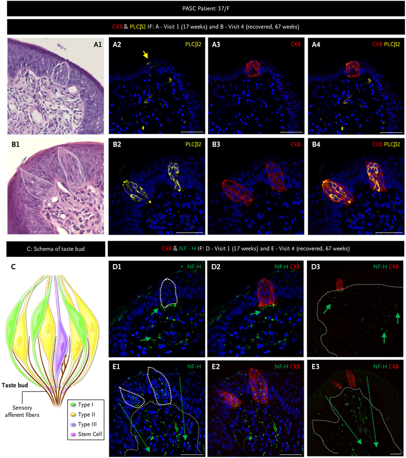
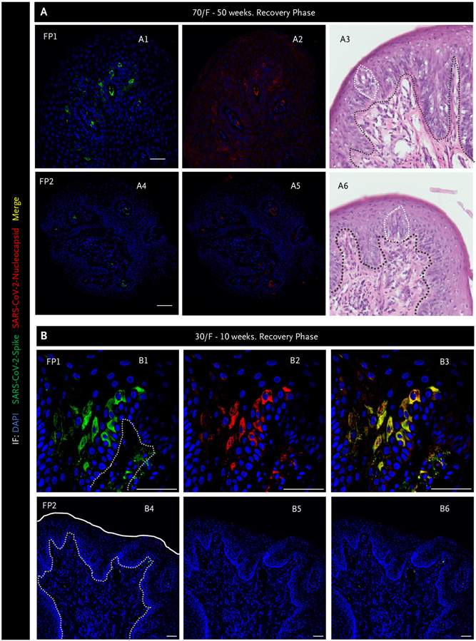
Results: In all patients, we found evidence of SARS-CoV-2, accompanying immune response and misshapen or absent taste buds with loss of intergemmal neurite fibers. Six patients reported normal taste perception by 6 months postinfection and were not further biopsied. In the remaining 10, the virus was eliminated in a seemingly random fashion from their fungiform papillae, but four patients still, by history, reported incomplete return to preinfection taste perception by the time we wrote this report.

Conclusions: Our data show a temporal association in patients between functional taste, taste papillae morphology, and the presence of SARS-CoV-2 and its associated immunological changes. (Funded by Intramural Research Program/National Institute on Aging/National Institute of Allergy and Infectious Diseases/National Institutes of Health; ClinicalTrials.gov numbers NCT03366168 and NCT04565067.).


Figures

Figure 1.
Figure 1.. Severe Acute Respiratory Syndrome Coronavirus 2 Persists in Fungiform Papillae from Postacute Sequelae of Covid-19 Patients.
Panel A1 shows a human tongue with a fungiform papilla outlined in a black circle. Panel A2 shows excised fungiform papillae (FP) under a dissection microscope. Panel A3 shows hematoxylin and eosin (H&E) staining of an fungiform papilla. The black dotted line delineates the epithelial layer from the lamina propria, whereas the blue dotted line delineates the epithelial suprabasal from basal cell layers. The red dotted line outlines a taste bud. The surface of the fungiform papilla is covered by a keratinized cell layer. Panel B presents representative images of immunofluorescent staining for severe acute respiratory syndrome coronavirus 2 (SARS-CoV-2) spike (green) and nucleocapsid (red) proteins in FP epithelium from two patients experiencing chronic taste disturbances post–SARS-CoV-2. Nuclei are stained in blue using 4′,6-diamidino-2-phenylindole (DAPI). The solid white line indicates the surface of FP, and the white dotted line demarcates the epithelium from lamina propria. Note consistent colocalization of spike (S) and nucleocapsid proteins, both of which are present in the FP epithelium and in a blood vessel in the lamina propria in Panel B2. Panels C to F, fluorescence in situ hybridization (FISH), demonstrate the presence of viral RNA using a probe for S (green dots) and a probe for the SARS-CoV-2 open reading frame 1 ab (ORF1ab) negative-strand RNA, produced when the virus is replicating (red dots). Inserts show a higher magnification of the areas marked by rectangles. In all instances, note the presence of S. The presence of ORF1ab indicates replication of the virus in FP. Scale bars indicate 50 μm. F denotes female; and M, male.
Figure 2.
Figure 2.. Fungiform Papillae in Postacute Sequelae Covid-19 Patients Are Characterized by the Presence of Activated CD8 T Cells and Cytokine Expression.
Panels A to D show fluorescence in situ hybridization (FISH) using probes for CD3 (red dots) and CD8α (purple dots) probes for cytotoxic CD8 T cells, in fungiform papillae (FP) of a healthy control (Panel A), and in patients with chronic taste disturbances (Panels B to D1) and after recovery (D2). White arrows indicate CD8 T cells located in the epithelial layer and lamina propria. The dotted white line demarcates the epithelial layer from the lamina propria. Panel E shows CD8 T cell quantification from FP sections of a healthy control (A) and three patients (B to D). Although formal morphometry was not done, there were visually greater numbers of CD8 T cells in the epithelial layer of FP from patients compared with panel A, whereas there was a return to control numbers after recovery (D1 vs. D2), for example. Panels F and G show FISH using probes to either granzyme B (GMZB) or perforin shown in green (where indicated) and CD3 (red dots) and CD8α (purple dots) in a 45-year-old female patient (F) at 40 weeks postinfection. Rectangles in panels F and G indicate the areas shown in higher magnification in F1 to F4 and G1 to G4, respectively. Panels H to L show FISH using probes for severe acute respiratory syndrome coronavirus 2 (SARS-CoV-2) spike (S, green dots) and the cytokine IL-1β (yellow dots) in FP of a healthy control (H), a patient with acute taste disturbances (I), and postacute sequelae of coronavirus disease 2019 (PASC) patients (J–L). The solid white line indicates the surface of FP, and the white dotted line demarcates the epithelial layer from the lamina propria. The green arrows indicate cells positive for S, and the yellow arrows indicate cells positive for IL-1β. Healthy FP controls do not express either S or IL-1β, whereas both are expressed in FP during acute infection and in PASC patients. Scale bars indicate 50 μm.
Figure 3.
Figure 3.. Taste Bud and Sensory Fiber Architecture Are Disrupted in Postacute Sequelae of Coronavirus Disease 2019 That Can Resolve Spontaneously.
We followed a 37-year-old female (F) with multiple biopsies from 17 weeks postinfection through to full recovery by 67 weeks using hematoxylin and eosin and immunofluorescent (IF) staining on adjacent fungiform papillae for the taste receptor cells marker cytokeratin 8 (CK8, red) present on all taste cell receptors; the type II taste cell receptor marker, phospholipase C beta-2 (PLCβ2, yellow); the taste cell receptor subtype that also has angiotensin-converting enzyme 2 (Panels A and B); and the nerve fiber marker neurofilament heavy (NF-H, green) (Panels D and E). Panel C schema shows taste cell receptor cell types and their afferent nerve fibers. Taste buds are composed of discreet clusters of taste cell receptors that include type I, II (the most numerous cell type in human fungiform papillae), and III, as well as a few stem cells that differentiate into mature taste cell receptors. The white dotted lines (A1, B1, D1, and E1) outline taste buds, and grey dotted lines (D3, E1, and E3) delineate the epithelial layer from the lamina propria. Nerve fibers run between taste cell receptors in taste buds, converging in the plexus beneath the taste bud and, from there, relay tastant information to the brain. At 17 weeks, a solitary disordered taste bud in 1/8 fungiform papillae was present, and it contained just one type II taste cell receptor (Panel A). By 67 weeks, fungiform papillae contains normal-appearing taste buds and type II taste cell receptors, normal orientation of the basal cell layer (Panel B), and nerve fibers that were disrupted and lacking in the taste bud at 17 weeks (Panel D) were now reinnervating the taste buds as well as the fungiform papillae lamina propria (Panel E). Scale bars indicate 50 μm. PASC denotes postacute sequelae of coronavirus disease 2019.
Figure 4.
Figure 4.. Severe Acute Respiratory Syndrome Coronavirus 2 Is Still Present in Some Fungiform Papillae as Taste Perception Recovers.
Immunofluorescence (IF) images for spike (green) and nucleocapsid (red) proteins and hematoxylin and eosin staining from two separate fungiform papillae (FP) (Panels A1 to A6). The black dotted line indicates the separation of the epithelial layer from lamina propria, whereas the white dotted line outlines taste buds. Panels A1 and A2 show one fungiform papilla (FP1) that still has virus present and has a disordered taste bud and basal cell layer (Panel A3), whereas Panels A4 and A5 show FP2 that does not contain virus and has a normal-appearing taste bud and stereotypical basal cell layer (Panel A6). Panel B has similar findings to Panel A. FP1 (Panels B1 to B3) contains virus, whereas the neighboring FP2 does not (Panel B4 to B6). The white line outlines the surface of the FP, and the white dotted lines delineate the epithelial layer from the lamina propria. However, no taste buds were present in either fungiform papilla. Note clearance of severe acute respiratory syndrome coronavirus 2 (SARS-CoV-2) does not occur simultaneously across all FP in an individual recovering taste perception but rather can occur randomly. Scale bars indicate 50 μm. DAPI denotes 4′,6-diamidino-2-phenylindole; and F, female.

References

    1. Doyle ME, Appleton A, Liu Q-R, Yao Q, Mazucanti CH, Egan JM. Human type II taste cells express angiotensin-converting enzyme 2 and are infected by severe acute respiratory syndrome coronavirus 2 (SARS-CoV-2). Am J Pathol 2021;191:1511–1519. DOI: 10.1016/j.ajpath.2021.05.010. - DOI - PMC - PubMed
    1. Robineau O, Zins M, Touvier M, et al. Long-lasting symptoms after an acute Covid-19 infection and factors associated with their resolution. JAMA Netw Open 2022;5:e2240985. DOI: 10.1001/jamanetworkopen.2022.40985. - DOI - PMC - PubMed
    1. Spielman AI, Pepino MY, Feldman R, Brand JG. Technique to collect fungiform (taste) papillae from human tongue. J Vis Exp 2010(42): e2201. DOI: 10.3791/2201. - DOI - PMC - PubMed
    1. Schindelin J, Arganda-Carreras I, Frise E, et al. Fiji: an open-source platform for biological-image analysis. Nat Methods 2012;9:676–682. DOI: 10.1038/nmeth.2019. - DOI - PMC - PubMed
    1. Mele D, Calastri A, Maiorano E, et al. High frequencies of functional virus-specific CD4+ T cells in SARS-CoV-2 subjects with olfactory and taste disorders. Front Immunol 2021;12:748881. DOI: 10.3389/fimmu.2021.748881. - DOI - PMC - PubMed

Publication types

Associated data