. 2024 Mar 1;326(3):C850-C865.
doi: 10.1152/ajpcell.00410.2023. Epub 2023 Dec 25.

WISP1 induces the expression of macrophage migration inhibitory factor in human lung fibroblasts through Src kinases and EGFR-activated signaling pathways

Affiliations

WISP1 induces the expression of macrophage migration inhibitory factor in human lung fibroblasts through Src kinases and EGFR-activated signaling pathways

Maria-Elpida Christopoulou et al. Am J Physiol Cell Physiol. .

Abstract

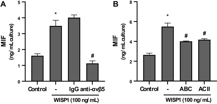
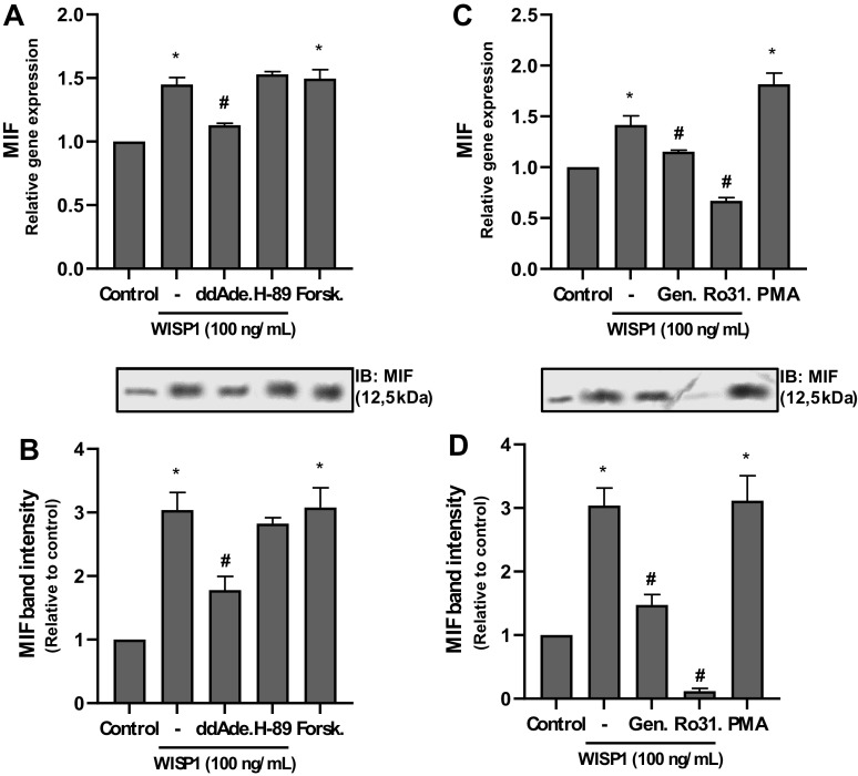
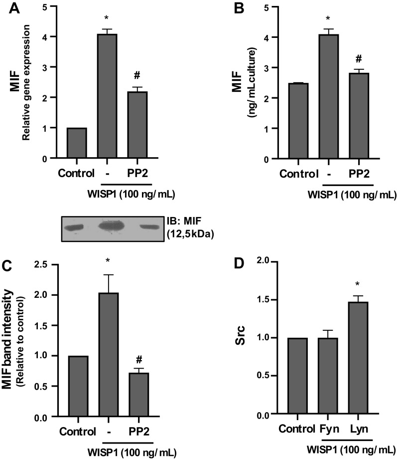
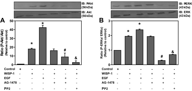
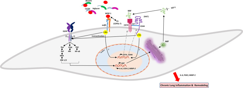
Wnt1-inducible signaling protein 1 (WISP1/CCN4) is a secreted matricellular protein that is implicated in lung and airway remodeling. The macrophage migration inhibitory factor (MIF) is a pleiotropic cytokine that has been associated with chronic lung diseases. In this study, we aimed to investigate the WISP1 signaling pathway and its ability to induce the expression of MIF in primary cultures of fibroblasts from normal human lungs (HLFs). Our results showed that WISP1 significantly stimulated the expression of MIF in a concentration- and time-dependent fashion. In WISP1-induced expression of MIF, αvβ5-integrin and chondroitin sulfate proteoglycans as well as Src tyrosine kinases, MAP kinases, phosphatidylinositol 3-kinase/Akt, PKC, and NF-κB were involved. WISP1-induced expression of MIF was attenuated in the presence of the Src kinase inhibitor PP2 or the MIF tautomerase activity inhibitor ISO-1. Moreover, WISP1 significantly increased the phosphorylation and activation of EGF receptor (EGFR) through transactivation by Src kinases. WISP1 also induced the expression of MIF receptor CD74 and coreceptor CD44, through which MIF exerts its effects on HLFs. In addition, it was found that MIF induced its own expression, as well as its receptors CD74/CD44, acting in an autocrine manner. Finally, WISP1-induced MIF promoted the expression of cyclooxygenase 2, prostaglandin E2, IL-6, and matrix metalloproteinase-2 demonstrating the regulatory role of WISP1-MIF axis in lung inflammation and remodeling involving mainly integrin αvβ5, Src kinases, PKC, NF-κB, and EGFR. The specific signaling pathways involved in WISP1-induced expression of MIF may prove to be excellent candidates for novel targets to control inflammation in chronic lung diseases.NEW & NOTEWORTHY The present study demonstrates for the first time that Wnt1-inducible signaling protein 1 (WISP1) regulates migration inhibitory factor (MIF) expression and activity and identifies the main signaling pathways involved. The newly discovered WISP1-MIF axis may drive lung inflammation and could result in the design of novel targeted therapies in inflammatory lung diseases.

Keywords: EGFR; MIF; Src kinases; WISP1/CCN4; lung inflammation.


Conflict of interest statement

No conflicts of interest, financial or otherwise, are declared by the authors.

Figures

None
Graphical abstract
Figure 1.
Figure 1.
Concentration- and time-dependent effects of Wnt1-inducible signaling protein 1 (WISP1) on the expression of migration inhibitory factor (MIF) in human lung fibroblasts (HLFs). AC: HLFs were cultured with different concentrations (0-100 ng/mL) of WISP1 for 48 h, and the expression of MIF at both the mRNA and protein levels was examined by quantitative PCR and ELISA/immunoblotting (IB), respectively. D: HLFs were cultured with WISP1 (100 ng/mL) for different time periods (0, 12, 24, and 48 h), and the secreted levels of MIF were determined by ELISA. Data represent the means ± SD (n = 3). *Statistically significant increase (P < 0.05) compared to control.
Figure 2.
Figure 2.
αvβ5-integrin and chondroitin sulfate proteoglycans are implicated in Wnt1-inducible signaling protein 1 (WISP1)-induced production of migration inhibitory factor (MIF) from human lung fibroblasts (HLFs). HLFs were pretreated for 30 minutes with anti-αvβ5-integrin antibody (6 µg/mL) (A) or for 2 h with chondroitinase ABC or chondroitinase AC II (3 units/mL) (B), followed by stimulation with WISP1 (100 ng/mL) for 48 h, and the levels of MIF in conditioned media were determined by ELISA. Data represent the means ± SD (n = 3). *Statistically significant increase (P < 0.05) compared to control. #Statistically significant decrease (P < 0.05) compared to WISP1-treated cells. IgG represents immunoglobulins from nonimmunized mouse.
Figure 3.
Figure 3.
cAMP pathway, PKC, and tyrosine kinases are implicated in Wnt1-inducible signaling protein 1 (WISP1)-induced expression of migration inhibitory factor (MIF) in human lung fibroblasts (HLFs). A and B: HLFs were pretreated for 30 minutes with adenylate cyclase inhibitor 2′,5′-dideoxy-adenosine (ddAde.; 10 µM) or PKA inhibitor H-89 (5 µM) followed by stimulation with WISP1 (100 ng/mL) for 48 or were stimulated with adenylate cyclase activator forskolin (Forsk.; 10 µM) alone for 48 h and the expression of MIF at both the mRNA and protein levels was examined by quantitative PCR (qPCR) and immunoblotting (IB), respectively. C and D: HLFs were pretreated for 30 minutes with the PKC inhibitor Ro31-8220 (Ro31.; 5 µM) or the tyrosine kinase inhibitor genistein (Gen.; 100 µM) followed by stimulation with WISP1 (100 ng/mL) for 48 h or were stimulated with PKC activator PMA (100 nM) alone for 48 h, and the expression of MIF at both the mRNA and protein levels was examined by qPCR and IB, respectively. Data represent the means ± SD (n = 3). *Statistically significant increase (P < 0.05) compared to control. #Statistically significant decrease (P < 0.05) compared to WISP1-treated cells.
Figure 4.
Figure 4.
Src kinases are implicated in Wnt1-inducible signaling protein 1 (WISP1)-induced expression of migration inhibitory factor (MIF) in human lung fibroblasts (HLFs). AC: HLFs were pretreated for 30 minutes with the Src kinase inhibitor PP2 (1 µM) followed by stimulation with WISP1 (100 ng/mL) for 48 h, and the expression of MIF at both the mRNA and protein levels was examined by quantitative PCR (qPCR) and ELISA/immunoblotting (IB), respectively. D: HLFs were treated with WISP-1 (100 ng/mL) for 48 h, and the mRNA expression of Src kinases Fyn and Lyn was examined by qPCR analysis. Data represent the means ± SD (n = 3). *Statistically significant increase (P < 0.05) compared to control. #Statistically significant decrease (P < 0.05) compared to WISP1-treated cells.
Figure 5.
Figure 5.
NF-κB is implicated in Wnt1-inducible signaling protein 1 (WISP1)-induced expression of migration inhibitory factor (MIF) in human lung fibroblasts (HLFs). A: HLFs were treated with WISP1 for the indicated time intervals and P-p65 was determined by immunoblotting (IB). BD: HLFs were pretreated for 30 minutes with the NF-κB activation inhibitor Bay-11-7082 (BAY; 5 µM) followed by stimulation with WISP1 (100 ng/mL) for 48 h, and the expression of MIF at both the mRNA and protein levels was examined by quantitative PCR and ELISA/IB, respectively. Data represent the means ± SD (n = 3). *Statistically significant increase (P < 0.05) compared to time zero (A) or control (B and C). #Statistically significant decrease (P < 0.05) compared to WISP1-treated cells.
Figure 6.
Figure 6.
Wnt1-inducible signaling protein 1 (WISP1) activates ERK1/2 and Akt. Human lung fibroblasts (HLFs) were pretreated for 30 minutes with the Src kinase inhibitor PP2 (1 µM) or with the EGF receptor tyrosine kinase activity inhibitor AG-1478 (1 µM) followed by stimulation with WISP1 or EGF (100 ng/mL) each for 7 minutes, and P-Akt (A) and P-ERK1/2 (B) were determined by immunoblotting (IB). Data represent the means ± SD (n = 3). *Statistically significant increase (P < 0.05) compared to control. #Statistically significant decrease (P < 0.05) compared to EGF-treated cells. &Statistically significant decrease (P < 0.05) compared to WISP1-treated cells.
Figure 7.
Figure 7.
MAP kinases (MAPKs) and phosphatidylinositol 3-kinase (PI3K) are implicated in Wnt1-inducible signaling protein 1 (WISP1)-induced expression of migration inhibitory factor (MIF) in human lung fibroblasts (HLFs). A–C: HLFs were pretreated for 30 minutes with the MAPK inhibitors U0126 (10 μM), SP600125 (SP; 20 μM), SB203580 (SB; 10 μM), and the PI3K inhibitor LY294002 (LY; 10 μΜ) followed by stimulation with WISP1 (100 ng/mL) for 48 h and the expression of MIF at both the mRNA and protein levels was examined by quantitative PCR and ELISA/immunoblotting (IB), respectively. Data represent the means ± SD (n = 3). *Statistically significant increase (P < 0.05) compared to control. #Statistically significant decrease (P < 0.05) compared to WISP1-treated cells.
Figure 8.
Figure 8.
EGF is able to induce the expression of migration inhibitory factor (MIF), and EGF receptor (EGFR) is activated by WISP1 through transactivation by Src kinases in human lung fibroblasts (HLFs). A: HLFs were stimulated with EGF (100 ng/mL) for 48 h, and the mRNA expression of MIF was examined by quantitative PCR. Data represent the means ± SD (n = 3). *Statistically significant increase (P < 0.05) compared to control. B: HLFs were pretreated for 30 minutes with the Src kinase inhibitor PP2 (1 µM) or with the EGFR tyrosine kinase activity inhibitor AG-1478 (1 µM) followed by stimulation with WISP1 (100 ng/mL) for 5 minutes. Then, cells were lysed and extracted, and the cell extracts were subjected to immunoprecipitation, using a polyclonal antibody against EGFR and a protein A agarose bead slurry. After centrifugation, the pellets were treated with Laemmli sample buffer and subjected to immunoblotting (IB) using mouse mAb against phosphotyrosine (P-Tyr) and rabbit mAb against EGFR. Data represent the means ± SD (n = 3) of the P-EGFR/total EGFR ratio. *Statistically significant increase (P < 0.05) compared to control. #Statistically significant decrease (P < 0.05) compared to WISP1-treated cells.
Figure 9.
Figure 9.
Migration inhibitory factor (MIF) is able to stimulate its own gene expression in human lung fibroblasts (HLFs). AC: HLFs were pretreated for 30 minutes with the MIF inhibitor ISO-1 (100 μM), followed by stimulation with WISP1 (100 ng/mL) for 48 h, and the expression of MIF at both the mRNA and protein levels was examined by quantitative PCR (qPCR) and ELISA/immunoblotting (IB), respectively. Data represent the means ± SD (n = 3). *Statistically significant increase (P < 0.05) compared to control. #Statistically significant decrease (P < 0.05) compared to WISP1-treated cells. D: HLFs were stimulated with recombinant MIF (rMIF) protein (100 ng/mL) for 48 h, and the mRNA expression of MIF was examined by qPCR. Data represent the means ± SD (n = 3). *Statistically significant increase (P < 0.05) compared to control.
Figure 10.
Figure 10.
Wnt1-inducible signaling protein 1 (WISP1) and migration inhibitory factor (MIF) induce the expression of CD74 and CD44 receptors in human lung fibroblasts (HLFs). HLFs were pretreated for 30 minutes with the Src kinase inhibitor PP2 (1 µM) or the MIF inhibitor ISO-1 (100 μM), followed by stimulation with WISP1 (100 ng/mL) for 48 h, or cells were stimulated with rMIF (100 ng/mL) alone for 48 h, and the expression of CD74 or CD44 at both the mRNA and protein levels was examined by quantitative PCR (A and B) and immunoblotting (IB; C and D), respectively. Data represent the means ± SD (n = 3). *Statistically significant increase (P < 0.05) compared to control. #Statistically significant decrease (P < 0.05) compared to WISP1-treated cells.
Figure 11.
Figure 11.
Wnt1-inducible signaling protein 1 (WISP1) induces the expression of cyclooxygenase (COX)-2, IL-6, and matrix metalloproteinase (MMP)-2 and the production of prostaglandin E2 (PGE2) from HLFs. Human lung fibroblasts (HLFs) were pretreated for 30 minutes with the Src kinase inhibitor PP2 (1 µM) or the MIF inhibitor ISO-1 (100 µM) followed by stimulation with WISP1 (100 ng/mL) for 48 h; the mRNA expression of COX-2, IL-6, and MMP-2 was examined by quantitative PCR (A, C, and E); and the levels of PGE2, IL-6, and pro-MMP-2 in conditioned media were determined by ELISA (B and D) and gelatin zymography (F), respectively. Data represent the means ± SD (n = 3). *Statistically significant increase (P < 0.05) compared to control. #Statistically significant decrease (P < 0.05) compared to WISP1-treated cells.
Figure 12.
Figure 12.
Wnt1-inducible signaling protein 1 (WISP1)-induced MIF promotes the migration of human lung fibroblasts (HLFs). HLFs were stimulated with migration inhibitory factor (MIF; 100 ng/mL) alone or they were pretreated for 30 minutes with the Src kinase inhibitor PP2 (1 µM) or the MIF inhibitor ISO-1 (100 μM), followed by stimulation with WISP1 (100 ng/mL) for 0, 24, and 48 h and the migration of cells was examined by the wound healing assay; 10% FCS was used as a positive control. Data represent the means area occupied by cells (%) ± SD (n = 3). *Statistically significant increase (P < 0.05) compared to control (0.1% FCS). #Statistically significant decrease (P < 0.05) compared to WISP1-treated cells.
Figure 13.
Figure 13.
Proposed model of the effects of Wnt1-inducible signaling protein 1 (WISP1) on the expression of migration inhibitory factor (MIF) and its receptors, as well as on the expression of cyclooxygenase (COX)-2, IL-6, and matrix metalloproteinase (MMP)-2, and production of prostaglandin E2 (PGE2) from human lung fibroblasts (HLFs). The effect of WISP-1 on the expression of MIF in HLFs with the complex interactions between WISP-1, αvβ5, Src kinases, EGF receptor (EGFR), and CD74/CD44 receptors, reveal intricate regulatory networks that could contribute to the expression of proinflamamtory molecules and remodeling enzymes secretion.

References

    1. Deng W, Fernandez A, McLaughlin SL, Klinke DJ 2nd.. WNT1-inducible signaling pathway protein 1 (WISP1/CCN4) stimulates melanoma invasion and metastasis by promoting the epithelial-mesenchymal transition. J Biol Chem 294: 5261–5280, 2019. doi:10.1074/jbc.RA118.006122. - DOI - PMC - PubMed
    1. Perbal B, Tweedie S, Bruford E. The official unified nomenclature adopted by the HGNC calls for the use of the acronyms, CCN1-6, and discontinuation in the use of CYR61, CTGF, NOV and WISP 1-3 respectively. J Cell Commun Signal 12: 625–629, 2018. [Erratum in J Cell Commun Signal 13: 435, 2019]. doi:10.1007/s12079-018-0491-1. - DOI - PMC - PubMed
    1. Holbourn KP, Acharya KR, Perbal B. The CCN family of proteins: structure-function relationships. Trends Biochem Sci 33: 461–473, 2008. doi:10.1016/j.tibs.2008.07.006. - DOI - PMC - PubMed
    1. Lau LF. Cell surface receptors for CCN proteins. J Cell Commun Signal 10: 121–127, 2016. doi:10.1007/s12079-016-0324-z. - DOI - PMC - PubMed
    1. Hou CH, Chiang YC, Fong YC, Tang CH. WISP-1 increases MMP-2 expression and cell motility in human chondrosarcoma cells. Biochem Pharmacol 81: 1286–1295, 2011. doi:10.1016/j.bcp.2011.03.016. - DOI - PubMed

Publication types