. 2024 Feb:340:199303.
doi: 10.1016/j.virusres.2023.199303. Epub 2023 Dec 30.

Inhibition of porcine deltacoronavirus entry and replication by Cepharanthine

Affiliations

Inhibition of porcine deltacoronavirus entry and replication by Cepharanthine

Yumei Sun et al. Virus Res. 2024 Feb.

Abstract

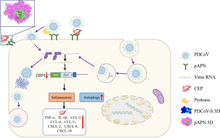
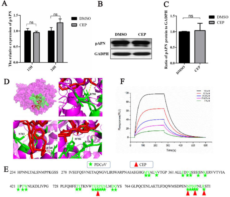
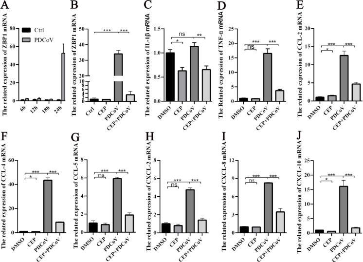
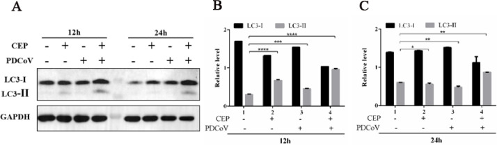
Porcine deltacoronavirus (PDCoV) is an emerging swine enteropathogenic coronavirus (CoV) that mainly causes acute diarrhea/vomiting, dehydration, and mortality in piglets, possessing economic losses and public health concerns. However, there are currently no proven effective antiviral agents against PDCoV. Cepharanthine (CEP) is a naturally occurring alkaloid used as a traditional remedy for radiation-induced symptoms, but its underlying mechanism of CEP against PDCoV has remained elusive. The aim of this study was to investigate the anti-PDCoV effects and mechanisms of CEP in LLC-PK1 cells. The results showed that the antiviral activity of CEP was based on direct action on cells, preventing the virus from attaching to host cells and virus replication. Importantly, Surface Plasmon Resonance (SPR) results showed that CEP has a moderate affinity to PDCoV receptor, porcine aminopeptidase N (pAPN) protein. AutoDock predicted that CEP can form hydrogen bonds with amino acid residues (R740, N783, and R790) in the binding regions of PDCoV and pAPN. In addition, RT-PCR results showed that CEP treatment could significantly reduce the transcription of ZBP1, cytokine (IL-1β and IFN-α) and chemokine genes (CCL-2, CCL-4, CCL-5, CXCL-2, CXCL-8, and CXCL-10) induced by PDCoV. Western blot analysis revealed that CEP could inhibit viral replication by inducing autophagy. In conclusion, our results suggest that the anti-PDCoV activity of CEP is not only relies on competing the virus binding with pAPN, but also affects the proliferation of the virus in vitro by downregulating the excessive immune response caused by the virus and inducing autophagy. CEP emerges as a promising candidate for potential anti-PDCoV therapeutic development.

Keywords: Antivirals; Cepharanthine; Entry; Porcine deltacoronavirus; Replication.


Conflict of interest statement

Declaration of Competing Interest The authors declare that they have no known competing financial interests or personal relationships that could have appeared to influence the work reported in this paper.

Figures

Image, graphical abstract
Graphical abstract
Fig 1
Fig. 1
Antiviral effect of CEP on PDCoV infection. The effect of CEP on LL-CPK1 cell viability were was detected using the CCK8 assay. (B) The effect of different concentrations of CEP on PDCoV-infected cells determined by Fluorescence microscopy. Green fluorescence represents PDCoV distribution; blue fluorescence represents nuclei from DAPI. (C) Flow cytometric analysis was used to detect infected cells in the presence or absence of CEP with PDCoV (MOI=0.1) for 24 h. (D) Quantitative analysis of the data shown in C. (E-F) The infected LLC-PK1 cells were treated with or without CEP for 24 h, and the viral RNA copies and viral titers in the supernatant were examined by qPCR and TCID50. The shown results are representative of one experiment out of at least three experiments. Error bars represent ±1 SD; ns, no significant difference; *p<0.05, **p<0.01, ***p<0.001 by Student's t-test.
Fig 2
Fig. 2
Effect of CEP on different stages of the PDCoV replication cycle. The effect of CEP on PDCoV adsorption. CEP and PDCoV (MOI=5) were incubated with precooled cells for 2 h at 4 °C. Cells were washed with cold PBS three times, and cells were continued to grow for 8 h; The effect of CEP on PDCoV entry. Precooled cells were infected with PDCoV for 2 h at 4 °C and incubated with CEP for 1 h at 37 °C after washing with PBS, and cells were continued to grow for 8 h; The effect of CEP on PDCoV replication. Cells were infected with PDCoV for 1 h at 37 °C after washing with PBS, and cells were continued to grow with CEP for 8 h at 37 °C. PDCoV-infected cells were analyzed using Fluorescence microscopy (A) and flow cytometric analysis (B). (C) Viral RNA was extracted for qPCR quantification. (D) Viral titers in the supernatant were examined by TCID50. DMSO was added as the positive control. The experiments were repeated at least three times. Error bars represent ±SD; ns, no significant difference; ***p<0.001 by Student's t-test.
Fig 3
Fig. 3
CEP indirectly inhibits viral proliferation by acting on cells. Cell pretreatment (Pre-cell): Cells were preincubated with CEP for 2 h at 37 °C and washed with PBS three times. The preincubated cells were infected with PDCoV at an MOI of 1 for 1 h; Virus pretreatment (Pre-virus): PDCoV was preincubated with CEP for 2 h at 37 °C and then diluted 1000 times and incubated with LLC-PK1 cells for 1 h; Co-treatment: PDCoV and CEP were simultaneously added to LLC-PK1 cells for 1 h. The unbound virus was washed away with PBS, and the cells were cultured at 37 °C for 8 h in fresh medium. (A-B) PDCoV infection was observed by fluorescence staining and analysed by flow cytometric analysis. (C) The RNAs levels of PDCoV in the culture supernatants were extracted and measured by qPCR. (D) Viral titers in the culture supernatants were calculated by TCID50. DMSO was added as the positive control. The data are representative of mean values from three independent experiments. Error bars represent±SD; ns, no significant difference; ***p<0.001 by Student's t-test.
Fig 4
Fig. 4
Effect of CEP on the relative expression of the pAPN gene. (A) The relative gene expression level of pAPN in CEP-incubated cells was determined by RT‒PCR. (B) Verification of pAPN receptor expression in CEP-incubated cells by WB. (C) Quantitative analysis of the data shown in B. DMSO was added as the positive control. (D) The upper left of picture: Overlay of PDCoV footprints on pAPN. pAPN is shown as a surface representation, colored in pink. Residues on pAPN contacting the PDCoV RBD are colored green. The lower left and right of three picture: Atomic details of the interaction between pAPN RBD and CEP. Contacting residues on proteins are represented as sticks, with nitrogen and oxygen atoms colored green. CEP is represented as sticks colored in red. (E) Sequence alignment of pAPN. Residues on pAPN binding to PDCoV RBD are marked in green according to the code of the key above the sequences. The amino acids of PAPN that bind to CEP are marked by red triangles. (F) Analysis of the binding of pAPN and CEP using surface plasmon resonance (SPR).The experiments were repeated at least three times. Error bars represent ±SD; ns, no significant difference; ***p<0.001 by Student's t-test.
Fig 5
Fig. 5
CEP reduces inflammatory signaling and necrosis caused by PDCoV infection. The transcription level of ZBP1 in PDCoV-infected cells was determined by RT-qPCR after 6 h, 12 h, 18 h and 24 h, respectively. (B-J) LLC-PK1 cells were inoculated with DMSO or CEP and mock-infected or infected with PDCoV (MOI=) for 24 h. The total RNA of PDCoV-infected cells was harvested, and the relative mRNA levels of the ZBP1 and indicated cytokine (IL-1β and IFN-α) and chemokine genes (CCL-2, CCL-4, CCL-5, CXCL-2, CXCL-8, and CXCL-10) were measured by RT‒qPCR. For quantitative RT‒PCR analysis, β-actin mRNA was used as an internal control. The experiments were repeated at least three times. Error bars represent ±SD; ns, no significant difference; *p< 0.05, **p< 0.01, ***p< 0.001, ***p< 0.001 by Student's t-test.
Fig 6
Fig. 6
CEP inhibits PDCoV infection in LLC-PK1 cells by inducing autophagy. CEP inhibits PDCoV infection by inducing autophagy, as determined by Western blot analysis. LLC-PK1 cells were incubated with DMSO or CEP and mock-infected or infected with PDCoV at an MOI of 1. The cells were further maintained in the presence of DMSO or CEP and fixed at 12 h of virus infection. Cellular lysates and viral lysates were prepared and subjected to immunoblotting using an antibody against LC3-I, LC3-II, or GAPDH. (B-C) The expression levels of each protein were quantitatively analysed by densitometry and expressed as density values relative to the GAPDH gene, and multiples of changes in the GAPDH ratio for each protein were plotted. Data indicate the representative mean values from three independent experiments, and error bars represent ±SD; ns, no significant difference; ***p<0.001 by Student's t-test.
Fig 7
Fig. 7
CEP Confers Broad-Spectrum Resistance to other coronaviruses (TGEV and MHV). LLC-PK1 cells were incubated with TGEV in the presence or absence of CEP for 24 h. The propagation of TGEV in porcine testis cells was observed by IFA (A), and the virus titer was measured in ST cells by TCID50 assay (B). LR7 cells were infected with MHV in the presence or absence of CEP for 24 h, MHV in cells was observed by immunofluorescence microscopy (C), and viral titers in cell supernatant were examined by TCID50 (D). The results are representative of one of at least three experiments. Error bars represent ±1 SD; ns, no significant difference; *p<0.05, **p<0.01, ***p<0.001 by Student's t-test.

Similar articles

Cited by

References

    1. Bailly C. Cepharanthine: an update of its mode of action, pharmacological properties and medical applications. Phytomedicine. 2019;62 - PMC - PubMed
    1. Chitsike L., Krstenansky J., Duerksen-Hughes P.J. Advances in Pharmacological and Pharmaceutical Sciences 2021. 2021. ACE2: S1 RBD interaction-targeted peptides and small molecules as potential COVID-19 therapeutics. - PMC - PubMed
    1. Cottam E.M., Maier H.J., Manifava M., Vaux L.C., Chandra-Schoenfelder P., Gerner W., Britton P., Ktistakis N.T., Wileman T. Coronavirus NSP6 proteins generate autophagosomes from the endoplasmic reticulum via an omegasome intermediate. Autophagy. 2011;7:1335–1347. - PMC - PubMed
    1. Cottam E.M., Whelband M.C., Wileman T. Coronavirus NSP6 restricts autophagosome expansion. Autophagy. 2014;10:1426–1441. - PMC - PubMed
    1. Duan C., Ge X., Wang J., Wei Z., Feng W.H., Wang J. Ergosterol peroxide exhibits antiviral and immunomodulatory abilities against porcine deltacoronavirus (PDCoV) via suppression of NF-kappaB and p38/MAPK signaling pathways in vitro. Int. Immunopharmacol. 2021;93 - PMC - PubMed

Supplementary concepts

LinkOut - more resources