Review
. 2024 Feb;11(8):e2306470.
doi: 10.1002/advs.202306470. Epub 2023 Dec 25.

Diffusion-Based 3D Bioprinting Strategies

Affiliations
Review

Diffusion-Based 3D Bioprinting Strategies

Betty Cai et al. Adv Sci (Weinh). 2024 Feb.

Abstract

3D bioprinting has enabled the fabrication of tissue-mimetic constructs with freeform designs that include living cells. In the development of new bioprinting techniques, the controlled use of diffusion has become an emerging strategy to tailor the properties and geometry of printed constructs. Specifically, the diffusion of molecules with specialized functions, including crosslinkers, catalysts, growth factors, or viscosity-modulating agents, across the interface of printed constructs will directly affect material properties such as microstructure, stiffness, and biochemistry, all of which can impact cell phenotype. For example, diffusion-induced gelation is employed to generate constructs with multiple materials, dynamic mechanical properties, and perfusable geometries. In general, these diffusion-based bioprinting strategies can be categorized into those based on inward diffusion (i.e., into the printed ink from the surrounding air, solution, or support bath), outward diffusion (i.e., from the printed ink into the surroundings), or diffusion within the printed construct (i.e., from one zone to another). This review provides an overview of recent advances in diffusion-based bioprinting strategies, discusses emerging methods to characterize and predict diffusion in bioprinting, and highlights promising next steps in applying diffusion-based strategies to overcome current limitations in biofabrication.

Keywords: bioprinting; diffusion; interfacial gelation; multi-material constructs; perfusable structures.


Conflict of interest statement

The authors declare no conflict of interest.

Figures
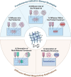
Figure 1
Figure 1
Summary of diffusion‐based bioprinting approaches and applications. Diffusion‐based bioprinting strategies can be subcategorized into those leveraging diffusion 1) into the printed ink, 2) out of the printed ink, and 3) within the printed construct. Applications of diffusion‐based bioprinting include 4) generation of multi‐material constructs via the interfacial diffusion of crosslinkers, and 5) generation of self‐supporting perfusable structures via diffusion‐induced gelation. For such approaches and applications, control of mass transport according to Fick's second law of diffusion (central schematic) is a key requirement.
Figure 2
Figure 2
Bioprinting strategies involving the inward diffusion of crosslinkers for in situ gelation. A) Extrusion bioprinting into a crosslinker‐containing medium results in crosslinker diffusion into the printed ink, facilitating in situ gelation. B) A section of a human coronary arterial tree was fabricated by extruding alginate into a calcium ion‐containing support bath. Reproduced under terms of the CC‐BY license.[ 25 ] Copyright 2015, The Authors, published by AAAS. C) Stable hydrogel fibers can be formed via coaxial bioprinting of a bioink in the inner nozzle and a crosslinker‐containing solution in the outer nozzle. Reproduced with permission.[ 16 ] Copyright 2016, Wiley‐VCH. D) Scaffolds with different patterns were fabricated by coaxial printing of a GelMA‐alginate ink and calcium chloride solution. Reproduced with permission.[ 18 ] Copyright 2017, Elsevier. E) A GelMA‐alginate scaffold containing endothelial cells (cytoskeleton in green; nuclei in blue) was fabricated by the coaxial extrusion of an endothelial cell‐laden GelMA‐alginate bioink and calcium chloride solution. Reproduced with permission.[ 18 ] Copyright 2017, Elsevier.
Figure 3
Figure 3
Bioprinting strategies involving the inward diffusion of molecules that trigger crosslinking or self‐assembly. A) A bioink containing polymers with phenolic hydroxyl moieties (polymer‐Ph) and horseradish peroxidase (HRP) was printed into air containing vaporized H2O2. During printing, H2O2 diffused into the ink and induced HRP‐catalyzed crosslinking, enabling the formation of stable constructs with a variety of geometries (left). Bioinks composed of hyaluronic acid (HA) and gelatin, both possessing phenolic hydroxyl (Ph) moieties, were printed using this peroxidase‐catalyzed technique. A 3D reconstruction of fluorescently labeled mouse fibroblasts encapsulated within the composite bioink showed cell spreading within the 3D matrix after three days of culture (right). Reproduced with permission.[ 35 ] Copyright 2018, IOP Publishing. B) A Pluronic F127‐dimethacrylate ink was mixed with an initiator (APS) and extruded into a support bath containing a catalyst (TEMED). The diffusion of catalyst into the printed ink enabled in situ polymerization inside the support bath, allowing the fabrication of complex 3D constructs with high stability and shape retention. Reproduced with permission.[ 36 ] Copyright 2017, American Chemical Society. C) A solution of amphiphiles in DMSO was extruded into a water bath. The diffusion of water into the printed ink facilitated rapid self‐assembly of amphiphiles into fibers, thus stabilizing the ink filament. Reproduced with permission.[ 38 ] Copyright 2020, Elsevier.
Figure 4
Figure 4
Bioprinting strategies involving post‐printing gelation through the inward diffusion of crosslinkers. Examples: A) A gelatin‐alginate bioink was extruded onto a cooled printing platform for the diffusion‐independent thermal gelation of gelatin. The construct was then transferred into a calcium chloride (CaCl2) solution, where the inward diffusion of Ca2+ led to second‐stage ionic crosslinking of alginate to form open‐porous scaffolds with varying infill patterns (strand‐to‐strand distances, DL : 2–4 mm). Reproduced under terms of the CC‐BY license.[ 42 ] Copyright 2016, The Authors, published by Springer Nature. B) Bioinks composed of ɑ‐cyclodextrin (ɑ‐CD), pegylated chitosan (CS‐mPEG), and gelatin formed a supramolecular hydrogel inside the print syringe. After printing, constructs were transferred into a β‐glycerophosphate (β‐GPS) solution for second‐stage crosslinking. Dual‐stage‐crosslinked lattices retained their shape for 21 days, while lattices with only one stage of crosslinking (by either β‐GPS or ɑ‐CD) collapsed within 30 min. Reproduced with permission.[ 46 ] Copyright 2020, KeAi Publishing.
Figure 5
Figure 5
Bioprinting strategies leveraging inward diffusion to introduce additional functionality. A) Printed methacrylated alginate (AA‐MA) films were crosslinked by light irradiation, then transferred into an aqueous medium with or without ions for spontaneous folding into tubes. For printed AA‐MA films, reversible folding/unfolding behavior was achieved by immersion in calcium chloride solution to trigger unfolding, followed by immersion in EDTA to trigger re‐folding. Reproduced with permission.[ 53 ] Copyright 2017, Wiley‐VCH. B) The diffusion of chitosan into printed methacrylated hyaluronic acid (HA‐MA) constructs resulted in shrinkage due to charge complexation. The degree of shrinkage was dependent on the immersion time in chitosan solution. Reproduced under terms of the CC‐BY license.[ 54 ] Copyright 2020, The Authors, published by Elsevier Nature. C) The diffusion of citrate ions into printed sacrificial gelatin‐chitosan inks was used to form a non‐sacrificial double‐network hydrogel. An inverted hollow pyramid was fabricated by sequentially patterning gelatin‐chitosan and citrate inks. Reproduced with permission.[ 50 ] Copyright 2020, IOP Publishing. D) Printed constructs composed of peptide‐conjugated HA were treated with complementary peptides, which diffused inward and subsequently associated with the hydrogel. To enable biomineralization, constructs were first incubated with a biotin‐labeled peptide (JR2KK‐Biotin), which recruited avidin‐modified proteins, then exposed to streptavidin‐modified alkaline phosphatase (streptavidin‐ALP) for ALP functionalization. The resultant ALP‐containing constructs exhibited mineral deposition in the presence of Ca2+. Reproduced under terms of the CC‐BY license.[ 55 ] Copyright 2020, The Authors, published by IOP Publishing.
Figure 6
Figure 6
Methods leveraging outward diffusion to alter the properties of bioprinted constructs post‐printing. A) A temporary viscosity modifier can be included to improve ink printability and stability. After crosslinking of the printed ink, the viscosity modifier diffuses out of the construct. B) The diffusion of methylcellulose (MC) out of an alginate‐MC construct over time was observed by staining MC (dark violet) using a chlorine‐zinc‐iodine solution. Reproduced with permission.[ 57 ] Copyright 2017, Wiley‐VCH. C) The mechanical properties of bioinks were modulated using small molecule catalysts and competitors, which diffused out of the bioink after printing to stabilize the printed construct. Hyaluronan and Elastin‐Like Protein (HELP) inks containing competitor and catalyst were printable while maintaining stability over two weeks. Reproduced under terms of the CC‐BY license.[ 62 ] Copyright 2023, The Authors, published by AAAS.
Figure 7
Figure 7
Bioprinting strategies leveraging diffusion within a printed construct. A) The diffusion of morphogens encapsulated within printed inks can be used to generate defined spatial gradients, which are initially discrete and become increasingly continuous over time within the printed construct. B) Spatial gradients of nerve growth factor (NGF) and glial cell line‐derived growth factor (GDNF) were generated by printing growth factor‐loaded hydrogels along a 3D‐printed nerve conduit (left). Diffusion of growth factors over time enabled the formation of continuous growth factor gradients, which were predicted by finite element modeling (right). Reproduced with permission.[ 65 ] Copyright 2015, Wiley‐VCH. C) The differentiation factors transforming growth factor‐β3 (TGF‐β3) and bone morphogenetic protein‐2 (BMP‐2) were included in zonally defined core compartments of a core‐shell‐printed structure to induce chondrogenic and osteogenic differentiation of shell‐encapsulated human chondrocytes (hChon) and pre‐osteoblasts (hOB), respectively, by diffusion from the core depot to the shell. Using this locally restricted diffusion strategy, the co‐differentiation of different cell types in multi‐zonal constructs can be achieved. Reproduced under terms of the CC‐BY license.[ 66 ] Copyright 2022, The Authors, published by IOP Publishing.
Figure 8
Figure 8
Bioprinting approaches leveraging diffusion to generate multi‐material constructs. A) Constructs with multiple cell types were fabricated by depositing a sacrificial (vascular) ink and a cell‐laden gelatin‐fibrinogen bioink, then casting a gelatin‐fibrinogen matrix over the printed inks. Crosslinking was facilitated by the diffusion of thrombin and transglutaminase from the cast matrix into the cell ink as well as the diffusion of thrombin from the vascular ink into the cast matrix. Reproduced with permission.[ 73 ] Copyright 2016, National Academy of Sciences. B) A construct demonstrated the patterning of three different cell types, including human umbilical vein endothelial cells (HUVECs), human neonatal dermal fibroblasts (hNDFs), and human mesenchymal stromal cells (hMSCs) within the printed vasculature, cast matrix, and cell ink, respectively. Reproduced with permission.[ 73 ] Copyright 2016, National Academy of Sciences. C) Multiple crosslinkers were included in a single embedding medium, allowing the fabrication of constructs integrating biopolymers with different crosslinking mechanisms. Reproduced with permission.[ 74 ] Copyright 2022, IOP Publishing. D) UNIversal Orthogonal Network (UNION) bioinks enabled multi‐material bioprinting based on the diffusion of a small molecule crosslinker from a support bath into the printed structure. Cohesive structures composed of gelatin (red) and PEG (blue) were fabricated using the UNION strategy. Reproduced with permission.[ 76 ] Copyright 2021, Wiley‐VCH.
Figure 9
Figure 9
Diffusion‐based coaxial bioprinting strategies to fabricate self‐supporting perfusable structures. A) Perfusable channels were generated by co‐extruding a crosslinker solution in the inner nozzle and a hydrogel precursor in the outer nozzle. Reproduced with permission.[ 78 ] Copyright 2014, Elsevier. B) Coaxial printing of a CaCl2‐containing core and an alginate shell was used to fabricate structures perfusable with yellow food dye. Both single‐ and multi‐layered perfusable structures were fabricated through continuous coaxial extrusion. Reproduced with permission.[ 79 ] Copyright 2013, IOP Publishing. C) Channels incorporating two cell types were fabricated via the co‐extrusion of a core sacrificial ink containing crosslinker and endothelial cells (HUVECs, red) and shell bioink of catechol‐modified GelMA (GelMA/C) containing smooth muscle cells (HCASMCs, green). Reproduced with permission.[ 81 ] Copyright 2019, IOP Publishing.
Figure 10
Figure 10
Diffusion‐based interfacial gelation methods to fabricate self‐supporting perfusable structures. A) Hollow structures were generated by extruding a photo‐crosslinkable bioink into a photoinitiator solution. Free radicals produced by a laser beam diffused into the printed filament, crosslinking an outer shell of the filament. Reproduced under terms of the CC‐BY license.[ 84 ] Copyright 2021, The Authors, published by Frontiers Media S.A. B) A branched, perfusable network was fabricated by extruding a polylysine (PLL) ink into an oxidized bacterial cellulose (oxBC) solution. PLL diffused across the interface between the ink and oxBC solution, enabling the formation of complex coacervates. Reproduced with permission.[ 86 ] Copyright 2023, Wiley‐VCH. C) In the GUIDE‐3DP strategy, a sacrificial ink containing a reaction‐initiator was extruded into a gel precursor support bath, after which the reaction‐initiator diffused into the gel precursor to enable crosslinking. GUIDE‐3DP is compatible with a variety of materials and enabled the fabrication of complex perfusable networks, such as a model of the coronary vasculature. Reproduced with permission.[ 87 ] Copyright 2023, Wiley‐VCH.
Figure 11
Figure 11
Experimental and computational characterization of diffusion in bioprinting. A) Fluorescence imaging was used to characterize the diffusion of a rhodamine‐based dye through a crosslinked Pluronic F127‐diacrylate hydrogel. The spatial peak variance σ 2 was calculated based on fluorescence intensity profiles, while the diffusion coefficient was determined from the slope of σ 2 as a function of time. Reproduced with permission.[ 102 ] Copyright 2011, Wiley‐VCH. B) Finite element modeling was used to predict the diffusion of core and sheath inks inside a coaxial nozzle. The concentration profiles of GelMA, which diffused outward from the core to the sheath, and glycerol, which diffused inward from the sheath to the core, were determined at varying distances along the nozzle. Reproduced with permission.[ 118 ] Copyright 2021, American Chemical Society. C) Finite element models were used to predict the diffusion of a photoinitiator from ink filaments printed into a GelMA support bath. The predicted concentration profiles were correlated with experimental results for a crosslinked bifurcated channel. Reproduced with permission.[ 87 ] Copyright 2023, Wiley‐VCH.

References

    1. Derakhshanfar S., Mbeleck R., Xu K., Zhang X., Zhong W., Xing M., Bioact Mater 2018, 3, 144. - PMC - PubMed
    1. Bernal P. N., Delrot P., Loterie D., Li Y., Malda J., Moser C., Levato R., Adv. Mater. 2019, 31, 1904209. - PubMed
    1. Größbacher G., Bartolf‐Kopp M., Gergely C., Bernal P. N., Florczak S., de Ruijter M., Rodriguez N. G., Groll J., Malda J., Jungst T., Levato R., Adv. Mater. 2023, 35, 2300756. - PubMed
    1. Murphy S. V., Atala A., Nat. Biotechnol. 2014, 32, 773. - PubMed
    1. Zhang Y. S., Haghiashtiani G., Hübscher T., Kelly D. J., Lee J. M., Lutolf M., McAlpine M. C., Yeong W. Y., Zenobi‐Wong M., Malda J., Nat. Rev. Methods Primers 2021, 1, 75.

Publication types

MeSH terms

LinkOut - more resources