. 2024 Mar;11(9):e2308686.
doi: 10.1002/advs.202308686. Epub 2023 Dec 25.

Hypertensive Pressure Mechanosensing Alone Triggers Lipid Droplet Accumulation and Transdifferentiation of Vascular Smooth Muscle Cells to Foam Cells

Affiliations

Hypertensive Pressure Mechanosensing Alone Triggers Lipid Droplet Accumulation and Transdifferentiation of Vascular Smooth Muscle Cells to Foam Cells

Pamela Swiatlowska et al. Adv Sci (Weinh). 2024 Mar.

Abstract

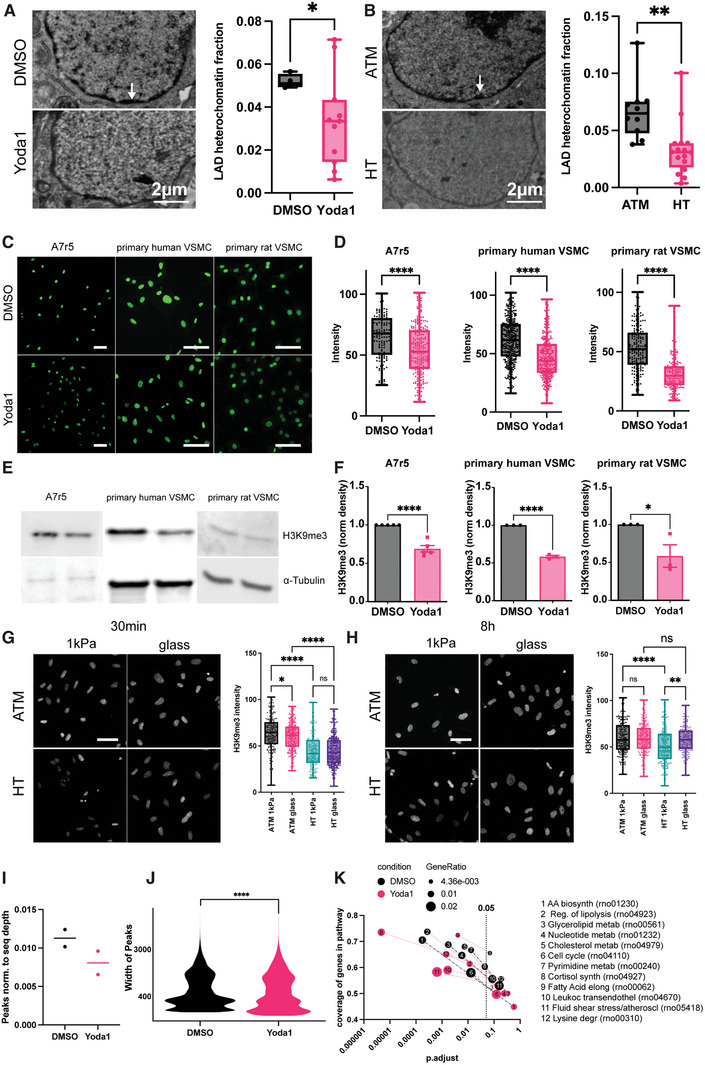
Arterial Vascular smooth muscle cells (VSMCs) play a central role in the onset and progression of atherosclerosis. Upon exposure to pathological stimuli, they can take on alternative phenotypes that, among others, have been described as macrophage like, or foam cells. VSMC foam cells make up >50% of all arterial foam cells and have been suggested to retain an even higher proportion of the cell stored lipid droplets, further leading to apoptosis, secondary necrosis, and an inflammatory response. However, the mechanism of VSMC foam cell formation is still unclear. Here, it is identified that mechanical stimulation through hypertensive pressure alone is sufficient for the phenotypic switch. Hyperspectral stimulated Raman scattering imaging demonstrates rapid lipid droplet formation and changes to lipid metabolism and changes are confirmed in ABCA1, KLF4, LDLR, and CD68 expression, cell proliferation, and migration. Further, a mechanosignaling route is identified involving Piezo1, phospholipid, and arachidonic acid signaling, as well as epigenetic regulation, whereby CUT&Tag epigenomic analysis confirms changes in the cells (lipid) metabolism and atherosclerotic pathways. Overall, the results show for the first time that VSMC foam cell formation can be triggered by mechanical stimulation alone, suggesting modulation of mechanosignaling can be harnessed as potential therapeutic strategy.

Keywords: atherosclerosis; foam cells; mechanosensing; pressure sensing; vascular smooth muscle cells.


Conflict of interest statement

The authors declare no conflict of interest.

Figures

Figure 1
Figure 1
Piezo1 localises to the VSMC nuclear lamina and controls calcium levels. Immunofluorescent micrographs shows enrichment of Piezo1 at the nuclear membrane of A7r5 VSMCs (A–C). A) Anti‐piezo1 staining shows a dotted appearance around the nuclear membrane (zoom in inset). B) Example line profile (line indicated in A) shows enrichment at the edges. C) Quantification of enrichment against cytoplasm and nuclear intensity. **** P<0.0001 from one‐sample t‐test, compared to a value of 1, i.e., no enrichment. D) The nuclear localisation is further confirmed using a Piezo1‐GFP transfection (zoom in inset); here co‐transfected with Tractin‐Tomato. E–J) Yoda1 treatment leads to rapid Ca2+ transients in A7r5 (E,F), primary human (G,H) and primary rat (I,J) VSMCs. Data from three independent repeats with n = 30–45 cells.
Figure 2
Figure 2
Chronic Yoda1 treatment supports transition to a foam cell phenotype. A‐B) Wound scratch assay shows reduced migration of Yoda1 treated A7r5 cells. C) Nanoindentation indicates a lower Young's Modulus after Yoda1 treatment. D,E) Click‐EdU assay indicates higher cell proliferation following Yoda1 incubation, which is reversed after simultaneous Dooku1 treatment. F,G) TUNEL staining indicates no significant changes to apoptosis after 8 h Yoda1 treatment. H–L). qPCR testing indicate increased CD68 (H), KLF4 (I), LDLR (J) and lower ABCA1 (K) transcription after 8‐hour Yoda1 treatment, while LGALS3 (L) showed no change at this time point. p‐values from unpaired two‐tailed t‐tests (B,C, H–L), or one‐way ANOVA with Tukey correction for multiple comparisons (E,G): * p<0.0332, ** p<0.0021, *** p<0.0002.
Figure 3
Figure 3
Hyperspectral stimulated Raman Scattering (hsSRS) indicates lipid droplet accumulation and lipid metabolic changes after pressure and Yoda stimulation. A,B) Acute Piezo1 activation and HT pressure stimulation leads to lipid accumulation in A7r5 cells. A) Lipid droplet segmentation from phasor blots (bottom row) overlaid in red over 2930cm‐1 image for overall cell outlines (green); B,C) hsSRS shows increase in total lipids after HT pressure and reduction in TAGs after Yoda1 treatment in lipid droplets (B) and cytoplasm (C). D) Quantification of area ratio of lipid droplets; E) Quantification of ratios of Total Lipids compared to Proteins, F) TAGs compared to total Lipids, and G) cholesterol esters (CEs) compared to total lipids. p‐values from one‐way ANOVA with Dunnet correction for multiple comparisons (against ATM): * p<0.0332, ** p<0.0021, **** p<0.0001. Only significant comparisons are shown.
Figure 4
Figure 4
cPLA2 accumulation and ROS upregulation after Piezo1 activation. A) Immunofluorescent micrographs demonstrating cPLA2 accumulation following Yoda1 treatment. B) example line profiles over nucleus. C) Quantification of enrichment against cytoplasm and nuclear intensity. Pooled data from three independent repeats. *** p<0.0002, **** p<0.0001; p‐values from one‐sample t‐test compared to 1 (black, i.e., 1 = no enrichment) or unpaired two‐tailed t‐test (red). D) Immunofluorescent staining of mouse tissues after artery ligation or sham control. E–H) A7R5 cells incubated with CellROX dye. Acute Piezo1 activation leads to increased ROS production (E, F), which is blocked by simultaneous LOX inhibition through NDGA treatment (G, H). Data from three independent repeats with n = 25–40 cells.
Figure 5
Figure 5
Hypertensive pressure and Piezo1 control epigenetic changes through H3K9me3. A,B) Piezo1 activation (A) and hypertensive pressure (B) decreases lamin‐associated domain heterochromatin in A7r5 cells, as assessed from transmission electron micrograph analysis. C,D) Decreased H3K9me3 histone modification level in A7r5, primary human and primary rat VSMC are detected on immunofluorescent staining and western blots (E,F, three independent repeats. N = 40‐90 cells). G) Acute hypertensive pressure reduces H3K9me3 level on 1 kPa and glass substrates, but fails to change the level on glass substrate at the chronic timepoint (H). I–K) CUT&Tag confirms a reduction in H3K9me3 peaks (I) and peak width (J). KEGG pathway analysis indicates changes to metabolism and cell cycle downstream of epigenetic regulation. * p<0.0332, ** p<0.0021, *** p<0.0002, **** p<0.0001; p‐values from unpaired two‐tailed t‐test (A,B,D,F, J) or one‐way ANOVA with Tukey correction for multiple comparisons. (G,H). Scale bars (C,G,H): 100 µm.
Figure 6
Figure 6
Model of hypertensive pressure dependent VSMC foam cell formation. HT pressure leads to opening of Piezo1 channels at the outer (ONM) and/or inner nuclear membrane (INM) and calcium influx into the nucleoplasm. Calcium leads to recruitment of cPLA2 to the INM, where it cleaves glycerophospholipids into lysophospholipids and free arachidonic acid (AA). Lysophospholipids stimulate lipid droplet budding from ER while AA stimulates translocation of lipoxygenases (LOX) from the nucleoplasm to the INM. LOX converts AA to leukotrienes and at the same time produces reactive oxygen species (ROS) as a by‐product. ROS increases cPLA2 activity and induces the demethylation of Histone 3 lysine 9, leading to transcription of genes involved in metabolism, lipid metabolism and atherosclerosis.

References

    1. Basatemur G. L., Jørgensen H. F., Clarke M. C. H., Bennett M. R., Mallat Z., Nat Rev Cardiol 2019, 16, 727. - PubMed
    1. Mietus‐Snyder M., Gowri M. S., Pitas R. E., J. Biol. Chem. 2000, 275, 17661. - PubMed
    1. Wissler R. W., Vesselinovitch D., Komatsu A., Ann. N. Y. Acad. Sci. 1990, 598, 418. - PubMed
    1. Davies J. D., Carpenter K. L. H., Challis I. R., Figg N. L., Mcnair R., Proudfoot D., Weissberg P. L., Shanahan C. M., J. Biol. Chem. 2005, 280, 3911. - PubMed
    1. Yan P., Xia C., Duan C., Li S., Mei Z., Int J Biol Sci 2011, 7, 937. - PMC - PubMed

Publication types

LinkOut - more resources