. 2023 Dec 11:14:1224383.
doi: 10.3389/fimmu.2023.1224383. eCollection 2023.

NLRP6 controls pulmonary inflammation from cigarette smoke in a gut microbiota-dependent manner

Affiliations

NLRP6 controls pulmonary inflammation from cigarette smoke in a gut microbiota-dependent manner

Mégane Nascimento et al. Front Immunol. .

Abstract

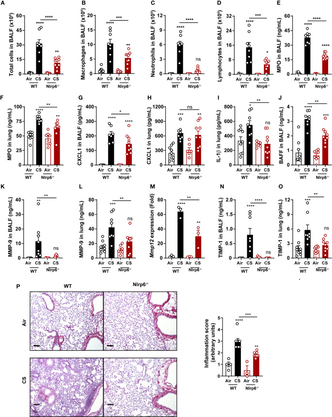
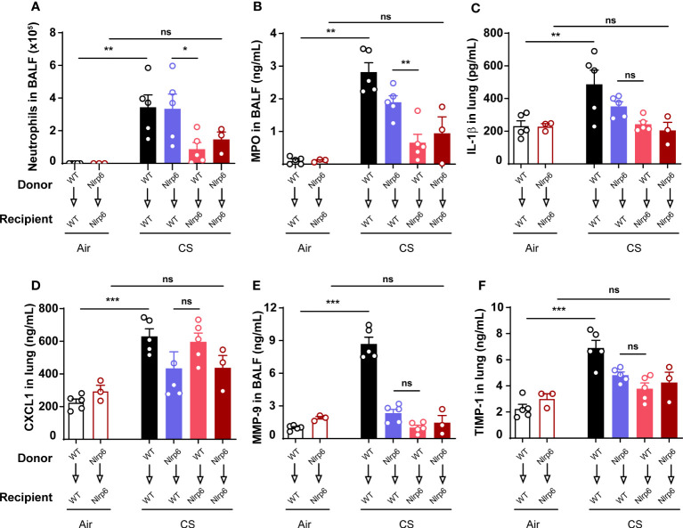
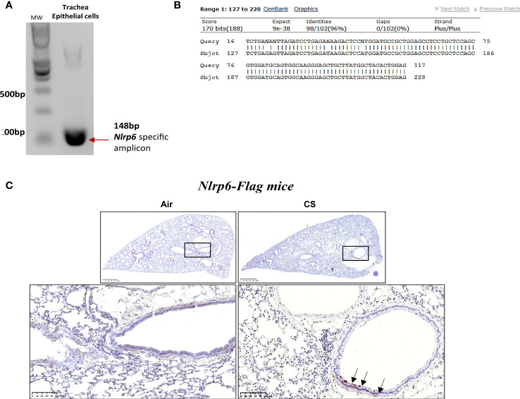
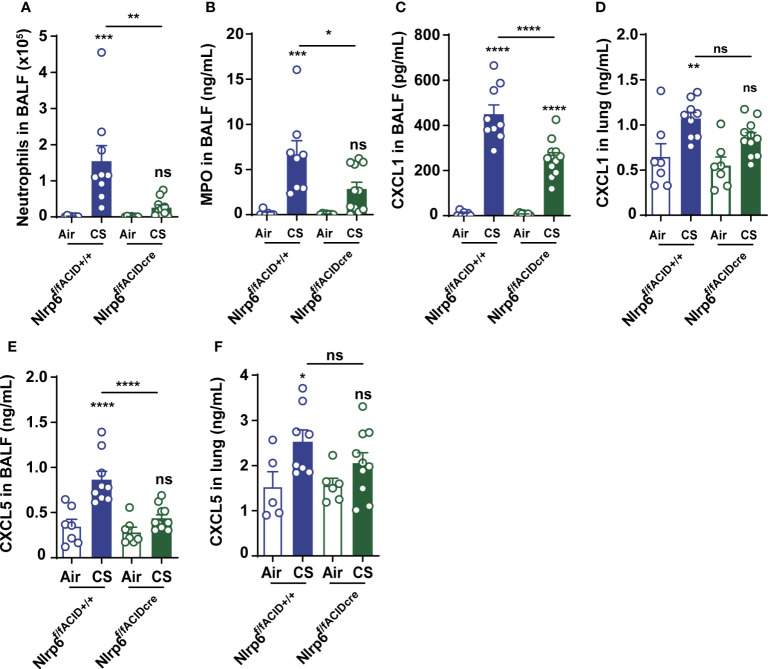
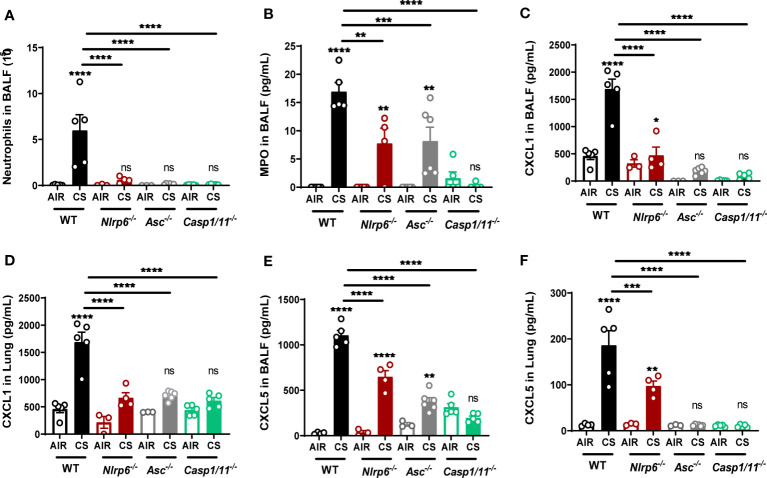
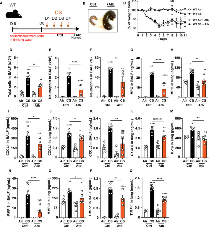
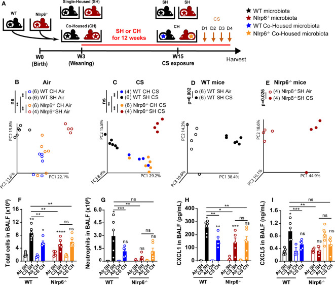
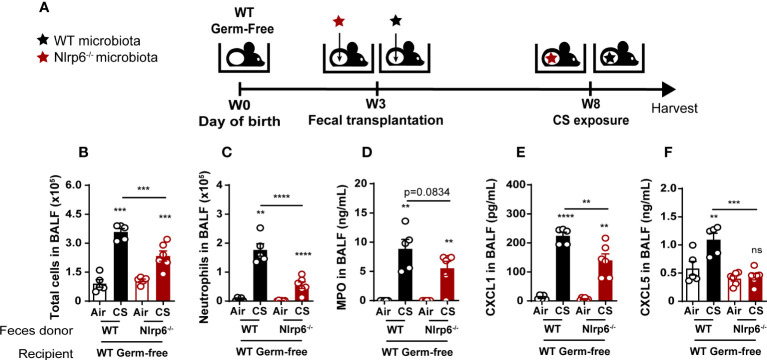
Chronic obstructive pulmonary disease (COPD) is a major health issue primarily caused by cigarette smoke (CS) and characterized by breathlessness and repeated airway inflammation. NLRP6 is a cytosolic innate receptor controlling intestinal inflammation and orchestrating the colonic host-microbial interface. However, its roles in the lungs remain largely unexplored. Using CS exposure models, our data show that airway inflammation is strongly impaired in Nlrp6-deficient mice with drastically fewer recruited neutrophils, a key cell subset in inflammation and COPD. We found that NLRP6 expression in lung epithelial cells is important to control airway and lung tissue inflammation in an inflammasome-dependent manner. Since gut-derived metabolites regulate NLRP6 inflammasome activation in intestinal epithelial cells, we investigated the link between NLRP6, CS-driven lung inflammation, and gut microbiota composition. We report that acute CS exposure alters gut microbiota in both wild-type (WT) and Nlrp6-deficient mice and that antibiotic treatment decreases CS-induced lung inflammation. In addition, gut microbiota transfer from dysbiotic Nlrp6-deficient mice to WT mice decreased airway lung inflammation in WT mice, highlighting an NLRP6-dependent gut-to-lung axis controlling pulmonary inflammation.

Keywords: NLRP6; cigarette smoke-exposure; gut microbiota; gut to lung axis; lung inflammation.


Conflict of interest statement

Author FT was employed by company ArtImmunne SAS. The remaining authors declare that the research was conducted in the absence of any commercial or financial relationships that could be construed as a potential conflict of interest.

Figures

Figure 1
Figure 1
NLRP6 deficiency impairs inflammation and remodeling factors to subchronic cigarette smoke exposure. C57BL/6 (WT) mice and Nlrp6-/- mice were exposed to air or cigarette smoke (CS) for 6 weeks. BALF and lungs were collected 16 hours after the last exposure. (A) Total cells, (B) macro- phages, (C) neutrophils and (D) lymphocytes were numerated in BALF. (E, F) Myeloperoxydase (MPO) and (G, H) CXCL1 levels were measured in BALF and lungs. (I) IL-1β and (J) BAFF protein levels were analyzed in lungs and BALF respectively. (K, L) MMP-9 levels in BALF and lungs were measured by ELISA. (M) Mmp12 mRNA expression was analyzed by qPCR in lung homogenates. (N, O) TIMP-1 levels in BALF and lungs were measured. (P) Lung sections were stained with Red Sirius and inflammation score was graphed ). Scale bar = 100μm. Data are representative of one experiment and are expressed as mean values ± SEM and n= 6-9 mice per group, ns, non-significant, *p < 0.05, **p < 0.01, ***p < 0.001, ****P<0.0001.
Figure 2
Figure 2
Nlrp6 expression in non-immune/resident cells is necessary for neutrophils influx upon CS exposure. WT (CD45.1) and Nlrp6-/- (CD45.2) recipient mice were sub-lethally irradiated, reconstituted by either WT or Nlrp6-/- BM cells and exposed to air or CS for 4 days. BALF and lungs were collected 16 hours after the last exposure. (A) Neutrophils were numerated in BALF. (B-D) MPO in BALF, IL-1β and CXCL1 levels in lungs, (E) remodeling factors MMP-9 in BALF and (F) TIMP-1 in lungs were assessed by ELISA. Data are representative of two experiments and are expressed as mean values ± SEM (n= 3-5 mice per group), ns, non-significant, *p < 0.05, **p < 0.01, ***p < 0.001.
Figure 3
Figure 3
Nlrp6 mRNA basal expression in trachea epithelial cells (in vitro) and NLRP6 induced protein expression in bronchial cells (in vivo) upon cigarette smoke exposure. Agarose DNA gel showing RT-PCR product of Nlrp6 sequence (A). The representative sequencing result of trachea epithelial cells detected band showing the correct Nlrp6 cDNA (B). Epithelial cells markers (Krt8 and Oclnl) were quantified by qPCR (C). FLAG immunohistochemistry on Nlrp6-FLAG-IRES-GFP mice exposed to cigarette smoke (CS) or air during 4 days CS exposure led to Nlrp6 expression in bronchial cells (D). Data are representative of 2 experiments and are expressed as mean values ± SEM.
Figure 4
Figure 4
NLRP6 expression in airway epithelial cells controls inflammation and CXCL5 secretion to CS. Mice deficient for Nlrp6 specifically in AEC (Nlrp6f/f Acid+/CRE) as well as their controls (Nlrp6fff/f Acid+/+) were exposed to air or CS for 4 days. BALF and lungs were collected 16 hours after the last exposure. (A) BALF Neutrophil counts. (B) BALF MPO levels. (C-F) CXCL1 and CXCL5 levels measured in BALF and lungs. Data are representative of three experiments and are expressed as mean values ± SEM (n= 7-9 mice per group), ns, non-significant, *p < 0.05, **p < 0.01, ***p < 0.001. ****P<0.0001.
Figure 5
Figure 5
Nlrp6 deficiency leads to tenuated lung inflammation acumulation of CXCL5 in bronchial. WT, ASC-/-, Asc-/-, Casp1/11-/- and Nlrp6-/- mice were exposed to air or CS for 4 days. BALF and lungs were collected 16 hours after the last exposure. (A) Neutrophil numbers and (B) MPO levels were analyzed in BALF. (C, D) CXCL1 and (E, F) CXCL5 levels were measured in BALF and lungs, respectively. Data are representative of three experiments and are expressed as mean values + SEM (n= 4-5 mice per group), ns, non-significant, *p < 0.05, **p < 0.01, ***p < 0.001, ****P<0.0001.
Figure 6
Figure 6
Oral antibiotic treatment of WT mice decreases pulmonary inflammation to cigarette smoke exposure. (A) WT mice were treated with antibiotic cocktail containing vancomycin, neomycin, ampicillin and me- tronidazole in drinking water for 12 days including the 4 days of CS exposure. BALF, lung and caecum were collected 16 hours after the last exposure. (B) Representative caecum picture from control (Ctrl) or treated (+Atb) mice. (C) Weight loss percentage (D). Total cells and (E) neutrophils numerated in BALF. (F) BALF neutrophil percentage. (G-L) MPO, CXCL1, CXCL5 measured in BALF and lungs. (M) IL-1β levels in lungs. (N-Q) Remodeling factors MMP-9 and TIMP-1 levels measured in BALF and lungs. Data are representative of two experiments and are expressed as mean values ± SEM (n= 5 mice per group), *p < 0.05, **p < 0.01, ***p < 0.001, ****P<0.0001.
Figure 7
Figure 7
Transfer of impaired cigarette smoke-induced airway inflammation of Nlrp6-/- mice to wild type mice by cohousing. (A) WT mice and Nlrp6-/- mice were single-housed (SH) or co-housed (CH) for 12 weeks and exposed for 4 days of CS. BALF, lung and caecum were collected 16 hours after the last exposure. Principal components analysis (PCA) analysis of WT and Nlrp6-/- SH or CH in (B) air or (C) CS conditions, (D) WT SH in air and CS conditions, and (E) Nlrp6-/- SH in air and CS conditions. (F) BALF total cells and (G) neutrophils. (H) BALF CXCL5 and (I) CXCL1 levels. Data are representative of tywo expirements and are expressed as mean values ± SEM and n= 6-9 mice per group, ns, non significant, group), *p < 0.05, **p < 0.01, ***p < 0.001, ****P < 0.0001.
Figure 8
Figure 8
Attenuation of CS-induced lung inflammation in WT germ-free mice after oral transplantation with fecal microbiota from Nlrp6-/- mice. (A) WT germ-free (GF) mice were colonized with WT or Nlrp6-/- microbiota at weaning and let 5 weeks for microbiota implantation for before CS exposure. (B) BALF total cell and (C) neutrophil counts. (D-F) BALF MPO, CXCL1 and CXCL5 levels. Data are representative of two experiments and are expressed as mean values ± SEM (n= 4-6 mice per group), ns, non-significant, **p < 0.01, ***p < 0.001, ****P<0.0001.

Similar articles

Cited by

References

    1. Barnes PJ. Mediators of chronic obstructive pulmonary disease. Pharmacol Rev (2004) 56:515–48. doi: 10.1124/pr.56.4.2 - DOI - PubMed
    1. Agusti A, Celli BR, Criner GJ, Halpin D, Anzueto A, Barnes P, et al. Global initiative for chronic obstructive lung disease 2023 report: GOLD executive summary. Am J Respir Crit Care Med (2023) 207:819–37. doi: 10.1164/rccm.202301-0106PP - DOI - PMC - PubMed
    1. Pauwels RA, Rabe KF. Burden and clinical features of chronic obstructive pulmonary disease (COPD). Lancet (2004) 364:613–20. doi: 10.1016/S0140-6736(04)16855-4 - DOI - PubMed
    1. Morrow JD, Make B, Regan E, Han M, Hersh CP, Tal-Singer R, et al. DNA methylation is predictive of mortality in current and former smokers. Am J Respir Crit Care Med (2020) 201(9):1099–109. doi: 10.1164/rccm.201902-0439OC - DOI - PMC - PubMed
    1. Brandsma CA, Van den Berge M, Hackett TL, Brusselle G, Timens W. Recent advances in chronic obstructive pulmonary disease pathogenesis: from disease mechanisms to precision medicine. J Pathol (2019) 250(5):624–35. doi: 10.1002/path.5364 - DOI - PMC - PubMed

Publication types

Substances