

Effects of sulfatide on peripheral nerves in metachromatic leukodystrophy
- PMID: 38146590
- PMCID: PMC10863914
- DOI: 10.1002/acn3.51954
Effects of sulfatide on peripheral nerves in metachromatic leukodystrophy
Abstract
Objective: To evaluate the longitudinal correlations between sulfatide/lysosulfatide levels and central and peripheral nervous system function in children with metachromatic leukodystrophy (MLD) and to explore the impact of intravenous recombinant human arylsulfatase A (rhASA) treatment on myelin turnover.
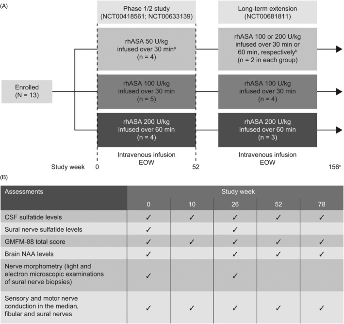
Methods: A Phase 1/2 study of intravenous rhASA investigated cerebrospinal fluid (CSF) and sural nerve sulfatide levels, 88-item Gross Motor Function Measure (GMFM-88) total score, sensory and motor nerve conduction, brain N-acetylaspartate (NAA) levels, and sural nerve histology in 13 children with MLD. Myelinated and unmyelinated nerves from an untreated MLD mouse model were also analyzed.
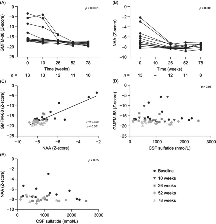
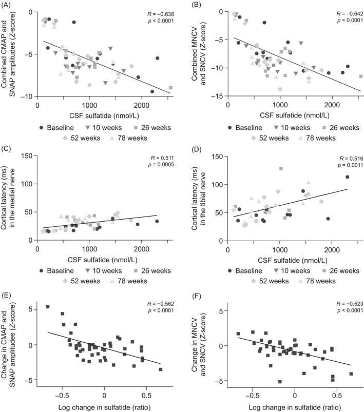
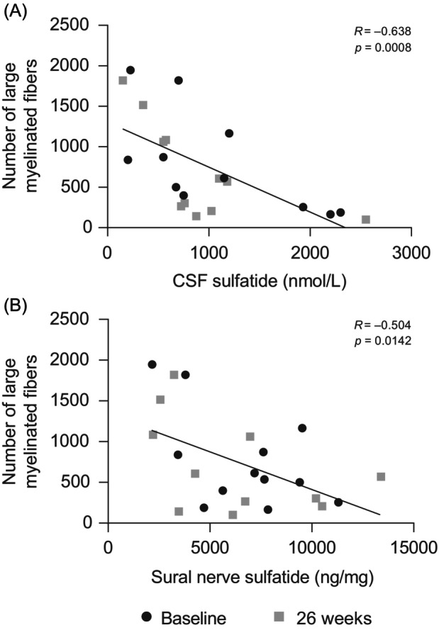
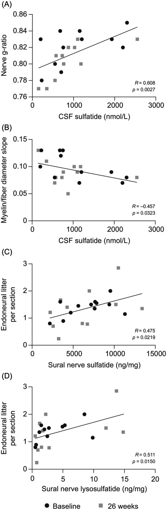
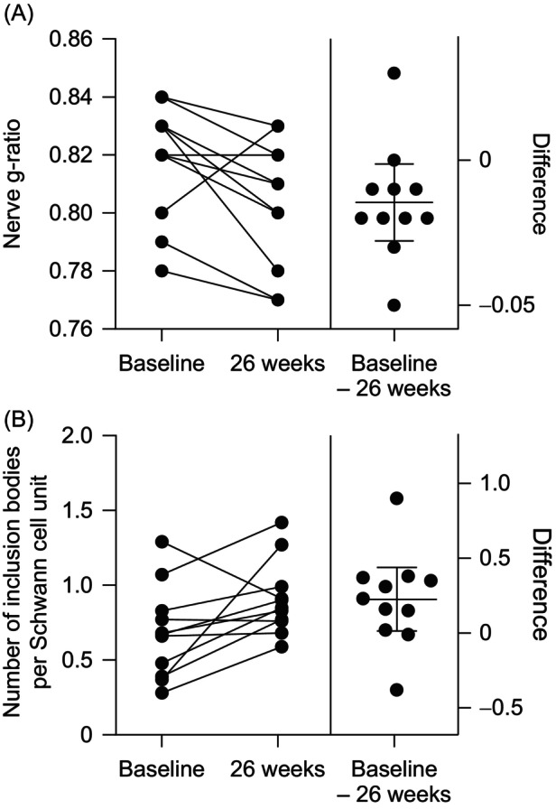
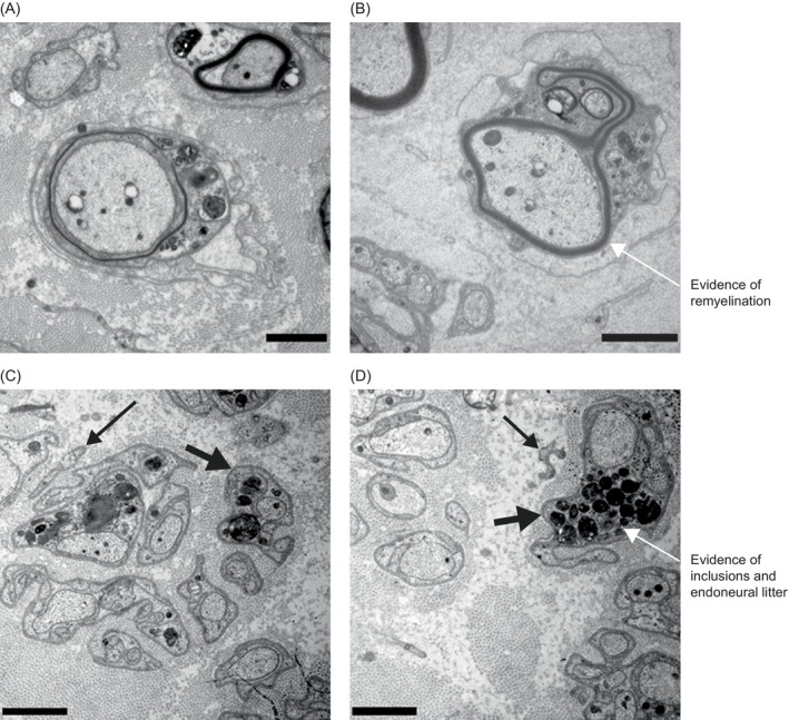
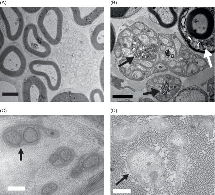
Results: CSF sulfatide levels correlated with neither Z-scores for GMFM-88 nor brain NAA levels; however, CSF sulfatide levels correlated negatively with Z-scores of nerve conduction parameters, number of large (≥7 μm) myelinated fibers, and myelin/fiber diameter slope, and positively with nerve g-ratios and cortical latencies of somatosensory-evoked potentials. Quantity of endoneural litter positively correlated with sural nerve sulfatide/lysosulfatide levels. CSF sulfatide levels decreased with continuous high-dose treatment; this change correlated with improved nerve conduction. At 26 weeks after treatment, nerve g-ratio decreased by 2%, and inclusion bodies per Schwann cell unit increased by 55%. In mice, abnormal sulfatide storage was observed in non-myelinating Schwann cells in Remak bundles of sciatic nerves but not in unmyelinated urethral nerves.
Interpretation: Lower sulfatide levels in the CSF and peripheral nerves correlate with better peripheral nerve function in children with MLD; intravenous rhASA treatment may reduce CSF sulfatide levels and enhance sulfatide/lysosulfatide processing and remyelination in peripheral nerves.
© 2023 Takeda Development Center Americas, Inc. Annals of Clinical and Translational Neurology published by Wiley Periodicals LLC on behalf of American Neurological Association.
Conflict of interest statement
M.H.F. has nothing to disclose. C.D. reports personal fees from University Hospital Copenhagen Rigshospitalet during the conduct of the study. S.G. reports an institutional research grant from Shire (a Takeda company) outside of the submitted work. He serves as an adviser for trials in MLD for Clario, Homology Medicines, and Passage Bio, but receives no personal payment related to this role. S.G. is a member of the European Reference Network for Rare Neurological Diseases, project ID 739510, and was partly supported by DFG grant GR 4688/2‐1. M.M. has nothing to disclose. D.A.H.W. is a full‐time employee of Takeda and a stockholder of Takeda Pharmaceutical Company Limited. C.J.M. is a full‐time employee of Takeda and a stockholder of Takeda Pharmaceutical Company Limited. I.K.‐M. reports grants from Shire (a Takeda company) during the conduct of the study. J.L. was a full‐time employee of Takeda and a stockholder of Takeda Pharmaceutical Company Limited at the time of the study. N.B. was a full‐time employee of Takeda and a stockholder of Takeda Pharmaceutical Company Limited at the time of the study. C.K. reports grants and personal fees from Shire (a Takeda company) and grants from Danish Medical Research during the conduct of the study. C.K. has also received royalties for teaching chapters from Gyldenal and FADL publishers.
Figures
Similar articles
-
Sulfatide levels correlate with severity of neuropathy in metachromatic leukodystrophy.Ann Clin Transl Neurol. 2015 May;2(5):518-33. doi: 10.1002/acn3.193. Epub 2015 Mar 27. Ann Clin Transl Neurol. 2015. PMID: 26000324 Free PMC article.
-
Safety of intrathecal delivery of recombinant human arylsulfatase A in children with metachromatic leukodystrophy: Results from a phase 1/2 clinical trial.Mol Genet Metab. 2020 Sep-Oct;131(1-2):235-244. doi: 10.1016/j.ymgme.2020.07.002. Epub 2020 Jul 16. Mol Genet Metab. 2020. PMID: 32792226 Clinical Trial.
-
Increasing sulfatide synthesis in myelin-forming cells of arylsulfatase A-deficient mice causes demyelination and neurological symptoms reminiscent of human metachromatic leukodystrophy.J Neurosci. 2007 Aug 29;27(35):9482-90. doi: 10.1523/JNEUROSCI.2287-07.2007. J Neurosci. 2007. PMID: 17728461 Free PMC article.
-
The role and metabolism of sulfatide in the nervous system.Mol Neurobiol. 2008 Apr-Jun;37(2-3):93-103. doi: 10.1007/s12035-008-8022-3. Epub 2008 May 9. Mol Neurobiol. 2008. PMID: 18465098 Review.
-
Sulfatide in health and disease. The evaluation of sulfatide in cerebrospinal fluid as a possible biomarker for neurodegeneration.Mol Cell Neurosci. 2021 Oct;116:103670. doi: 10.1016/j.mcn.2021.103670. Epub 2021 Sep 23. Mol Cell Neurosci. 2021. PMID: 34562592 Review.
Cited by
-
Anti-sulfatide antibodies in neurological disorders: should we test?J Neurol. 2024 Dec;271(12):7613-7618. doi: 10.1007/s00415-024-12668-8. Epub 2024 Sep 3. J Neurol. 2024. PMID: 39225745 Free PMC article.
-
Effective gene therapy for metachromatic leukodystrophy achieved with minimal lentiviral genomic integrations.Mol Ther Nucleic Acids. 2025 Jan 25;36(1):102464. doi: 10.1016/j.omtn.2025.102464. eCollection 2025 Mar 11. Mol Ther Nucleic Acids. 2025. PMID: 40171445 Free PMC article.
-
Characterization of gallbladder disease in metachromatic leukodystrophy across the lifespan.Mol Genet Metab. 2025 Jan;144(1):109003. doi: 10.1016/j.ymgme.2024.109003. Epub 2024 Dec 22. Mol Genet Metab. 2025. PMID: 39733668
References
-
- Gieselmann V. Metachromatic leukodystrophy: genetics, pathogenesis and therapeutic options. Acta Paediatr. 2008;97(s457):15‐21. - PubMed
-
- van Rappard DF, Boelens JJ, Wolf NI. Metachromatic leukodystrophy: disease spectrum and approaches for treatment. Best Pract Res Clin Endocrinol Metab. 2015;29(2):261‐273. - PubMed
-
- Taylor CM, Marta CB, Bansal R, Pfeiffer SE. Chapter 3 – the transport, assembly, and function of myelin lipids. In: Lazzarini RA, Griffin JW, Lassman H, Nave K‐A, Miller R, Trapp BD, eds. Myelin Biology and Disorders. Academic Press; 2004:57‐88.
-
- Grassi S, Prioni S, Cabitta L, Aureli M, Sonnino S, Prinetti A. The role of 3‐O‐sulfogalactosylceramide, sulfatide, in the lateral organization of myelin membrane. Neurochem Res. 2016;41(1–2):130‐143. - PubMed
Publication types
MeSH terms
Substances
Grants and funding
LinkOut - more resources
Full Text Sources
Medical
