. 2024 Jan 2;63(1):42-52.
doi: 10.1021/acs.biochem.3c00404. Epub 2023 Dec 26.

Crystal Structure and Enzymology of Solanum tuberosum Inositol Tris/Tetrakisphosphate Kinase 1 (St ITPK1)

Affiliations

Crystal Structure and Enzymology of Solanum tuberosum Inositol Tris/Tetrakisphosphate Kinase 1 (St ITPK1)

Hayley L Whitfield et al. Biochemistry. .

Abstract

Inositol phosphates and their pyrophosphorylated derivatives are responsive to the phosphate supply and are agents of phosphate homeostasis and other aspects of physiology. It seems likely that the enzymes that interconvert these signals work against the prevailing milieu of mixed populations of competing substrates and products. The synthesis of inositol pyrophosphates is mediated in plants by two classes of ATP-grasp fold kinase: PPIP5 kinases, known as VIH, and members of the inositol tris/tetrakisphosphate kinase (ITPK) family, specifically ITPK1/2. A molecular explanation of the contribution of ITPK1/2 to inositol pyrophosphate synthesis and turnover in plants is incomplete: the absence of nucleotide in published crystal structures limits the explanation of phosphotransfer reactions, and little is known of the affinity of potential substrates and competitors for ITPK1. Herein, we describe a complex of ADP and StITPK1 at 2.26 Å resolution and use a simple fluorescence polarization approach to compare the affinity of binding of diverse inositol phosphates, inositol pyrophosphates, and analogues. By simple HPLC, we reveal the novel catalytic capability of ITPK1 for different inositol pyrophosphates and show Ins(3,4,5,6)P4 to be a potent inhibitor of the inositol pyrophosphate-synthesizing activity of ITPK1. We further describe the exquisite specificity of ITPK1 for the myo-isomer among naturally occurring inositol hexakisphosphates.


Conflict of interest statement

The authors declare no competing financial interest.

Figures
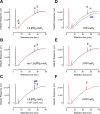
Figure 1
Figure 1
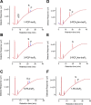
StITPK1 is a reversible inositol pyrophosphate-ADP phosphotransferase. HPLC resolution of products of 12h reaction of StITPK with ADP and (A) 1,5-[PP]2-InsP4; (B) rac-1,5-[PP]2-InsP4. Products of the dephosphorylation of 1,5-[PP]2-InsP4 coelute with 1-PP-InsP5, (C). Substrates are indicated, S and products, P. Chromatograms of reactions without enzyme are shown in red, with enzyme in black and chromatograms of standards are shown in blue. The position of elution of ATP formed by phosphotransfer to ADP is shown in panel A. ADP elutes in the solvent front. The standards showing the elution position of PP-InsP5 in C contain ATP. The ATP peaks in all other panels are products of phosphotransfer from substrate to ADP. HPLC of products of reaction of StITPK1 with ATP and 1-PP-InsP5, 3-PP-InsP5, or 5-PP-InsP5 are shown in D, E and F. Here, chromatograms of 3 h incubations with enzyme are shown in black, and 12 h incubations are shown in blue. The position of a 1,5-[PP]2-InsP4 standard (trace offset on the y-axis) is shown in panel D. The HPLC column was eluted with a gradient of HCl.
Figure 2
Figure 2
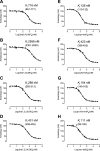
High-affinity binding of inositol phosphates to StITPK1. Displacement of 2-FAM-InsP5 with (A) Ins(3,4,5,6)P4; (B) Ins(1,4,5,6)P4; (C) Ins(1,2,3,5,6)P5; (D) Ins(1,2,3,4,5)P5; (E) myo-InsP6; (F) scyllo-InsP6; (G) d-chiro-InsP6; and (H) neo-InsP6. Data are the means and standard deviations of four replicate measurements; Ki (nM) with confidence interval (nM) in parentheses.
Figure 3
Figure 3
Overview of the crystal structure of StITPK1. (A) Cartoon representation of the structure of StITPK1, colored by secondary structure (α-helix red, β-sheet yellow, and coil green). Broken lines in the backbone trace indicate residues that were unresolved in the model due to disorder. Bound nucleotide is shown in stick format with coloring as follows: carbon-green, oxygen-red, nitrogen-blue, and phosphorus-orange. (B) Molecular surface representation of the structure of StITPK1 colored by subdomain. Subdomains are the kinase N-terminal domain (light blue), kinase central domain (lime green) and kinase central domain (sand). Here and in panel D, the polypeptide connecting the central and C-terminal domains is colored dark blue. Bound ADP is shown in atom sphere format. (C) Molecular surface representation of the structure of AtITPK4 (PDB: 7PUP). Coloring as in panel C except that the additional HAD domain found in this enzyme is colored pink and the tab insertion unique to ITPK4s is colored magenta. (D) Molecular surface of StITPK1 colored by electrostatic potential (red-acidic, blue-basic). The orientation of the molecule is the same as that in panel A. In panels B–D, bound ADP is shown in atom sphere format and colored according to that in panel A.
Figure 4
Figure 4
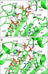
Prediction of the binding modes of an enantiomeric pair of substrates in the active site of StITPK1. (A) Closeup view of the energy minimized predicted binding mode of the poor substrate, Ins(1,4,5,6)P4, in the kinase domain active site. The enzyme is shown in the cartoon format and colored green. The substrate and active site residues (labeled) with which it forms polar interactions are shown in the stick format with carbon colored green, oxygen red, nitrogen blue, and phosphorus orange. Magnesium ions are shown as dark green spheres. Polar interactions are indicated by black dashed lines. Specificity subsites are labeled A–F (magenta font) such that the hydroxyl group positioned to accept the γ-phosphate of ATP by in-line transfer (the hydroxyl attached to carbon 3 of the inositol ring, in this case) occupies subsite A and the remaining subsites are arrayed in an anticlockwise sense when observed from the viewpoint adopted in this figure. (B) View of the energy minimized predicted binding mode of the good substrate, Ins(3,4,5,6)P4, in the kinase domain active site. The hydroxyl attached to carbon 1 of the inositol ring, in this case, occupies subsite A. Display format and coloring as in panel A.
Figure 5
Figure 5
Phosphorylation of inositol pyrophosphate analogues by StITPK1. HPLC resolution of products of reaction of StITPK1 with ATP and (A) 1-PCP-Ins(2,3,4,5,6)P5 [1-PCP-InsP5]; (B) 3-PCP-Ins(1,2,4,5,6)P5 [3-PCP-InsP5]; (C) 5-PA-Ins(1,2,3,4,6)P5 [5-PA-InsP5]; (D) 5-PCH2Am-Ins(1,2,3,4,6)P5 [5-PCH2Am-InsP5]; (E) 5-PCF2Am-Ins(1,2,3,4,6)P5 [5-PCF2Am-InsP5]; (F) 1-PA-Ins(2,3,4,5,6)P5 [1-PA-InsP5]. Substrates are indicated, S; products, P. For all panels, chromatograms of reactions without enzyme are shown in red, 3 h incubations with enzyme are shown in black, and 12 h incubations in blue. The position of elution of ATP is shown in panel A. The HPLC column was eluted with a gradient of HCl.
Figure 6
Figure 6
Inhibition of StITPK1 phosphokinase activity by inositol pyrophosphate analogues. (A) Ins(1,2,3,4,5)P5 5-phosphokinase activity; (B) InsP6 5-phosphokinase activity. Reactions were performed for 2 h at 30 °C with 3 μM StITPK1, 0.5 mM ATP, and 1 mM inositol phosphate in the absence or presence of competitor. The extent of inhibition at inhibitor concentration (mM) indicated by number in each column was estimated from the integrated peak areas of substrate and product peaks resolved by HPLC (example HPLC traces are shown in Figure S10). Significant difference at p = 0.05 between inhibitor treatments (by one-way ANOVA and Tukey’s multiple comparisons test) is indicated by the absence of a common letter.

References

    1. Parmar P. N.; Brearley C. A. Identification of 3- and 4-phosphorylated phosphoinositides and inositol phosphates in stomatal guard cells. Plant Journal 1993, 4 (2), 255–263. 10.1046/j.1365-313X.1993.04020255.x. - DOI
    1. Brearley C. A.; Hanke D. E. Inositol phosphates in barley (Hordeum vulgare L.) aleurone tissue are stereochemically similar to the products of breakdown of InsP6 in vitro by wheat-bran phytase. Biochem. J. 1996, 318 (Pt 1), 279–86. 10.1042/bj3180279. - DOI - PMC - PubMed
    1. Riemer E.; Pullagurla N. J.; Yadav R.; Rana P.; Jessen H. J.; Kamleitner M.; Schaaf G.; Laha D. Regulation of plant biotic interactions and abiotic stress responses by inositol polyphosphates. Front Plant Sci. 2022, 13, 94451510.3389/fpls.2022.944515. - DOI - PMC - PubMed
    1. Stevenson-Paulik J.; Bastidas R. J.; Chiou S. T.; Frye R. A.; York J. D. Generation of phytate-free seeds in Arabidopsis through disruption of inositol polyphosphate kinases. Proc. Natl. Acad. Sci. U. S. A. 2005, 102 (35), 12612–7. 10.1073/pnas.0504172102. - DOI - PMC - PubMed
    1. Wild R.; Gerasimaite R.; Jung J. Y.; Truffault V.; Pavlovic I.; Schmidt A.; Saiardi A.; Jessen H. J.; Poirier Y.; Hothorn M.; Mayer A. Control of eukaryotic phosphate homeostasis by inositol polyphosphate sensor domains. Science 2016, 352 (6288), 986–90. 10.1126/science.aad9858. - DOI - PubMed

Publication types