

Cytotoxic lesions of the corpus callosum: a systematic review
- PMID: 38147170
- PMCID: PMC11213749
- DOI: 10.1007/s00330-023-10524-3
Cytotoxic lesions of the corpus callosum: a systematic review
Abstract
Objectives: Cytotoxic lesions of the corpus callosum (CLOCC) are a common magnetic resonance imaging (MRI) finding associated with various systemic diseases including COVID-19. Although an increasing number of such cases is reported in the literature, there is a lack of systematic evidence summarizing the etiology and neuroimaging findings of these lesions. Thus, the aim of this systematic review was to synthesize the applied nomenclature, neuroimaging and clinical features, and differential diagnoses as well as associated disease entities of CLOCC.
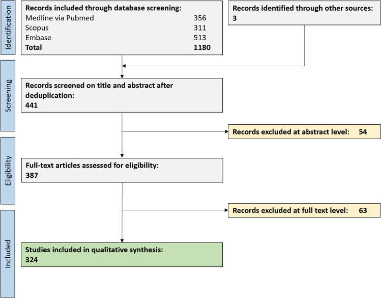
Materials and methods: A comprehensive literature search in three biomedical databases identified 441 references, out of which 324 were eligible for a narrative summary including a total of 1353 patients.
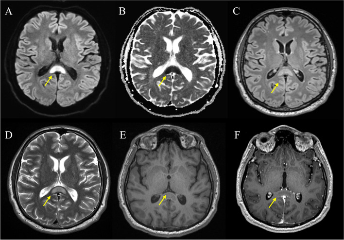
Results: Our PRISMA-conform systematic review identifies a broad panel of disease entities which are associated with CLOCC, among them toxic/drug-treatment-associated, infectious (viral, bacterial), vascular, metabolic, traumatic, and neoplastic entities in both adult and pediatric individuals. On MRI, CLOCC show typical high T2 signal, low T1 signal, restricted diffusion, and lack of contrast enhancement. The majority of the lesions were reversible within the follow-up period (median follow-up 3 weeks). Interestingly, even though CLOCC were mostly associated with symptoms of the underlying disease, in exceptional cases, CLOCC were associated with callosal neurological symptoms. Of note, employed nomenclature for CLOCC was highly inconsistent.
Conclusions: Our study provides high-level evidence for clinical and imaging features of CLOCC as well as associated disease entities.
Clinical relevance statement: Our study provides high-level evidence on MRI features of CLOCC as well as a comprehensive list of disease entities potentially associated with CLOCC. Together, this will facilitate rigorous diagnostic workup of suspected CLOCC cases.
Key points: • Cytotoxic lesions of the corpus callosum (CLOCC) are a frequent MRI feature associated with various systemic diseases. • Cytotoxic lesions of the corpus callosum show a highly homogenous MRI presentation and temporal dynamics. • This comprehensive overview will benefit (neuro)radiologists during diagnostic workup.
Keywords: COVID-19; Central nervous system diseases; Corpus callosum; Magnetic resonance imaging; Systematic review.
© 2023. The Author(s).
Conflict of interest statement
The authors of this manuscript declare no relationships with any companies, whose products or services may be related to the subject matter of the article.
Figures
References
Publication types
MeSH terms
Grants and funding
LinkOut - more resources
Full Text Sources
Medical
