. 2024 Jan 2;121(1):e2308706120.
doi: 10.1073/pnas.2308706120. Epub 2023 Dec 26.

Social anxiety disorder-associated gut microbiota increases social fear

Affiliations

Social anxiety disorder-associated gut microbiota increases social fear

Nathaniel L Ritz et al. Proc Natl Acad Sci U S A. .

Erratum in

Abstract

Social anxiety disorder (SAD) is a crippling psychiatric disorder characterized by intense fear or anxiety in social situations and their avoidance. However, the underlying biology of SAD is unclear and better treatments are needed. Recently, the gut microbiota has emerged as a key regulator of both brain and behaviour, especially those related to social function. Moreover, increasing data supports a role for immune function and oxytocin signalling in social responses. To investigate whether the gut microbiota plays a causal role in modulating behaviours relevant to SAD, we transplanted the microbiota from SAD patients, which was identified by 16S rRNA sequencing to be of a differential composition compared to healthy controls, to mice. Although the mice that received the SAD microbiota had normal behaviours across a battery of tests designed to assess depression and general anxiety-like behaviours, they had a specific heightened sensitivity to social fear, a model of SAD. This distinct heightened social fear response was coupled with changes in central and peripheral immune function and oxytocin expression in the bed nucleus of the stria terminalis. This work demonstrates an interkingdom basis for social fear responses and posits the microbiome as a potential therapeutic target for SAD.

Keywords: faecal transplant; microbiome; microbiota-gut-brain axis; social phobia.


Conflict of interest statement

Competing interests statement:The authors declare no competing interest.

Figures
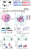
Fig. 1.
Fig. 1.
Human gut microbiota from social anxiety disorder (SAD) promotes social fear behaviour in mice but does not alter other general social behaviours. (A) In order to test whether the microbiota plays a causal role in SAD, the microbiota of individuals with SAD was transferred by faecal microbiota transplantation (FMT) to rodent recipients to assess phenotypic characteristics compared to FMT from healthy control. (B) Study diagram and human donor and rodent recipient group legend, male (m) and female (f). (C) Experimental design: animals first received an antibiotic cocktail (Abx) for 1 wk to deplete the resident microbiota, before receiving FMTs, and behavioural assessment (elevated plus maze—EPM, three-chamber test—3CT, carmine red gastrointestinal transit and motility test—CR, social fear conditioning—SFC, and forced swim test—FST). Faecal samples were collected and microbiota analysed at the beginning and end of study. Corticosterone was measured before and after the FST. At the end of the study animals were killed, tissue was collected; then flow cytometry, and ileal explants were stimulated with lipopolysaccharide, concanavalin A, and CD3/CD28. (D) Aitchison beta diversity of SAD human faecal microbiota clustered differentially compared to healthy controls (R2 = 0.03477, P < 0.05, 1,000 permutations). Donors (indicated by the faecal symbol) were selected based on microbiota, Liebowitz Social Anxiety Scale score, age, body mass index, sex, and an absence of psychotropic or microbiota medications to treat SAD. (E) Murine faecal bacteriome beta diversity was significantly different between SAD and HC groups at the end of the study (R2 = 0.02767, P < 0.001, 1,000 permutations). (F) Diagram represents social fear conditioning and social fear extinction, which takes place over two days. Social fear behaviour is increased in mice that receive SAD FMT (W(1) = 3.239, P < 0.05). Social fear extinction data showing interaction percent with either nonsocial stimulus (ns1-3) or six novel social stimuli (s1-6). Transparent lines represent each individual donor group (SAD5 and HC3 n = 5; other groups n = 6) while bold lines show mean ± SE of the total HC and SAD groups (n = 35 per group). (G) Diagrams represent the three-chamber social interaction test. Following habituation, sociability was assessed by preference for a novel social stimulus over an inanimate, nonsocial object (rubber duck), then social novelty preference was assessed by preference for a novel conspecific over the now-familiar conspecific used in the sociability phase. There were no significant group differences in sociability; both groups spent significantly more time interacting with the social (Soc) stimulus over the nonsocial (Non) stimulus (t(68) = 12.01, 11.64, P < 0.001). (H) There were no significant group differences in social novelty preference; both groups spent significantly more time interacting with the novel (Nov) over the familiar (Fam) social stimulus (t(68) = 3.158, P < 0.01; t(68) = 2.513, P < 0.05) (n = 35 per group).
Fig. 2.
Fig. 2.
SAD FMT modulates basal stress hormone corticosterone plasma levels. (A) Basal plasma corticosterone is significantly reduced in the SAD group compared to HC (t(64) = −1.858, P < 0.05) (n = 35 HC, n = 33 SAD). (B) The stress response timeline measured by plasma corticosterone collected at 30-min intervals over 90 min. The forced swim test (stress stimulus) took place from 5 min to 11 min (total duration of stress was 6 min). There was a significantly lower concentration of plasma corticosterone at the basal timepoint (0 min) in the SAD compared to HC group and no significant effects at the 30-, 60-, or 90-min time points.
Fig. 3.
Fig. 3.
SAD FMT modulates ileal IL-17A, immune cell population composition, and gene expression markers related to neuroinflammation and blood-brain barrier integrity. (A) SAD FMT reduces IL-17A cytokines produced by ileal explants following stimulation with vehicle (buffer; t(32) = −1.804, P = 0.081), lipopolysaccharide (LPS; t(30) = −2.297, P < 0.05), concanavalin A (ConA; t(32) = −2.457, P < 0.05) and CD3/CD28 (t(30) = −1.813, P = 0.100) (n = 16–18 per group). (B) F4/80+ macrophages in the mesenteric lymph nodes (MLNs) were significantly reduced in the SAD group compared to the HC group (t(30) = −2.602, P < 0.05) (n = 17 HC, n = 15 SAD). (C) There was a trend for increased CD4+ T helper cells in the MLNs of the SAD group compared to the HC group (t(10) = 1.953, P = 0.079) (n = 17 HC, n = 18 SAD). (D) The CD44 median fluorescent intensity was reduced in blood CD4+ T helper cells in the SAD group compared to the HC group (t(34) = −2.049, P < 0.05) (n = 18 per group). (E) Neuroinflammatory- and neuroimmune-related gene expression measured in the bed nucleus of the stria terminalis (BNST), medial amygdala (MeA), and medial prefrontal cortex (MePFC). In the BNST, arginase 1 (Arg1) was reduced by SAD microbiota transfer (t(43) = −3.927, P < 0.001). In the MeA, there were reductions in expression of tumour necrosis factor alpha (TNF-α; t (35) = −2.081, P < 0.05), interleukin 10 (Il-10; t(36) = −2.342, P < 0.05), Arg1 (t(43) = −3.241, P < 0.01), and chemokine C-X-C ligand 15 (Cxcl15; t(38) = −2.137, P < 0.05). In the MePFC, there were reductions in Toll-like receptor 4 (Tlr4; t(40) = −2.839, P < 0.05) and Arg1 (t(42) = −3.55, P < 0.001) following SAD microbiota transfer (n = 17–23 per group). (F) Blood–brain barrier–related gene expression measured in the BNST, MeA, and MePFC. In the BNST, there was a trend for reduced occludin (Ocln) in the SAD microbiota transfer recipient group (t(43) = −1.901, P = 0.06). In the MeA, there were reductions in expression of tight junction protein 1 (Tjp1; t(43) = −3.033, P < 0.01), claudin 5 (Cldn5; t(41) = −2.601, P < 0.05), and Ocln (t(42) = −1.811, P = 0.08). In the MePFC, there was a reduction of expression of Tjp1 (t(41) = −2.473, P < 0.05) following SAD microbiota transfer (n = 20–23 per group).
Fig. 4.
Fig. 4.
SAD FMT modulates neurons containing oxytocin in the bed nucleus of the stria terminalis (BNST) and oxytocin- and vasopressin-related genes in the medial amygdala (MeA) and medial prefrontal cortex (MePFC). Representative illustration of the bed nucleus of the stria terminalis (BNST) (17) and immunohistochemistry microscopy images of oxytocin (Oxt) staining in BNST neurons from HC (A) and SAD (B) FMT recipients (0.02 and -0.08mm from Bregma; NeuN Alexa555, Oxt Alexa488). (C) There was a significant reduction in the number of Oxt containing neurons in the BNST of the SAD group compared to the HC group (t(17) = −2.369, P < 0.05) (n = 10 HC, n = 9 SAD). (D) BNST, MeA, and MePFC expression of oxytocin- and vasopressin-related genes in the HC and SAD microbiota recipient groups. There was a trend for reduced vasopressin receptor 1b (Avpr1b) in the BNST (t(41) = −2.115, P = 0.06) (n = 22 per group). In the MeA there were reductions in oxytocin (Oxt; t(34) = −2.455, P < 0.05), oxytocin receptor (Oxtr; t(40) = −2.143, P < 0.05), and vasopressin receptor 1a (Avpr1a; t(41) = −1.895, P = 0.07; n = 20–22 per group). In the MePFC there were reductions in Oxtr (t(40) = −2.419, P < 0.05) and Avpr1a (t(42) = −3.184, P < 0.01; n = 21–22 per group).

References

    1. Stein M. B., Kean Y. M., Disability and quality of life in social phobia: Epidemiologic findings. Am. J. Psychiatry 157, 1606–1613 (2000). - PubMed
    1. Keller M., The lifelong course of social anxiety disorder: A clinical perspective. Acta Psychiatr. Scand. 108, 85–94 (2003). - PubMed
    1. Baldwin D. S., et al. , Evidence-based pharmacological treatment of anxiety disorders, post-traumatic stress disorder and obsessive-compulsive disorder: A revision of the 2005 guidelines from the British Association for Psychopharmacology. J. Psychopharmacol. 28, 403–439 (2014). - PubMed
    1. Patel A., Knapp M., Henderson J., Baldwin D., The economic consequences of social phobia. J. Affect. Disord. 68, 221–233 (2002). - PubMed
    1. Stein M. B., Stein D. J., Social anxiety disorder. The Lancet 371, 1115–1125 (2008). - PubMed

LinkOut - more resources