. 2023 Nov-Dec;175(6):e14079.
doi: 10.1111/ppl.14079.

Same but different - pseudo-pectin in the charophytic alga Chlorokybus atmophyticus

Affiliations

Same but different - pseudo-pectin in the charophytic alga Chlorokybus atmophyticus

Marie N Rapin et al. Physiol Plant. 2023 Nov-Dec.

Abstract

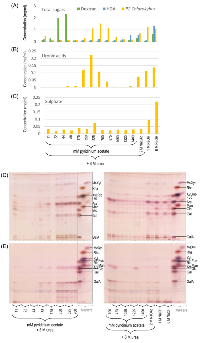
All land-plant cell walls possess hemicelluloses, cellulose and anionic pectin. The walls of their cousins, the charophytic algae, exhibit some similarities to land plants' but also major differences. Charophyte 'pectins' are extractable by conventional land-plant methods, although they differ significantly in composition. Here, we explore 'pectins' of an early-diverging charophyte, Chlorokybus atmophyticus, characterising the anionic polysaccharides that may be comparable to 'pectins' in other streptophytes. Chlorokybus 'pectin' was anionic and upon acid hydrolysis gave GlcA, GalA and sulphate, plus neutral sugars (Ara≈Glc>Gal>Xyl); Rha was undetectable. Most Gal was the l-enantiomer. A relatively acid-resistant disaccharide was characterised as β-d-GlcA-(1→4)-l-Gal. Two Chlorokybus 'pectin' fractions, separable by anion-exchange chromatography, had similar sugar compositions but different sulphate-ester contents. No sugars were released from Chlorokybus 'pectin' by several endo-hydrolases [(1,5)-α-l-arabinanase, (1,4)-β-d-galactanase, (1,4)-β-d-xylanase, endo-polygalacturonase] and exo-hydrolases [α- and β-d-galactosidases, α-(1,6)-d-xylosidase]. 'Driselase', which hydrolyses most land-plant cell wall polysaccharides to mono- and disaccharides, released no sugars except traces of starch-derived Glc. Thus, the Ara, Gal, Xyl and GalA of Chlorokybus 'pectin' were not non-reducing termini with configurations familiar from land-plant polysaccharides (α-l-Araf, α- and β-d-Galp, α- and β-d-Xylp and α-d-GalpA), nor mid-chain residues of α-(1→5)-l-arabinan, β-(1→4)-d-galactan, β-(1→4)-d-xylan or α-(1→4)-d-galacturonan. In conclusion, Chlorokybus possesses anionic 'pectic' polysaccharides, possibly fulfilling pectic roles but differing fundamentally from land-plant pectin. Thus, the evolution of land-plant pectin since the last common ancestor of Chlorokybus and land plants is a long and meandering path involving loss of sulphate, most l-Gal and most d-GlcA; re-configuration of Ara, Xyl and GalA; and gain of Rha.


Figures

FIG. 1
FIG. 1
The green plant lineage. The images show the six species studied in this paper. The names of relevant streptophyte classes are shown, with relevant phyla in bold text and less formal names in italics. Classes and phyla that are not studied in this paper are written vertically. The tree is after Li et al. (2020), with broadly indicative dates from Leliaert et al. (2011). Photo credits: Ulva, Gabriele Kothe‐Heinrich; Anthoceros, H. Bernd; Chlorokybus, Iker Irisarri; Coleochaete and Klebsormidium, CCAP Culture Collection; Chara, Cambridge University Botanic Garden.
FIG. 2
FIG. 2
Ratio of polysaccharide fractions in six plant species. (A) Proportions of the different polysaccharide extracts, in various plants, relative to the initial AIR quantity. Owing to the small scale of the Chlorokybus sample, the Wash and α‐Cellulose fractions were not separated and are both presented as part of the Wash fraction. (B) Susceptibility of pectin to extraction (see text). The experiment was run in triplicate; error bars are SD, n = 3.
FIG. 3
FIG. 3
Relative quantities of sugar residues in six polysaccharide extracts from each of six plants. Quantification of hydrolysates prepared as in Fig. S1. The histograms show TFA hydrolysis products of polysaccharide fractions from (A,B) untreated AIR and (C) α‐amylase‐treated AIR. Chromatography solvents (always with two ascents) were: A, BAW; B,C, EPAW. Each TLC loading was derived from 15 μg of the polysaccharide fraction. Polymer fractions were: P1 and P2, ‘pectins’; Ha and Hb, hemicelluloses a and b; W, mildly acidic wash after alkaline extraction; αC, residual ‘α‐cellulose’. Sugars in the hydrolysates are colour‐coded. There are slight differences between the BAW and EPAW runs owing to differences in resolving power of the two solvent systems. Three aliquots of each AIR preparation were extracted to produce the listed polymer fractions, then three aliquots of each of the extracts were separately acid‐hydrolysed and analysed by TLC. Error bars are SD, with n = 3 (referring to the number of AIR preparations). ‘Unknown’ sugars could not be confidently identified with the markers.
FIG. 4
FIG. 4
Endopolygalacturonase digestion of ‘pectic’ polysaccharides from six species of the Viridiplantae. AIR was de‐esterified with NaOH then digested with EPG, and the water‐soluble products were analysed by TLC on aluminium‐backed silica‐gel plates in BAW (2:1:1; single ascent). EPG alone gave no detectable spots. We loaded two replicate digests. Aqueous suspensions of the AIRs (without EPG treatment) were also loaded, demonstrating the absence of pre‐existing oligogalacturonides. MM, marker mixture of GalA and its disaccharide.
FIG. 5
FIG. 5
The galactose in Chlorokybus pectin and in its aldobiouronic acid (A1) are the l‐enantiomer. (A) Whole Chlorokybus pectin P2 was acid‐hydrolysed then treated (Y), or not (N, incubated in enzyme buffer), with d‐galactose oxidase. Products were analysed by TLC (in EPAW). For comparison, commercial l‐ and d‐galactose were treated, or not, with d‐galactose oxidase. (B) Purified A1 (see Fig. 6A) was re‐hydrolysed with TFA (A1,h), then treated with d‐galactose oxidase (A1,h,o), and mixed with galactose oxidase‐treated d‐Gal (A1,h,o + d‐Gal,o). A sample of oxidase‐treated d‐Gal (d‐Gal,o) was run for comparison. The TLC was developed in BAW with one ascent. Abbreviations: h, hydrolysed in TFA; o, incubated with galactose oxidase.
FIG. 6
FIG. 6
Chromatography of acid hydrolysate of pectin 2 from Chlorokybus. The pectin was hydrolysed with 2 M TFA for 1 h at 120°C. (A) Preparative paper chromatography of the total hydrolysate. Chromatography solvent BAW (12:3:5), run for 24 h. The fringes of the preparative loading were stained with aniline hydrogen‐phthalate, then the unstained portion of interesting compounds (not shown here in full), A1, A2 and M1, were eluted for further analysis. The dashed black line indicates the position of Xyl on the paper, as a visual guide. (B,C) TLC of small portions of A1, A2 and M1 from (A). Markers included an Anthoceros pectin hydrolysate containing the aldobiouronic acid α‐d‐GlcA‐(1→3)‐l‐Gal (Popper et al., 2003). The solvents were (B) BAW (4:1:1) and (C) EPAW (6:3:1:1).
FIG. 7
FIG. 7
Enzymic digests of Chlorokybus ‘pectin’. (A) ‘Pectin’ fraction P2 from Chlorokybus was treated with a series of endo‐ and exo‐hydrolases. N = no enzyme (pectin incubated alone for 72 h). (B) Substrate‐less controls of the same enzyme collection. Incubation conditions were as in Table S1. The TLC plates in were developed twice in BAW.
FIG. 8
FIG. 8
Fractionation of intact Chlorokybus P2 ‘pectin’ on anion‐exchange chromatography. Prior to analyses, the urea was removed by dialysis. (A) Profile of total sugars, measured by a thymol dot‐blot assay. Dextran and homogalacturonan (‘HGA’) were run on the column as respectively neutral and anionic standards. (B) Profile of uronic acid residues, measured by m‐hydroxybiphenyl assay. (C) Sulphate profile, measured by a barium acetate assay complete acid hydrolysis of the fractions. (D) TLC of fully hydrolysed fractions from Chlorokybus P2 (2 M TFA, 120°C, 1 h). (E) TLC of mildly hydrolysed fractions (0.1 M TFA, 100°C, 2 h). For (D) and (E), each fraction comprised two samples collected successively in each indicated eluent. TLC plates were developed in EPAW for (D), and in BAW for (E). The marker mixtures (in grey rectangles) interrupt the sequence of fractions.

References

    1. Albersheim P., Darvill A., Roberts K., Sederoff R., and Staehelin A. (2011) Plant Cell Walls. Garland Science, New York: Taylor and Francis Group.
    1. Anderson C.T. (2016) ‘We be jammin’: an update on pectin biosynthesis, trafficking and dynamics, Journal of Experimental Botany, 67, 495–502. - PubMed
    1. Aquino R.S., Grativol C., and Mourão P.A.S. (2011) Rising from the sea: correlations between sulfated polysaccharides and salinity in plants, PLoS ONE, 6, e 18862. - PMC - PubMed
    1. Begum R.A. and Fry S.C. (2022) Boron bridging of rhamnogalacturonan‐II in Rosa and arabidopsis cell cultures occurs mainly in the endo‐membrane system and continues at a reduced rate after secretion, Annals of Botany, 130, 703–715. - PMC - PubMed
    1. Bischoff H.W. and Bold H.C. (1963) Some soil algae from Enchanted Rock and related algal species. Phycological Studies IV. Univ. Texas Publ., 6318,1‐95.

LinkOut - more resources