Review
. 2024 Dec:60:1-14.
doi: 10.1016/j.jdsr.2023.11.002. Epub 2023 Dec 9.

Current status of Er:YAG laser in periodontal surgery

Affiliations
Review

Current status of Er:YAG laser in periodontal surgery

Akira Aoki et al. Jpn Dent Sci Rev. 2024 Dec.

Abstract

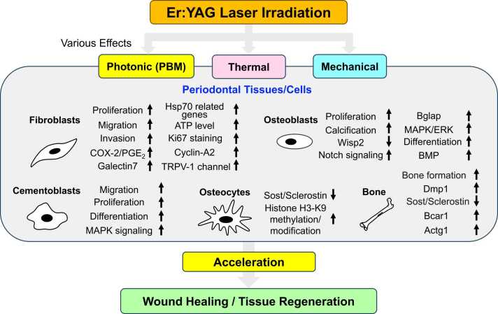
Lasers have numerous advantageous tissue interactions such as ablation or vaporization, hemostasis, bacterial killing, as well as biological effects, which induce various beneficial therapeutic effects and biological responses in the tissues. Thus, lasers are considered an effective and suitable device for treating a variety of inflammatory and infectious conditions of periodontal disease. Among various laser systems, the Er:YAG laser, which can be effectively and safely used in both soft and hard tissues with minimal thermal side effects, has been attracting much attention in periodontal therapy. This laser can effectively and precisely debride the diseased root surface including calculus removal, ablate diseased connective tissues within the bone defects, and stimulate the irradiated surrounding periodontal tissues during surgery, resulting in favorable wound healing as well as regeneration of periodontal tissues. The safe and effective performance of Er:YAG laser-assisted periodontal surgery has been reported with comparable and occasionally superior clinical outcomes compared to conventional surgery. This article explains the characteristics of the Er:YAG laser and introduces its applications in periodontal surgery including conventional flap surgery, regenerative surgery, and flapless surgery, based on scientific evidence from currently available basic and clinical studies as well as cases reports.

Keywords: Lasers; Periodontal debridement; Periodontal surgery; Periodontitis; Regeneration; Wound healing.


Conflict of interest statement

none.

Figures

Fig. 1
Fig. 1
Biological effects of Er:YAG laser. photonic, thermal, and mechanical stimuli induced by Er:YAG laser produce various advantageous biological effects on periodontal cells/tissues.
Fig. 2
Fig. 2
Application of Er:YAG laser in periodontal flap surgery in dog. Histological photomicrographs of mesio-distal sections of furcation immediately (A, B) and 12 weeks after surgery (C, D). The debridement was performed above the notch (arrow heads) using a hand curette (A) or Er:YAG laser (B). After 12 weeks, in both laser (C) and curette (D) sites, periodontal tissue attachment with bone formation was observed. The newly-formed bone (NB) extended along the dental root surface (D) in the defect. Note the greater new bone formation in the laser-treated site than the curette-treated site (Azan stain).
Fig. 3
Fig. 3
Periodontal flap surgery (open flap debridement) using Er:YAG laser. A 63-year-old female. Before surgery (A), a 9-mm deep PPD with bleeding on probing remained at the distal site of the mandibular right canine after initial treatment. Granulation tissue removal and root surface debridement were effectively and safely achieved by Er:YAG laser alone at 30 Hz and 80 mJ/pulse (panel setting) using an 80° curved and 600 µm diameter tip in contact mode under saline water spray (B). After complete degranulation, a three–wall, large and deep vertical bone defect with a 7-mm depth was observed and no visible major thermal damage such as carbonization on the laser treated root and bone surfaces was detected. The inner surface of flaps was also ablated with the Er:YAG laser at 30 Hz and 40 mJ/pulse to decontaminate the surface with diseased granulation tissue and stimulate the gingival flap tissue (C). After suturing (D). Wound healing was uneventful without any clinical complications at 1 week (E). Although gingival recession was observed, finally the probing pocket depth decreased to 2 mm without BOP at 14 years following surgery. A 7 mm of pocket reduction and 5 mm of clinical attachment gain were obtained (F). On radiographs, the vertical bone defect (arrow head) on the distal site observed at the first visit (G) was successfully repaired by apparent bone regeneration at 1 (H) and 8 (I), and 17 years (J). No adverse side effects were observed in the irradiated bone tissue (case by A. A.).
Fig. 4
Fig. 4
Schematic Illustration of the procedures of Er:YAG laser-assisted bone regenerative therapy (Er-LBRT). Before surgery (A). After conventional mechanical debridement, thorough debridement of the root surface and the bone defect was performed by applying Er:YAG laser with saline spray in the contact mode (B). Completion of debridement of root surfaces and bone defects (C). Application of enamel matrix derivative (D). After bone grafting into the defect (E), the grafted bone surface was irradiated without saline spray in a defocused mode to form a blood clot on the grafted bone surface.
Fig. 5
Fig. 5
Er:YAG laser-assisted bone regenerative therapy. A 46-year-old female. Before surgery. The mesial PPD 5–6 mm (CAL 9 mm) for the first premolar and mesial PPD 6–7 mm (CAL 9 mm) for the second premolar (A). After debridement with a curette of the coronal area of the bone defects, remaining granulation tissue and calculus were thoroughly removed using Er:YAG laser at 20 Hz and 70 mJ/pulse (panel setting) with water spray (B). After debridement, a three-wall angular bone defect in the apical area and a one-wall angular bone defect in the coronal area were observed in the mesial region of the first premolar. Granulation tissue in microconcavities on the inner surface of the bone defects had been removed, and more bleeding was observed than is caused by conventional mechanical debridement (C). The root surface was treated with ethylene-diamenetetraacetic acid and irrigated with saline, followed by EMD application to the root surface and autogenous bone graft placement into the bone defect (D). Er:YAG laser defocused irradiation without saline was applied to the grafted bone surface to enhance blood clot formation (E). At 12 months after surgery, the papilla and marginal gingiva were slightly recessed, but no interdental concavity or flap dehiscence had occurred during the healing process. Significant pocket reduction was observed: the mesial PPD 2 mm (CAL 3–4 mm) for the first premolar and mesial PPD 2–3 mm (CAL 3 mm) for the second premolar (F). At reentry surgery after periodontal regenerative treatment, dramatic bone regeneration included even the one-wall component of the defect in the interdental alveolar bone (G). A radiograph before surgery. Deep angular bone defects (arrow head) were noted in the mesial regions of the first and second premolars on preoperative radiographs (H). A radiograph taken 12 months after surgery showed that alveolar bone regeneration had reached the alveolar crest, including the one-wall defect region (I). (case by Y. T.)
Fig. 6
Fig. 6
Periodontal surgical procedure with a modified minimally invasive surgical technique using the Er:YAG laser. A 57-year-old female. After non-surgical periodontal treatment, a PPD of 5 mm mesio-buccal and 6 mm mesio-palatal remained on the maxillary right premolar (A). To minimize surgical invasion, only the buccal gingiva was elevated without elevating the interdental papillae following a modified minimally invasive surgical technique (M-MIST) design. The Er:YAG laser was used to perform gingival incision, removal of granulation tissue, and debridement of the root surface at 20 Hz and 80 mJ/pulse (panel setting) under water spray (B). Subsequently, defocused irradiation without water spray was performed on the surface of the clots (C, D). The flap was repositioned and sutured (E). At re-evaluation, 11 months postoperatively, A PPD of 2 mm and CAL gain of 4 mm were observed (F). A 2-mm PPD has been maintained for seven years postoperatively (G). On radiological examination, preoperatively, a deep intrabony defect (arrow head) was found at the mesial site of the first premolar (H). At 11 months postoperatively, radiopacity had increased from the bottom of the defect (I), and after three years, the defect was observed to appear to be filled with bony tissue (J). The newly formed tissue appearance was maintained after seven years (K). (case by K. M.)
Fig. 7
Fig. 7
Schematic illustration of the procedures of Er:YAG laser-assisted comprehensive periodontal pocket therapy (Er-LCPT). Advanced periodontal pocket showing a deep periodontal pocket with vertical bone resorption (A). Laser-assisted debridement following mechanical instrumentation of the diseased root surface for removal of subgingival calculus deposits and decontamination/detoxification of the root surface (B, C). Ablation of lining epithelium and diseased connective tissue on the inner surface of the gingival tissue as well as diseased connective tissue in the vertical bone defect during pocket irradiation for comprehensive treatment in combination with mini-curettes and Er:YAG laser. This aims to thoroughly decontaminate the whole pocket and induce increased bleeding in the bone defect from the bone surface (which may be advantageous for tissue regeneration) (D, E). Expected simultaneous thermal and photobiomodulation (PBM) effects activating the surrounding gingival and bone tissues by low-level laser penetration during high-level laser irradiation within the pocket (F). Laser ablation of the inflamed epithelial tissue on the external gingival surface. Depending on the case, the underlying connective tissue is also ablated to some extent helping in pocket depth reduction. Removal of epithelial tissue also helps prevent down-growth of epithelial tissue due to the immediate collapse of the epithelial end at the pocket entrance into the hollow space of the debrided pocket. Exposure of connective tissue delays epithelial tissue migration from the external surface into the pocket, and production of an ablated, rough soft tissue surface enhances retention of the blood clot formed at the pocket entrance, thereby assuring sealing of the pocket entrance. At the same time, stimulation of the surrounding gingival tissue from the external surface is expected by simultaneous PBM effect. It is acceptable to perform removal of external epithelial tissue as well as connective tissue before debriding the inside of the pocket (G). Blood clot (BC) coagulation at the pocket entrance by defocused irradiation without water spray to stabilize the BC formation and seal the pocket entrance. Use of diode or Nd:YAG lasers may be more advantageous for blood coagulation at deeper sites. It also intends to activate the blood clot and surrounding gingival tissue via the external surface (H). Favorable pocket healing with gingival tissue attachment and bone tissue regeneration (I). E: enamel of tooth crown, D: dentin of tooth root, SC: subgingival calculus, B: alveolar bone, G: gingival tissue, L: laser tip.
Fig. 8
Fig. 8
Er:YAG laser-assisted comprehensive periodontal pocket therapy (Er-LCPT). A 58-year-old male. Before treatment (A), a 13-mm deep PPD (CAL 15 mm) with bleeding on probing (BOP) was detected at the distal site of the mandibular right canine. First, endodontic treatment was performed due to the presence of a perio-endo lesion. Initial periodontal therapy using an Er:YAG laser was performed prior to the planned regenerative surgery. The root surface was debrided by curette, ultrasonic scaler, and Er:YAG laser, and the inner surface of the gingival wall and bone defect was debrided by curette, micro-bone curette, and Er:YAG laser. Granulation tissue removal, root surface and bone defect debridement, and epithelial tissue removal were effectively and safely performed by Er:YAG laser at 30 Hz and 60–80 mJ/pulse (panel setting) in contact mode under water spray with 80° curved contact tips of diameter 400 and 600 µm. The buccal view immediately after pocket treatment as well as removal of external epithelial tissue shows bleeding without major thermal changes (B). Then, the pocket entrance as well as the surrounding gingival tissue were irradiated in non-contact, defocused mode without water spray and the blood was coagulated and slightly carbonized (C). The coagulated blood was stable after mouth rinsing and the pocket entrance was effectively sealed (D). After 1 week (E), wound healing was favorable and epithelialization was completed. Then, wound healing progressed uneventfully without any clinical complications. At five months, the gingival recession progressed slightly and the 6-mm PPD (CAL 8 mm) with BOP still remained; however, around 9 months the PPD was reduced to 3 mm (CAL 6 mm) without BOP and regenerative surgical therapy was postponed. Supportive therapy was initiated. Resin splinting was performed after 2 years. After 14 years (F), the condition was still maintained and finally the PPD reduced to 2 mm (CAL 6 mm) without BOP. A 11-mm PPD reduction and 9 mm CAL gain were obtained. Dental radiographs show that the original bone resorption (arrow head) at the first visit was severe and horizontal (G). After 8 months (H), 5 years (I), and 14 years (J). Bone regeneration gradually progressed and the bone defect was successfully repaired to some extent but the vertical increase was limited; however, no adverse side effects are observed in the irradiated bone tissue (case by A. A.).

Similar articles

Cited by

References

    1. Tribble G.D., Lamont R.J. Bacterial invasion of epithelial cells and spreading in periodontal tissue. Periodontol 2000. 2010;52(1):68–83. - PMC - PubMed
    1. Feres M., Figueiredo L.C., Soares G.M., Faveri M. Systemic antibiotics in the treatment of periodontitis. Periodontol 2000. 2015;67(1):131–186. - PubMed
    1. Graziani F., Karapetsa D., Alonso B., Herrera D. Nonsurgical and surgical treatment of periodontitis: how many options for one disease? Periodontol 2000. 2017;75(1):152–188. - PubMed
    1. Pick R.M., Pecaro B.C., Silberman C.J. The laser gingivectomy. The use of the CO2 laser for the removal of phenytoin hyperplasia. J Periodo. 1985;56(8):492–496. - PubMed
    1. Pick R.M., Pecaro B.C. Use of the CO2 laser in soft tissue dental surgery. Lasers Surg Med. 1987;7(2):207–213. - PubMed

LinkOut - more resources