. 2023 Dec 6;6(24):22939-22946.
doi: 10.1021/acsanm.3c04300. eCollection 2023 Dec 22.

Method to Regulate Monocyte Function by Silencing HIF-1α mRNA in a Model of Retinal Neovascularization

Affiliations

Method to Regulate Monocyte Function by Silencing HIF-1α mRNA in a Model of Retinal Neovascularization

Rita E Atalor et al. ACS Appl Nano Mater. .

Abstract

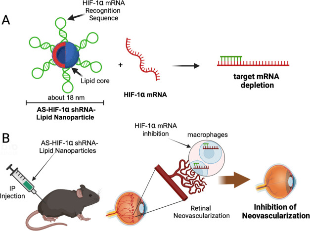
Circulating monocytes migrate into the retina in response to inflammation and neovascularization. Furthermore, under inflammatory conditions such as diabetes, healthy monocytes become activated in the circulation. However, the contribution of activated monocytes to neovascularization is largely unknown. HIF-1α has been shown to contribute to the pathogenesis of neovascularization. We describe here the synthesis of a hybrid nanomaterial for targeted delivery and gene silencing in activated monocytes that are associated with pathological neovascularization. To test the gene silencing ability of AS-shRNA-lipids in vitro, we used the probe to inhibit HIF-1α mRNA induced in mouse monocytes by exposing them to hypoxia. In addition, we tested AS-shRNA-lipids for inhibition of neovascularization in vivo using the mouse model of oxygen-induced retinopathy (OIR). Significant reduction of neovascularization was achieved in mouse OIR by targeting activated monocytes using intraperitoneal injections of AS-shRNA-lipids. Expression of HIF-1α and CD14 mRNA were both inhibited in circulating cells, suggesting normalization of the activated monocytes in P17 OIR animals treated with AS-shRNA-lipids. We hypothesized that inhibition of HIF-1α mRNA in activated monocytes may have a direct impact on VEGF expression in the retinal tissues in vivo. We observed that VEGF mRNA expression was inhibited in P17 retinal tissues after systemic treatment with HIF-1α-targeted AS-shRNA-lipids. These findings may provide a framework for a strategy to inhibit retinal neovascularization by targeting circulating activated monocytes.


Conflict of interest statement

The authors declare no competing financial interest.

Figures

Figure 1
Figure 1
Schematic drawing and hybridization motif of shRNA-lipid conjugates. (A) Design and synthesis of HIF-1α mRNA-targeted antisense shRNA-lipid conjugates (AS-HIF-1α-shRNA-lipid) for targeted delivery and gene silencing in vivo. AS-shRNA-lipid conjugates are designed for transfection-free delivery of shRNA to activated monocytes and inhibition of HIF-1α mRNA. AS-shRNA-lipid conjugates are designed by incorporating an antisense sequence complementary to HIF-1α mRNA and are stabilized using 2′-MeO nucleotides. A nonsense conjugate (NS-shRNA-lipid) was also designed from scrambled sequence of the HIF-1α mRNA recognition sequence. Physical properties of the freshly conjugated shRNA-lipids were analyzed using transmission electron microscopy (TEM) and dynamic light scattering (DLS) measurements as shown in Figure S2. (B) After systemic injections (intraperitoneally), the AS-shRNA-lipid allows efficient internalization of the conjugates by activated monocytes. Upon hybridization to the target sequence, depletion of target mRNA leads to inhibition of retinal neovascularization.
Figure 2
Figure 2
Monocyte activation and inhibition of HIF-1α mRNA. (A) Graphical representation of monocyte activation and HIF-1α mRNA inhibition using AS-shRNA-lipids. (B) HIF-1α mRNA was induced in monocytes by treating under hypoxic conditions. Expression of HIF-1α mRNA was inhibited significantly using AS-shRNA-lipids. (C) Under hypoxic conditions, monocytes get activated as monitored by CD14 expression and normalized using AS-shRNA-lipids. Monocyte cultures were treated with PMA for 78 h to induce activation. Next, AS-shRNA-lipid was added and cells were incubated for 4 h at 1% oxygen (hypoxia) and 21% oxygen (normoxia). qPCR data analysis showed both HIF-1α and CD14 expressions were significantly decreased in AS-shRNA-lipid-treated monocytes compared to PBS control. Hypoxia has little effect on CD14 expression in cultured monocytes. These data repetitive of three independent experiments with n = 3 for each samples size for statistical analysis.
Figure 3
Figure 3
Quantification of avascular and neovascular areas in P17 mouse OIR retinas after inhibition of HIF-1α mRNA in activated monocytes via intraperitoneal injections of shRNA-lipids. The OIR pups were injected with AS(NS)-shRNA-lipid on P12, P13, and P15 posthyperoxic treatment, and tissues were collected at P17. (A–C) Retinal tissues were stained with IB4 to analyze % avascular areas (white) and % NV areas (yellow). Panels (D–F) are zoomed images of panels (A–C), respectively. (G, H) The AS-shRNA-lipid significantly inhibited (over 90%) NV areas compared to controls, though changes in avascular areas were minimal. For each group, a total of 12 pups were used.
Figure 4
Figure 4
Expression of HIF-1α and CD14 mRNA in monocytes and HIF-1α and VEGF mRNA in retinal tissues from mouse OIR at P17 after treatment with PBS or AS-shRNA-lipids. (A, B) HIF-1α mRNA and CD14 mRNA expressions were significantly lower (P < 0.0001) in monocytes from OIR mice treated with the AS-shRNA-lipid compared to untreated OIR control mice. (C, D) Both HIF-1α mRNA and VEGF mRNA expressions were significantly decreased (P < 0.0001) in As-shRNA-lipid-treated mice compared to control mice. For each group, a total of 12 pups were used with pooled tissues from 3 pups for each experiment (n = 4).

Similar articles

Cited by

References

    1. Penn J. S.; Madan A.; Caldwell R. B.; Bartoli M.; Caldwell R. W.; Hartnett M. E. Vascular endothelial growth factor in eye disease. Prog. Retinal Eye Res. 2008, 27 (4), 331–371. 10.1016/j.preteyeres.2008.05.001. - DOI - PMC - PubMed
    1. Campbell M.; Humphries P.. THE BLOOD-RETINA BARRIER Tight Junctions and Barrier Modulation. In Advances in Experimental Medicine and Biology; Springer, 2013; Vol. 763, pp 70–84. - PubMed
    1. Gao X.; Wang Y. S.; Li X. Q.; Hou H. Y.; Su J. B.; Yao L. B.; Zhang J. Macrophages promote vasculogenesis of retinal neovascularization in an oxygen-induced retinopathy model in mice. Cell Tissue Res. 2016, 364 (3), 599–610. 10.1007/s00441-015-2353-y. - DOI - PubMed
    1. Uddin M. I.; Kilburn T. C.; Duvall C. L.; Penn J. S. Visualizing HIF-1 alpha mRNA in a Subpopulation of Bone Marrow-Derived Cells to Predict Retinal Neovascularization. ACS Chem. Biol. 2020, 15 (11), 3004–3012. 10.1021/acschembio.0c00662. - DOI - PMC - PubMed
    1. Davies M.; Eubanks J.; Powers M. Microglia and macrophages are increased in response to ischemia-induced retinopathy in the mouse retina. Mol. Vision 2006, 12 (53–54), 467–477. - PubMed

LinkOut - more resources