

ROS-scavenging materials for skin wound healing: advancements and applications
- PMID: 38149175
- PMCID: PMC10749972
- DOI: 10.3389/fbioe.2023.1304835
ROS-scavenging materials for skin wound healing: advancements and applications
Abstract
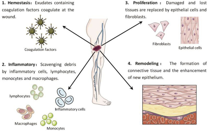
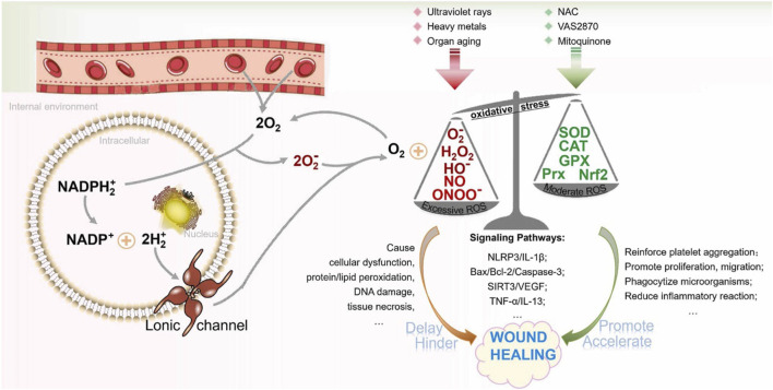
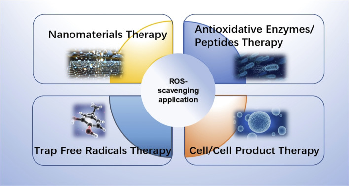
The intricate healing process of skin wounds includes a variety of cellular and molecular events. Wound healing heavily relies on reactive oxygen species (ROS), which are essential for controlling various processes, including inflammation, cell growth, angiogenesis, granulation, and the formation of extracellular matrix. Nevertheless, an overabundance of reactive oxygen species (ROS) caused by extended oxidative pressure may result in the postponement or failure of wound healing. It is crucial to comprehend the function of reactive oxygen species (ROS) and create biomaterials that efficiently eliminate ROS to enhance the healing process of skin wounds. In this study, a thorough examination is presented on the role of reactive oxygen species (ROS) in the process of wound healing, along with an exploration of the existing knowledge regarding biomaterials employed for ROS elimination. In addition, the article covers different techniques and substances used in the management of skin wound. The future prospects and clinical applications of enhanced biomaterials are also emphasized, highlighting the potential of biomaterials that scavenge active oxygen to promote skin repair. This article seeks to enhance the understanding of the complex processes of ROS in the healing of wounds and the application of ROS-scavenging materials. Its objective is to create novel strategies for effective treatment skin wounds.
Keywords: biomaterials; oxidative stress; reactive oxygen species; skin wound; wound healing.
Copyright © 2023 Dong and Wang.
Conflict of interest statement
The authors declare that the research was conducted in the absence of any commercial or financial relationships that could be construed as a potential conflict of interest.
Figures
Similar articles
-
In situ forming and reactive oxygen species-scavenging gelatin hydrogels for enhancing wound healing efficacy.Acta Biomater. 2020 Feb;103:142-152. doi: 10.1016/j.actbio.2019.12.009. Epub 2019 Dec 14. Acta Biomater. 2020. PMID: 31846801
-
Recent advances in reactive oxygen species scavenging nanomaterials for wound healing.Exploration (Beijing). 2024 Jan 17;4(3):20230066. doi: 10.1002/EXP.20230066. eCollection 2024 Jun. Exploration (Beijing). 2024. PMID: 38939866 Free PMC article. Review.
-
Advances and Challenges in Immune-Modulatory Biomaterials for Wound Healing Applications.Pharmaceutics. 2024 Jul 26;16(8):990. doi: 10.3390/pharmaceutics16080990. Pharmaceutics. 2024. PMID: 39204335 Free PMC article. Review.
-
Epigallocatechin Gallate: The Emerging Wound Healing Potential of Multifunctional Biomaterials for Future Precision Medicine Treatment Strategies.Polymers (Basel). 2021 Oct 23;13(21):3656. doi: 10.3390/polym13213656. Polymers (Basel). 2021. PMID: 34771213 Free PMC article. Review.
-
Reactive oxygen species-degradable polythioketal urethane foam dressings to promote porcine skin wound repair.Sci Transl Med. 2022 Apr 20;14(641):eabm6586. doi: 10.1126/scitranslmed.abm6586. Epub 2022 Apr 20. Sci Transl Med. 2022. PMID: 35442705 Free PMC article.
Cited by
-
Multifunctional layered microneedle patches enable transdermal angiogenesis and immunomodulation for scarless healing of thermal burn injuries.Mater Today Bio. 2024 Nov 22;29:101359. doi: 10.1016/j.mtbio.2024.101359. eCollection 2024 Dec. Mater Today Bio. 2024. PMID: 39655166 Free PMC article.
-
Sea food by-products valorization for biomedical applications: evaluation of their wound regeneration capabilities in an Ex vivo skin model.Front Vet Sci. 2024 Nov 18;11:1491385. doi: 10.3389/fvets.2024.1491385. eCollection 2024. Front Vet Sci. 2024. PMID: 39660177 Free PMC article.
-
Synergistic Combinations of Native Australian Plants For Skin Inflammation and Wound Healing.Biomedicines. 2025 Jul 17;13(7):1754. doi: 10.3390/biomedicines13071754. Biomedicines. 2025. PMID: 40722824 Free PMC article.
-
Harnessing Cannabis sativa Oil for Enhanced Skin Wound Healing: The Role of Reactive Oxygen Species Regulation.Pharmaceutics. 2024 Sep 30;16(10):1277. doi: 10.3390/pharmaceutics16101277. Pharmaceutics. 2024. PMID: 39458608 Free PMC article. Review.
-
Probiotic active gel promotes diabetic wound healing through continuous local glucose consumption and antioxidant.J Nanobiotechnology. 2025 Jan 30;23(1):62. doi: 10.1186/s12951-025-03115-5. J Nanobiotechnology. 2025. PMID: 39885505 Free PMC article.
References
Publication types
LinkOut - more resources
Full Text Sources