. 2023 Dec 27:12:RP88008.
doi: 10.7554/eLife.88008.

Vertical transmission of maternal DNA through extracellular vesicles associates with altered embryo bioenergetics during the periconception period

Affiliations

Vertical transmission of maternal DNA through extracellular vesicles associates with altered embryo bioenergetics during the periconception period

David Bolumar et al. Elife. .

Abstract

The transmission of DNA through extracellular vesicles (EVs) represents a novel genetic material transfer mechanism that may impact genome evolution and tumorigenesis. We aimed to investigate the potential for vertical DNA transmission within maternal endometrial EVs to the pre-implantation embryo and describe any effect on embryo bioenergetics. We discovered that the human endometrium secretes all three general subtypes of EV - apoptotic bodies (ABs), microvesicles (MVs), and exosomes (EXOs) - into the human endometrial fluid (EF) within the uterine cavity. EVs become uniformly secreted into the EF during the menstrual cycle, with the proportion of different EV populations remaining constant; however, MVs contain significantly higher levels of mitochondrial (mt)DNA than ABs or EXOs. During the window of implantation, MVs contain an eleven-fold higher level of mtDNA when compared to cells-of-origin within the receptive endometrium, which possesses a lower mtDNA content and displays the upregulated expression of mitophagy-related genes. Furthermore, we demonstrate the internalization of EV-derived nuclear-encoded (n)DNA/mtDNA by trophoblast cells of murine embryos, which associates with a reduction in mitochondrial respiration and ATP production. These findings suggest that the maternal endometrium suffers a reduction in mtDNA content during the preconceptional period, that nDNA/mtDNA become packaged into secreted EVs that the embryo uptakes, and that the transfer of DNA to the embryo within EVs occurs alongside the modulation of bioenergetics during implantation.

Keywords: developmental biology; endometrium; exosomes; extracellular vesicles; human; maternal-embryonic crosstalk; medicine; metabolism; mitochondrial DNA; mouse.


Conflict of interest statement

DB, JM, JG, AO, IM, AM, CM, AD, PF, MC, JE, DG, CS, FV No competing interests declared

Figures
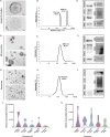
Figure 1.
Figure 1.. Characterization of endometrial fluid-derived extracellular vesicles.
(A–I) Analysis of ABs, MVs, and EXOs isolated from human EF samples: morphology by TEM (A, D, and G), size distribution by DLS (B, E, and H), and protein marker expression by Western blotting (C, F, and I). TEM images obtained using two different protocols for an external (deposition processing, upper images) or internal (ultrathin slide processing, lower images) view of EVs. Size distribution analyzed in a single EF sample by DLS during the receptive phase for (B) ABs, (E) MVs, and (H) EXOs. Graphs show the average size distribution and percentage of total particles contained within the populations. Specific protein markers analyzed by Western blotting for (C) ABs, (F) MVs, and (I) EXOs. Analyzed markers (and associated molecular mass) were calnexin (90–100 kDa), calreticulin (60 kDa), VDAC1 (31 kDa), ARF6 (18 kDa), CD9 (24 kDa), CD63 (30–60 kDa), and TSG101 (45–50 kDa). (J and K) Particle concentration and size distribution measured by NTA for (J) MVs and (K) EXOs secreted throughout the menstrual cycle. One-way ANOVA and Kruskal-Wallis rank sum tests performed to compare the distinct menstrual cycle phases - no significant differences were observed.
Figure 1—figure supplement 1.
Figure 1—figure supplement 1.. Transmission electron micrograph analysis of human endometrial fluid-derived extracellular vesicle morphology.
TEM micrographs show morphological details of human EF (A and B) ABs, (C) MVs, and (D) EXOs. Images obtained via two different protocols to obtain an external (deposition processing) and internal (ultrathin slide processing) view. Isolated ABs displayed a wide range of vesicle sizes (>1 µm to <50 nm) with heterogeneous content composition, including membranous structures within ABs (B3). Image B2 corresponds to a higher magnification of image B1. MVs were considerably more abundant, with sizes from 200 to 700 nm (C1 and C2) with highly electron-dense heterogeneous contents (C3 and C4). EXOs displayed a similar aspect and abundance to MVs (D) with sizes of 40–160 nm but more homogeneous structures.
Figure 1—figure supplement 2.
Figure 1—figure supplement 2.. Size distribution of human endometrial fluid-derived microvesicles and exosomes measured by nanoparticle tracking analysis.
Size patterning and total particle concentration of isolated (A) MVs and (B) EXOs obtained from a single human EF sample isolated during the receptive phase of the menstrual cycle. The standard error of five measurements shown as the grey area in each graph.
Figure 1—figure supplement 3.
Figure 1—figure supplement 3.. Microvesicle and exosome dynamics in endometrial fluid samples isolated during the menstrual cycle.
Particle concentration and size distribution by NTA for (A) MVs and (B) EXOs analyzed throughout the menstrual cycle.
Figure 2.
Figure 2.. DNA sequencing analysis and coding sequence comparisons of human endometrial fluid-derived extracellular vesicle populations.
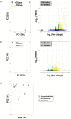
(A and B) Volcano plots comparing DNA sequence enrichment between ABs, MVs, and EXOs. Only MVs show significant sequence enrichment compared to ABs and EXOs. (C) Specific gene ID DNA sequences encapsulated within MVs compared to ABs and EXOs, which are mainly mitochondrial DNA.
Figure 2—figure supplement 1.
Figure 2—figure supplement 1.. Effect of DNase treatment in sequencing analysis and coding sequences comparison between human endometrial fluid-derived extracellular vesicle populations.
Sequencing analysis results for (A) ABs and (B) EXOs treated with (+DNase) and without (- DNase) DNase type I. (A and B; Left Panels) PCA analyses show grouping due to DNase treatment for both ABs and EXOs. (A and B; Right Panels) Volcano plots show the significant enrichment of DNA sequences in treated EVs (purple dots in volcano plots) versus untreated EVs (yellow dots in volcano plots). (C) Principal component analysis shows EV sample grouping due to specificity in coding-gene sequences.
Figure 3.
Figure 3.. Quantification of mitochondrial DNA in human endometrial tissues and human endometrial fluid-derived microvesicles.
(A) Relative mtDNA/nDNA ratio calculated from endometrial biopsies from donors undergoing HRT in pre-receptive (P+2), receptive (P+5), and post-receptive (P+8) periods, n=70. (B) Gene expression analysis of endometrial biopsies for nuclear genes coding for mitophagy- and mtDNA packing-related proteins (upper panel) and for genes coding for proteins related to mitochondrial function (lower panel), n=66. (C) Quantification of relative mtDNA copy number packed into MVs isolated from the EF in pre-receptive, receptive, and post-receptive periods, n=20. One-way ANOVA and Kruskal-Wallis rank sum tests performed to compare the distinct periods - no significant differences were observed. *p<0.05, ***p<0.001.
Figure 4.
Figure 4.. Internalization of endometrial extracellular vesicle-derived DNA by cells of the murine embryo.
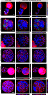
Confocal images show hatched embryos after co-culture with EdU-tagged ABs, MVs, and EXOs isolated from Ishikawa cell supernatants. Embryo membranes were visualized with Wheat germ agglutinin (WGA) in red, embryo nuclei with DAPI, and EdU-tagged transferred DNA in green. Zoomed images taken from the areas demarcated by white boxes in merge images. Cell-free DNA and residual small-sized EVs were used as control conditions (Neg). Scale bar in zoom = 20 µm.
Figure 4—figure supplement 1.
Figure 4—figure supplement 1.. Characterization of EdU-tagged DNA incorporation into extracellular vesicles isolated from Ishikawa cells.
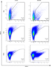
EXOs, MVs, and ABs isolated from non-EdU-tagged (Control) and EdU-tagged DNA (EdU-DNA) were analyzed by flow cytometry for complexity (SSC-A) and EdU-488 staining (FITC-A). Gates show positive EdU-488 EVs percentage in the distinct populations.
Figure 4—figure supplement 2.
Figure 4—figure supplement 2.. Z-stack/orthogonal sections of murine embryos co-cultured with extracellular vesicles containing EdU-tagged DNA.
Images show hatching/hatched embryos after co-culture with EdU-tagged EVs (ABs, MVs, and EXOs). White arrows show the co-localization of tagged DNA and nuclei, indicating the nuclear location of extracellular vesicle-derived DNA. Embryo membranes are visualized with WGA in red, embryo nuclei with DAPI, and EdU-tagged transferred DNA in green. Cell-free DNA and residual small-sized EVs were used as control conditions (Neg). Bars = 20 µm. Red lines indicate the intersection for orthogonal image acquisition.
Figure 4—figure supplement 3.
Figure 4—figure supplement 3.. Detection of exogenous mitochondrial DNA in mouse embryos.
Embryos co-incubated with 10 µM of an ATP8 DNA sequence (mtDNA fragment) conjugated with Biotin overnight ATP8-Biotin DNA were detected with streptavidin-Cy3 (Green) and nuclei counterstained with DAPI (Blue). Negative control embryos incubated without the ATP8-Biotin show the background signal after streptavidin-Cy3 staining.
Figure 5.
Figure 5.. Mitochondrial function in embryos incubated with human endometrial fluid-derived extracellular vesicles.
(A) Murine embryo ATP content (n=60) after overnight co-incubation with the EF-derived EV populations (phase IV or receptive phase of the natural menstrual cycle n=5). ‘All EVs’ indicates a combination of ABs, MVs, and EXOs. Embryos not incubated with EVs used as a control condition (Cnt). (B) OCR was recorded on a Seahorse instrument before and after drug injection (timing indicated on the graph). Blocked embryos used as an additional negative control (Neg Cnt), total embryos used (n=720). (C) Basal respiration [(Last rate measurement before the first injection)-(minimum rate measurement after Rotenone/antimycin A injection)] and (D) Maximal respiration [(Maximal rate measurement after FCCP injection)-(minimum rate measurement after Rotenone/antimycin A injection)] shown for each condition. One-way ANOVA and Tukey comparison post-hoc performed - no significant differences between conditions were observed (excluding the Neg Cnt condition). *p<0.05, ***p<0.001.

Update of

  • doi: 10.1101/2023.04.21.537765
  • doi: 10.7554/eLife.88008.1
  • doi: 10.7554/eLife.88008.2
  • doi: 10.7554/eLife.88008.3

References

    1. Antonyak MA, Li B, Boroughs LK, Johnson JL, Druso JE, Bryant KL, Holowka DA, Cerione RA. Cancer cell-derived microvesicles induce transformation by transferring tissue transglutaminase and fibronectin to recipient cells. PNAS. 2011;108:4852–4857. doi: 10.1073/pnas.1017667108. - DOI - PMC - PubMed
    1. Ashary N, Tiwari A, Modi D. Embryo implantation: war in times of love. Endocrinology. 2018;159:1188–1198. doi: 10.1210/en.2017-03082. - DOI - PubMed
    1. Bergsmedh A, Szeles A, Henriksson M, Bratt A, Folkman MJ, Spetz AL, Holmgren L. Horizontal transfer of oncogenes by uptake of apoptotic bodies. PNAS. 2001;98:6407–6411. doi: 10.1073/pnas.101129998. - DOI - PMC - PubMed
    1. Bidarimath M, Khalaj K, Kridli RT, Kan FWK, Koti M, Tayade C. Extracellular vesicle mediated intercellular communication at the porcine maternal-fetal interface: A new paradigm for conceptus-endometrial cross-talk. Scientific Reports. 2017;7:40476. doi: 10.1038/srep40476. - DOI - PMC - PubMed
    1. Burns G, Brooks K, Wildung M, Navakanitworakul R, Christenson LK, Spencer TE. Extracellular vesicles in luminal fluid of the ovine uterus. PLOS ONE. 2014;9:e90913. doi: 10.1371/journal.pone.0090913. - DOI - PMC - PubMed

Substances