

Evaluating the Irritant Factors of Silicone and Hydrocolloid Skin Contact Adhesives Using Trans-Epidermal Water Loss, Protein Stripping, Erythema, and Ease of Removal
- PMID: 38150300
- PMCID: PMC10792606
- DOI: 10.1021/acsabm.3c00874
Evaluating the Irritant Factors of Silicone and Hydrocolloid Skin Contact Adhesives Using Trans-Epidermal Water Loss, Protein Stripping, Erythema, and Ease of Removal
Abstract
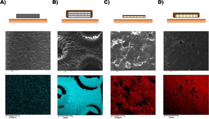
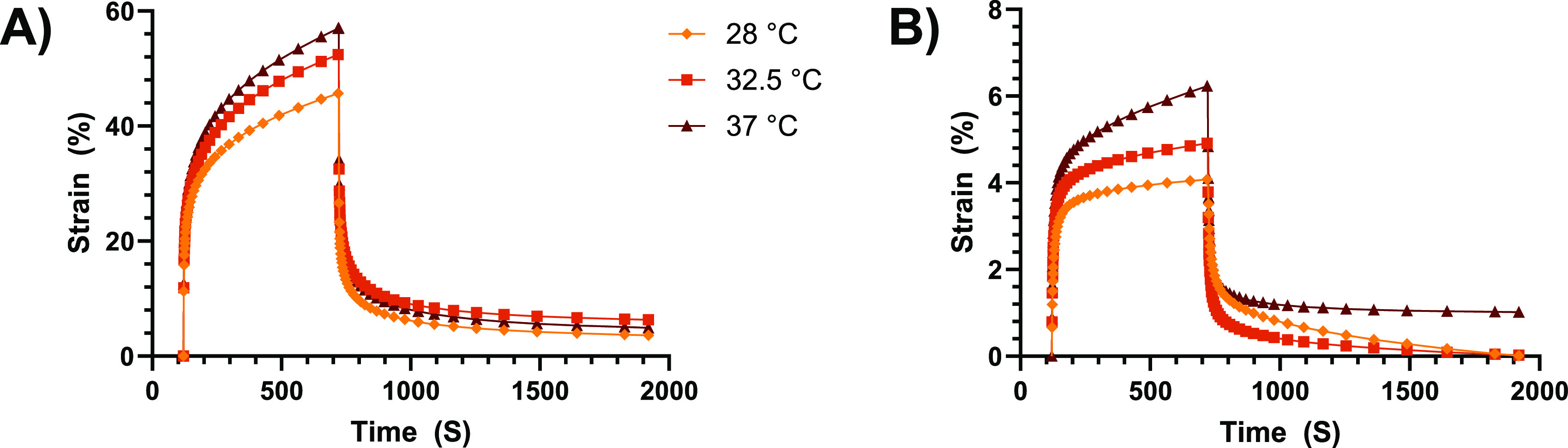
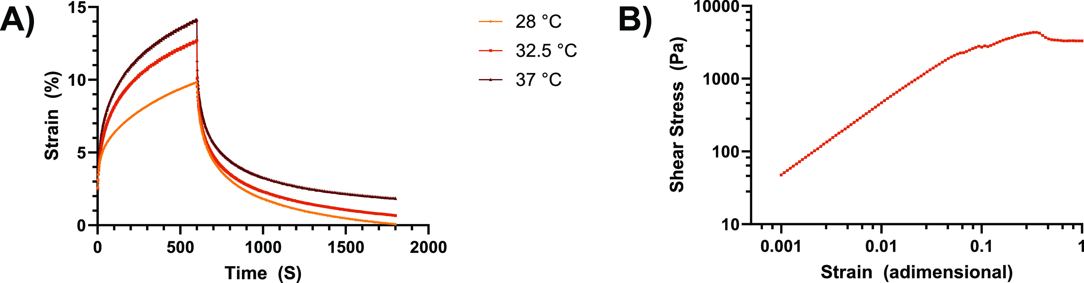
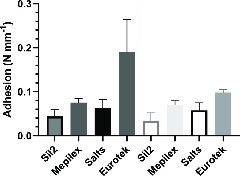
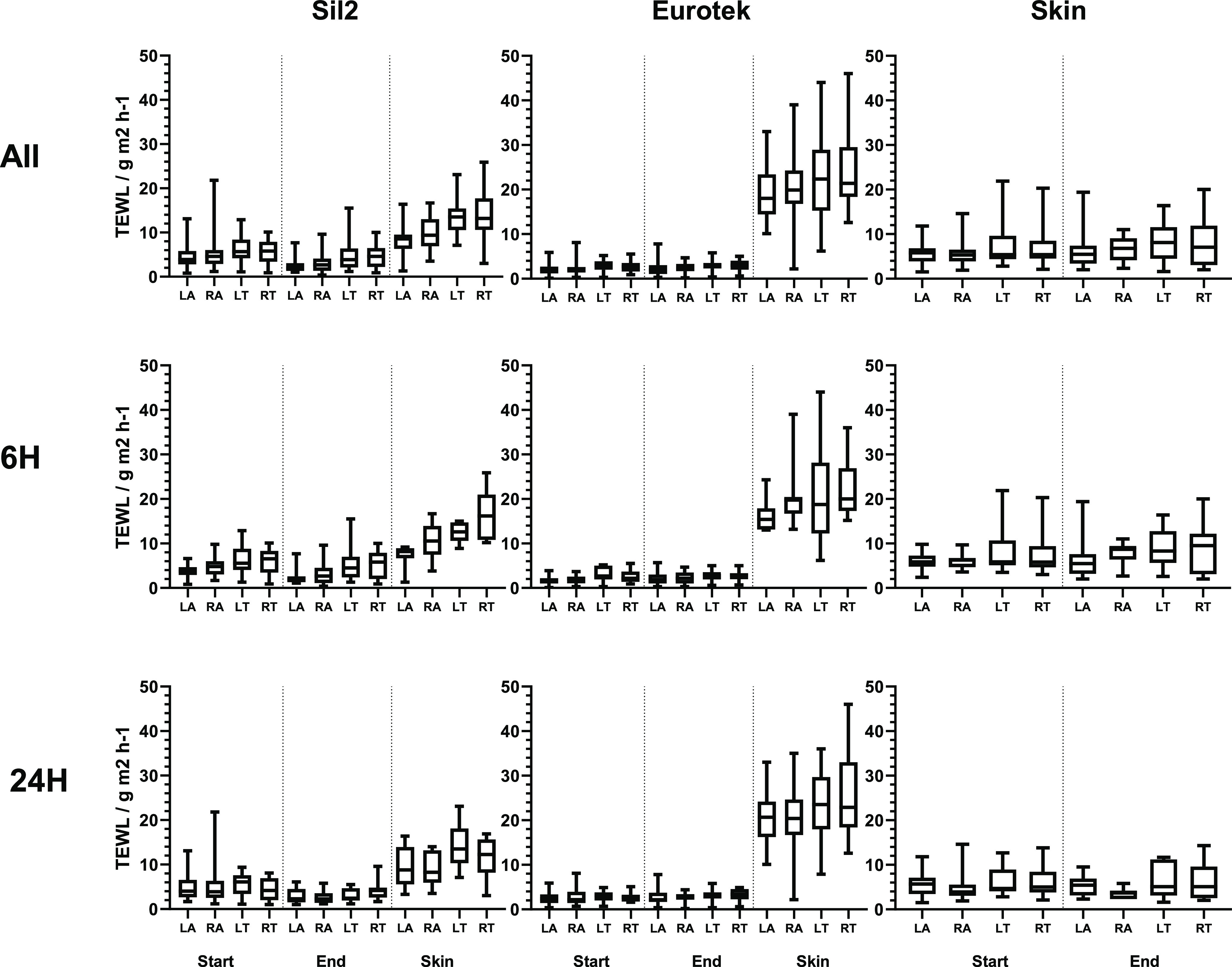
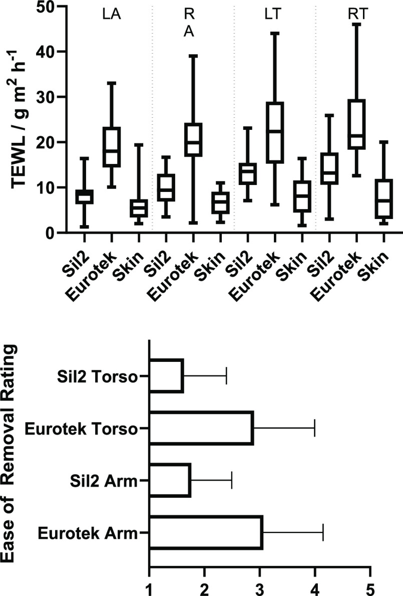
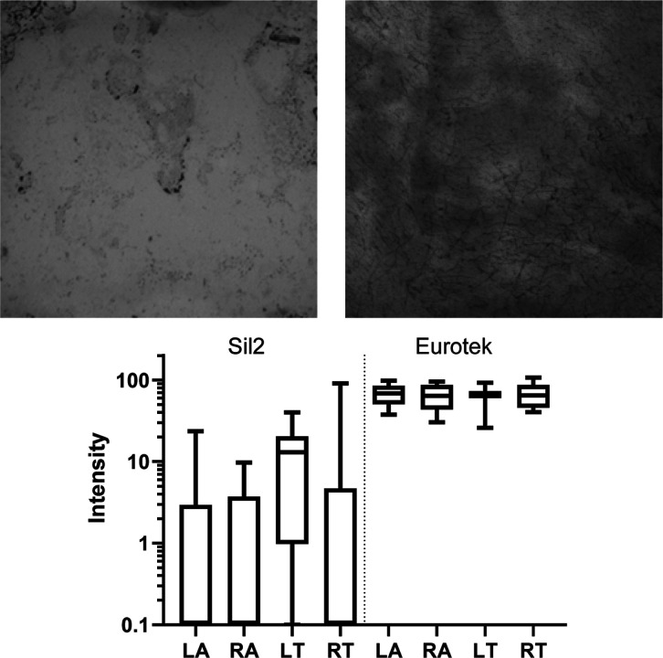
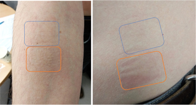
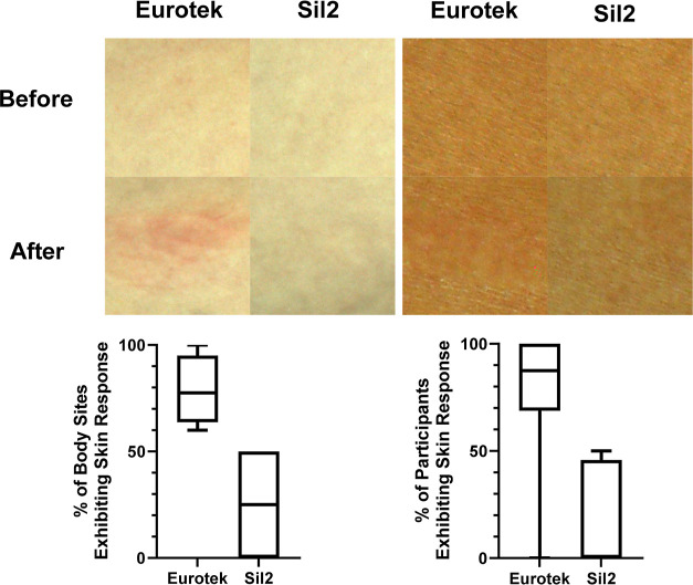
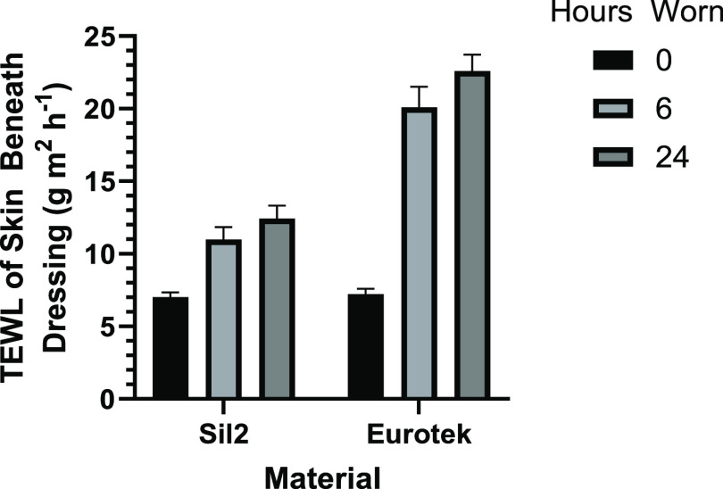
A composite silicone skin adhesive material was designed to improve its water vapor permeability to offer advantages to wearer comfort compared to existing skin adhesive dressings available (including perforated silicone and hydrocolloid products). The chemical and mechanical properties of this novel dressing were analyzed to show that it has a high creep compliance, offering anisotropic elasticity that is likely to place less stress on the skin. A participant study was carried out in which 31 participants wore a novel silicone skin adhesive (Sil2) and a hydrocolloid competitor and were monitored for physiological response to the dressings. Trans-epidermal water loss (TEWL) was measured pre- and postwear to determine impairment of skin barrier function. Sil2 exhibited a higher vapor permeability than the hydrocolloid dressings during wear. Peel strength measurements and dye counter staining of the removed dressings showed that the hydrocolloid had a higher adhesion to the participants' skin, resulting in a greater removal of proteins from the stratum corneum and a higher pain rating from participants on removal. Once the dressings were removed, TEWL of the participants skin beneath the Sil2 was close to normal in comparison to the hydrocolloid dressings that showed an increase in skin TEWL, indicating that the skin had been highly occluded. Analysis of the skin immediately after removal showed a higher incidence of erythema following application of hydrocolloid dressings (>60%) compared to Sil2, (<30%). In summary, this modified silicone formulation demonstrates superior skin protection properties compared to hydrocolloid dressings and is more suitable for use as a skin adhesive.
Keywords: TEWL; adhesive; peel strength; silicone; skin contact; vapor permeability.
Conflict of interest statement
The authors declare no competing financial interest.
Figures
Similar articles
-
Effects of adhesive dressings on the stratum corneum of the skin.J Wound Care. 2001 Feb;10(2):7-10. doi: 10.12968/jowc.2001.10.2.26054. J Wound Care. 2001. PMID: 12964220 Clinical Trial.
-
An evaluation of the skin stripping of wound dressing adhesives.J Wound Care. 2011 Sep;20(9):412, 414, 416-22. doi: 10.12968/jowc.2011.20.9.412. J Wound Care. 2011. PMID: 22068140 Clinical Trial.
-
Performance and safety of transparent postoperative dressings with silicone adhesive in daily practice on fragile skin.J Wound Care. 2024 Nov 2;33(11):824-832. doi: 10.12968/jowc.2024.0308. J Wound Care. 2024. PMID: 39480729
-
Removal of adhesive wound dressing and its effects on the stratum corneum of the skin: comparison of eight different adhesive wound dressings.Int Wound J. 2014 Feb;11(1):50-4. doi: 10.1111/j.1742-481X.2012.01061.x. Epub 2012 Aug 7. Int Wound J. 2014. PMID: 22883604 Free PMC article.
-
Mechanical properties of tissue adhesives used for retaining extraoral silicone prostheses in maxillofacial defects: A systematic review.J Prosthet Dent. 2024 Apr 22:S0022-3913(24)00223-3. doi: 10.1016/j.prosdent.2024.03.029. Online ahead of print. J Prosthet Dent. 2024. PMID: 38653691 Review.
Cited by
-
Comparative Efficacy of Silicone Sheets and Hyperbaric Oxygen Therapy in Post-Surgical Scar Prevention: A Prospective Observational Study.Int J Med Sci. 2025 Feb 11;22(5):1167-1175. doi: 10.7150/ijms.108397. eCollection 2025. Int J Med Sci. 2025. PMID: 40027186 Free PMC article. Clinical Trial.
-
Development of a Method for Blocking Polysodiumoxy(methyl)siloxane Obtained in an Alcohol Medium.Polymers (Basel). 2025 Jul 24;17(15):2023. doi: 10.3390/polym17152023. Polymers (Basel). 2025. PMID: 40808073 Free PMC article.
References
-
- Rippon M.; Ousey K.; Rogers A.; Atkin L. Wound Hydration versus Maceration: Understanding the Differences. Wounds 2016, 12 (3), 62–68.
-
- Vowden K.; Vowden P. Wound Dressings: Principles and Practice. Surgery 2017, 35 (9), 489–494. 10.1016/j.mpsur.2017.06.005. - DOI
Publication types
MeSH terms
Substances
LinkOut - more resources
Full Text Sources