. 2024 Apr 1;326(4):L440-L457.
doi: 10.1152/ajplung.00273.2023. Epub 2023 Dec 27.

Hemopexin reverses activation of lung eIF2α and decreases mitochondrial injury in chlorine-exposed mice

Affiliations

Hemopexin reverses activation of lung eIF2α and decreases mitochondrial injury in chlorine-exposed mice

Sadis Matalon et al. Am J Physiol Lung Cell Mol Physiol. .

Abstract

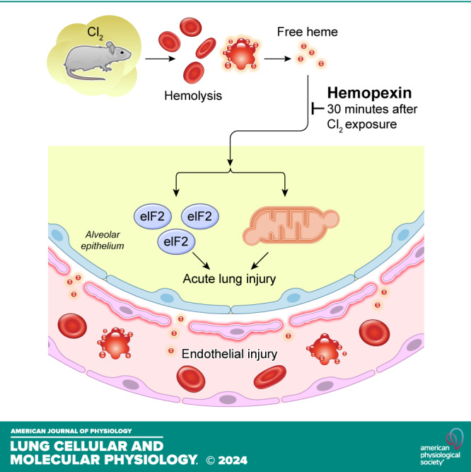
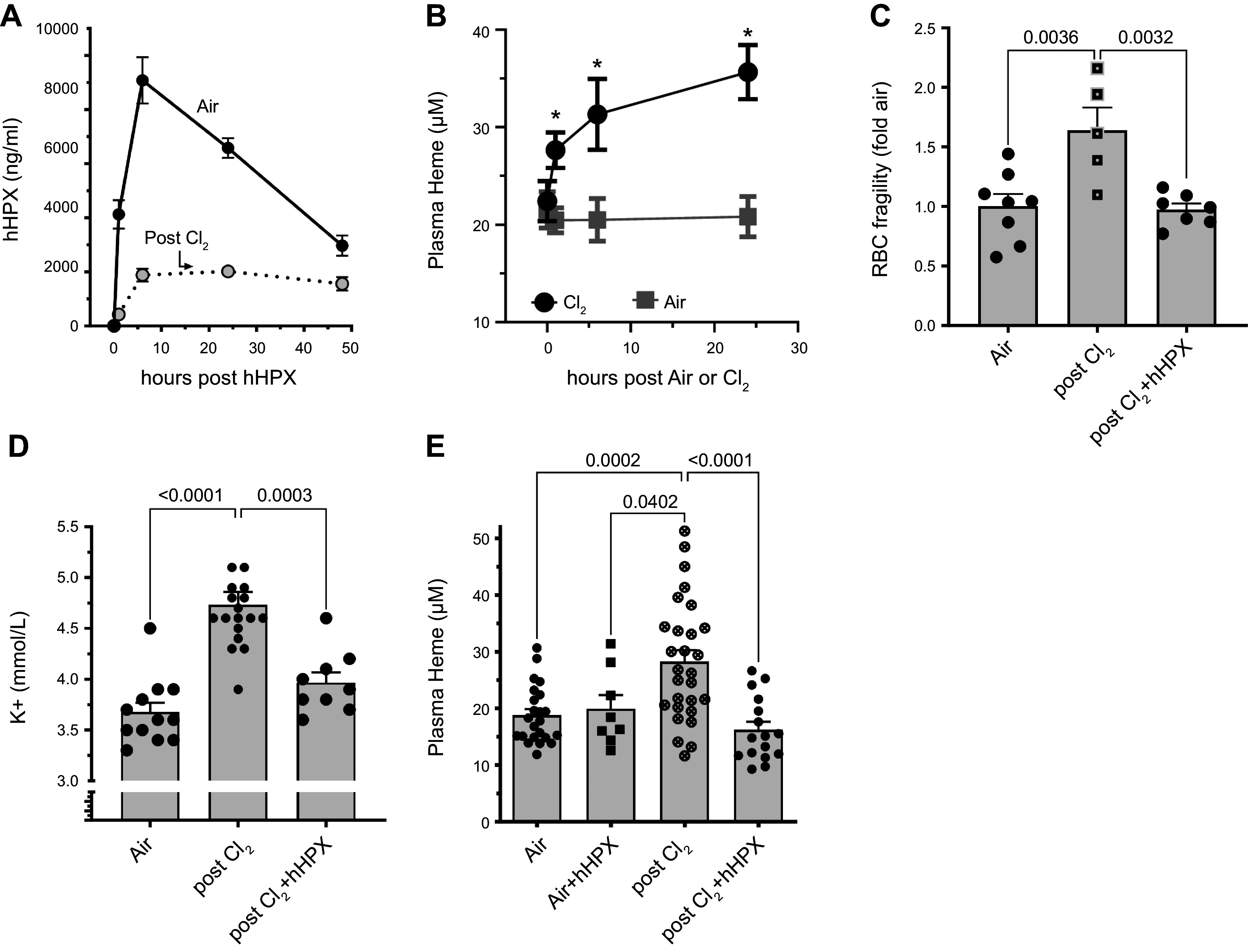
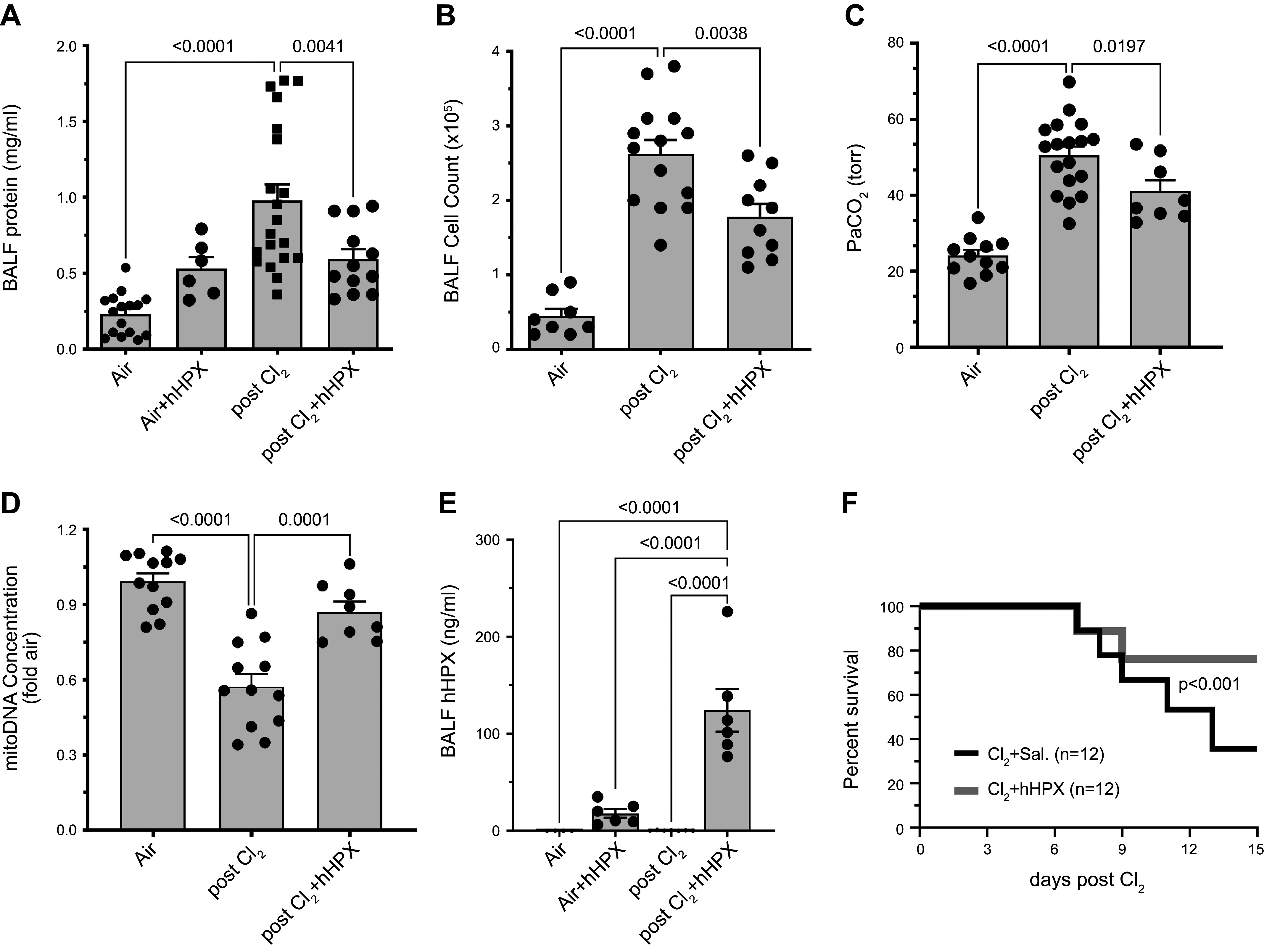
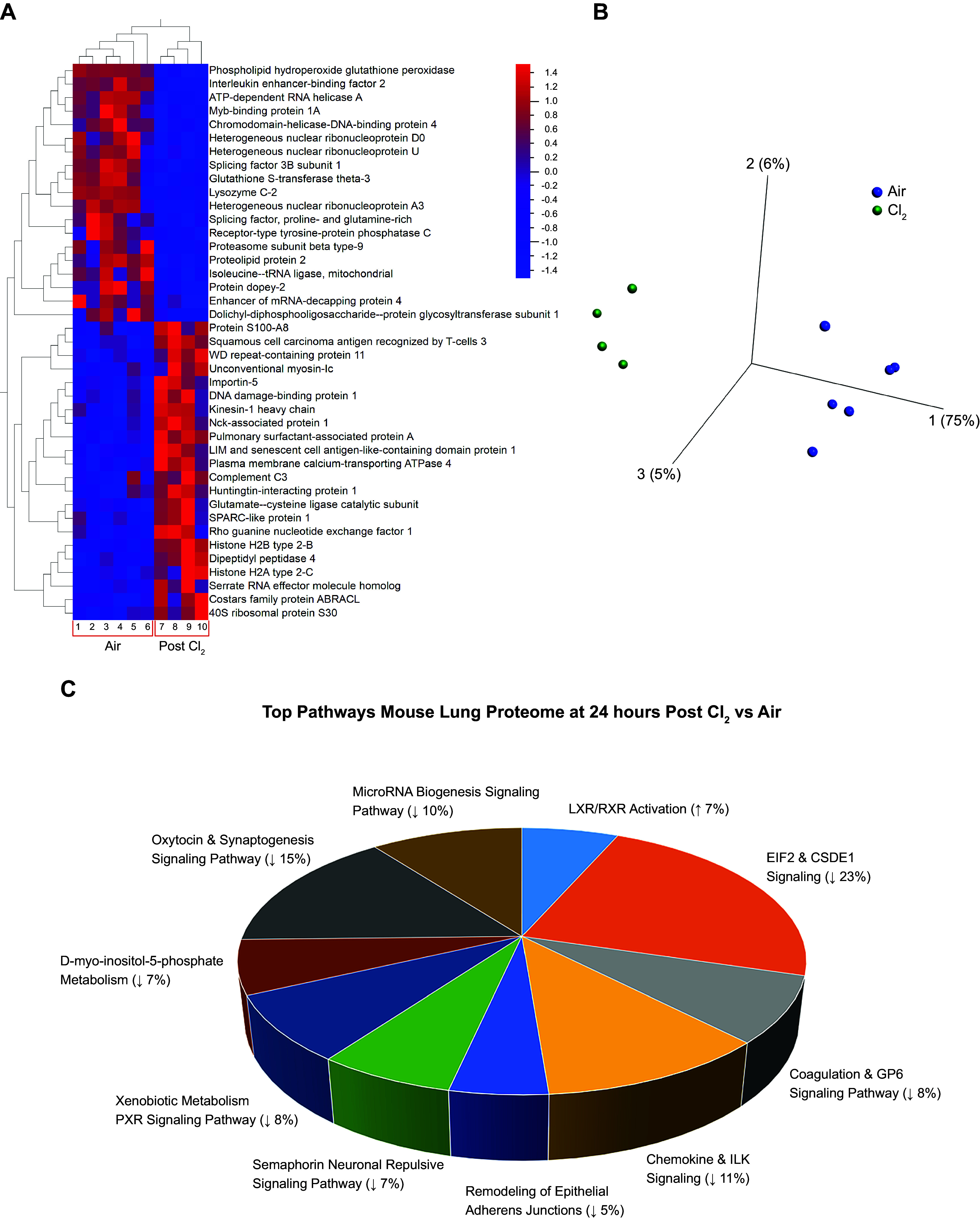
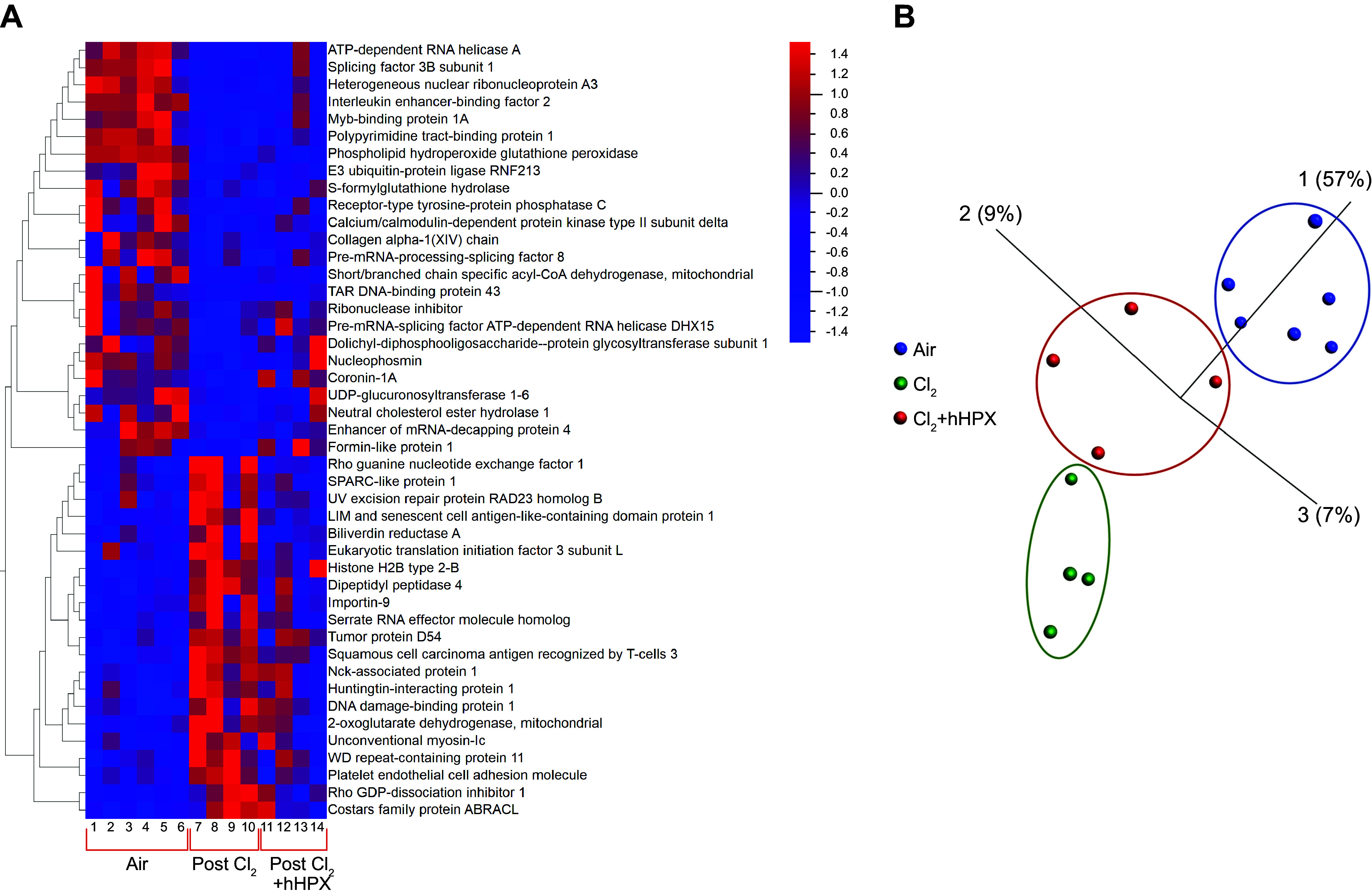
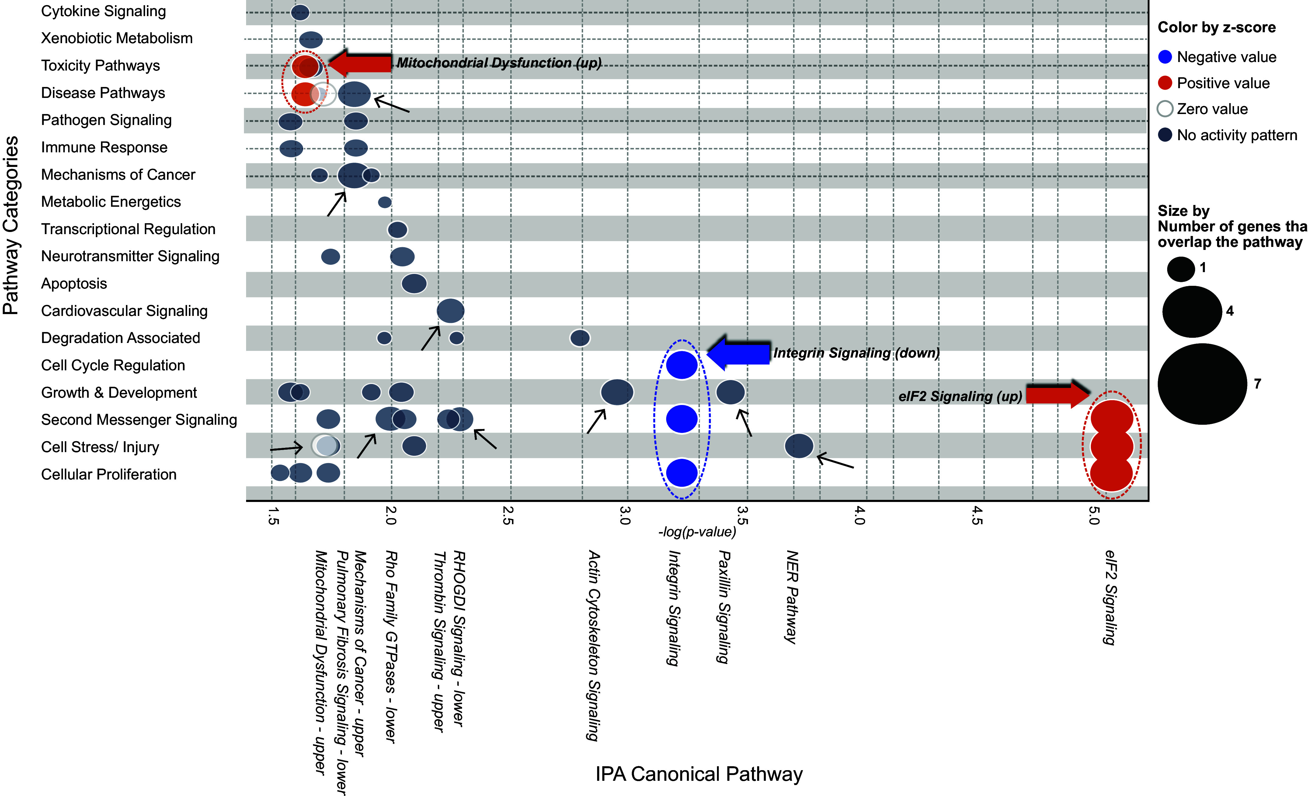
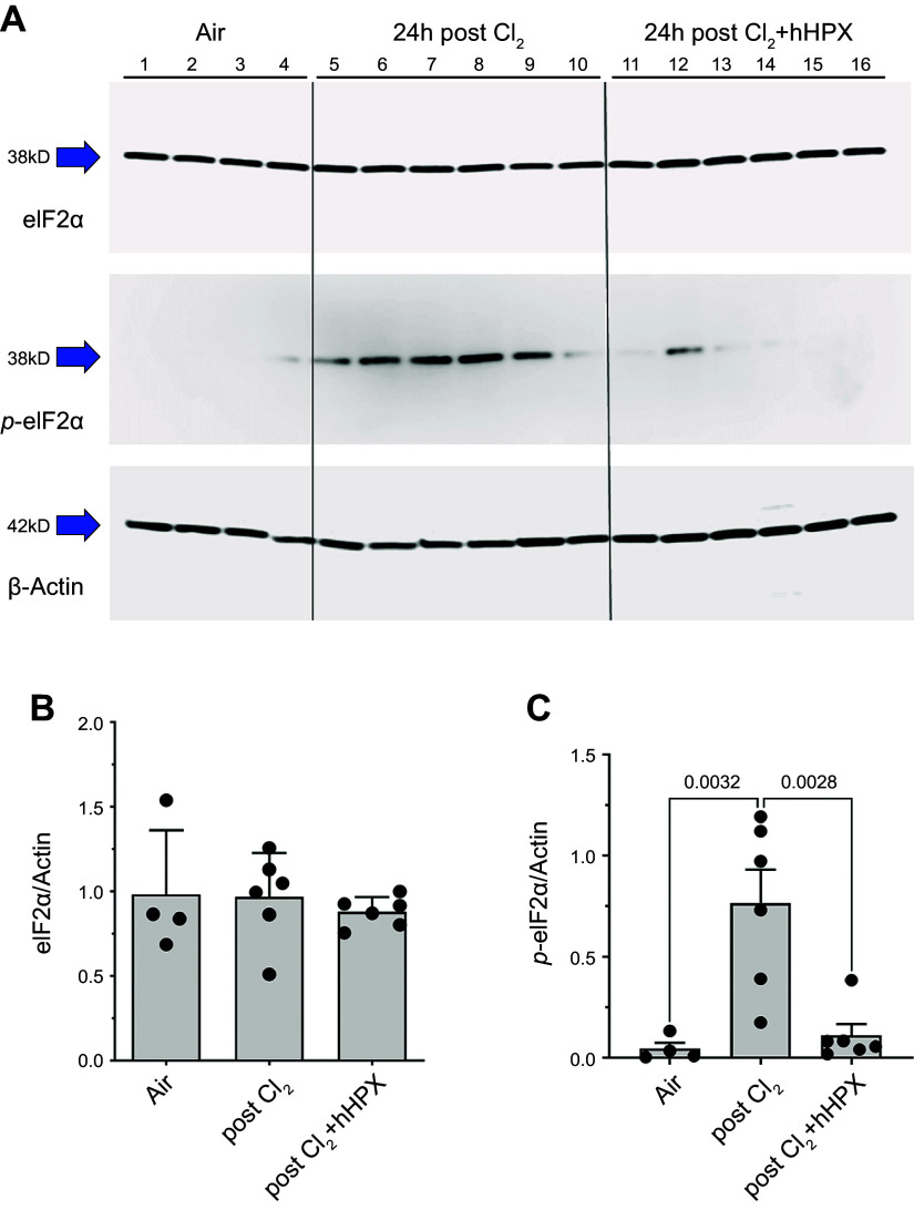
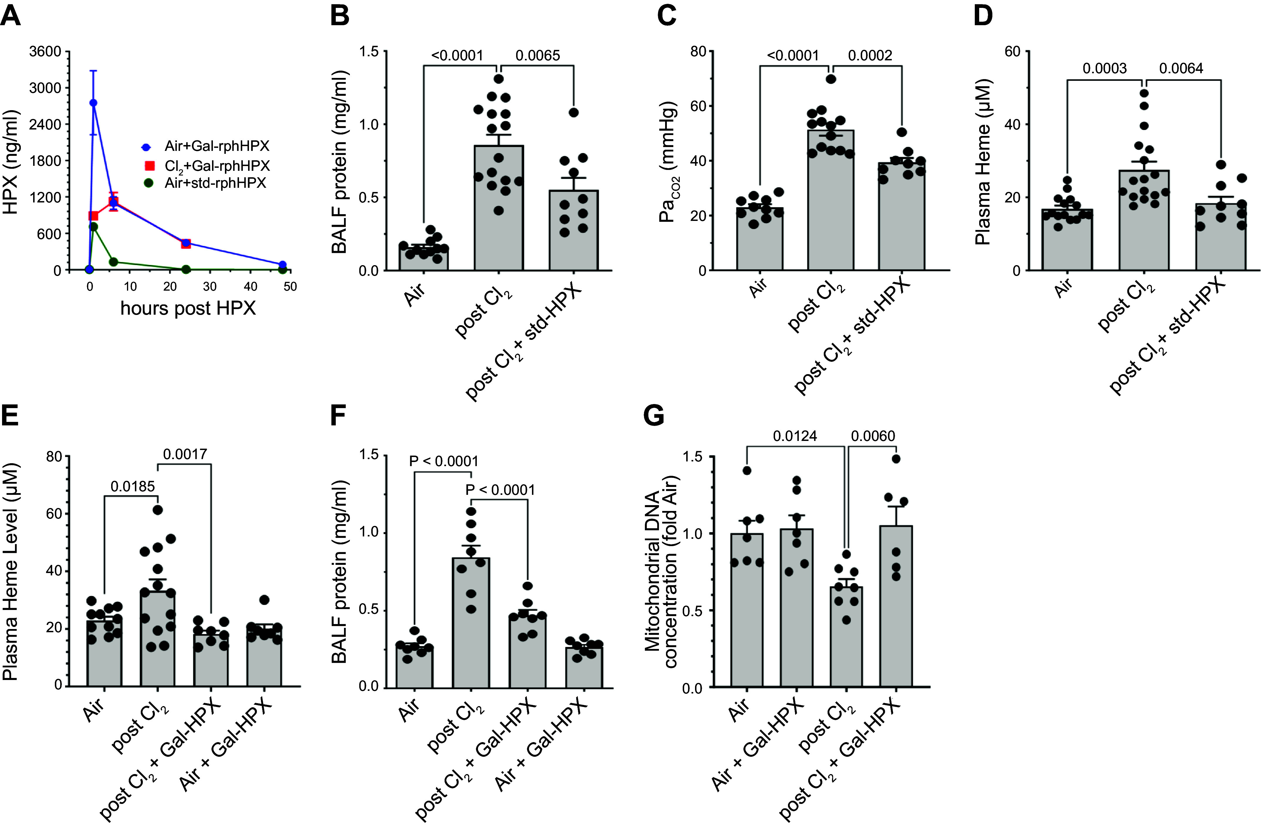
We assessed the mechanisms by which nonencapsulated heme, released in the plasma of mice after exposure to chlorine (Cl2) gas, resulted in the initiation and propagation of acute lung injury. We exposed adult male and female C57BL/6 mice to Cl2 (500 ppm for 30 min), returned them to room air, and injected them intramuscularly with either human hemopexin (hHPX; 5 µg/g BW in 50-µL saline) or vehicle at 1 h post-exposure. Upon return to room air, Cl2-exposed mice, injected with vehicle, developed respiratory acidosis, increased concentrations of plasma proteins in the alveolar space, lung mitochondrial DNA injury, increased levels of free plasma heme, and major alterations of their lung proteome. hHPX injection mice mitigated the onset and development of lung and mitochondrial injury and the increase of plasma heme, reversed the Cl2-induced changes in 83 of 237 proteins in the lung proteome at 24 h post-exposure, and improved survival at 15 days post-exposure. Systems biology analysis of the lung global proteomics data showed that hHPX reversed changes in a number of key pathways including elF2 signaling, verified by Western blotting measurements. Recombinant human hemopexin, generated in tobacco plants, injected at 1 h post-Cl2 exposure, was equally effective in reversing acute lung and mtDNA injury. The results of this study offer new insights as to the mechanisms by which exposure to Cl2 results in acute lung injury and the therapeutic effects of hemopexin.NEW & NOTEWORTHY Herein, we demonstrate that exposure of mice to chlorine gas causes significant changes in the lung proteome 24 h post-exposure. Systems biology analysis of the proteomic data is consistent with damage to mitochondria and activation of eIF2, the master regulator of transcription and protein translation. Post-exposure injection of hemopexin, which scavenges free heme, attenuated mtDNA injury, eIF2α phosphorylation, decreased lung injury, and increased survival.

Keywords: lung injury; mitochondrial DNA; proteomics; recombinant hemopexin.


Conflict of interest statement

S. Matalon and T. Jilling are Inventors in a US Provisional Patent Application #62/896,427, “Use of Hemopexin as a Treatment for Pulmonary Injury” [filed September 5, 2019; Inventors: Dr. Sadis Matalon (Primary), Dr. Saurabh Aggarwal, Dr. Tamas Jilling, Dr. Rakesh Patel]. S. Matalon is the Editor in Chief of Physiological Reviews for which he receives an honorarium from the American Physiological Society. None of the other authors has any conflicts of interest, financial or otherwise, to disclose.

Figures

None
Graphical abstract
Figure 1.
Figure 1.
Injection of human hemopexin (hHPX) decreased heme levels and red blood cell (RBC) fragility after chlorine (Cl2). A: C57BL/6 mice were exposed to air (solid circles) or Cl2 (500 ppm for 30 min; open circles). Twenty minutes after exposure, they were injected intramuscularly with human hemopexin (hHPX; 5 µg/g BW in 50 µL sterile saline). Mice were then euthanized at 1, 6, 24, or 48 h post-exposure and the concentration of hHPX in their plasmas was measured by ELISA as described in the materials and methods; n = 3–5 for each condition. Values are means ± 1 SE. Corresponding mean values at each time point are significantly different from each other with the unpaired t test (P < 0.001). B: C57BL/6 mice were exposed to air or Cl2 (500 ppm for 30 min) and returned to room air. Blood samples were drawn at prior to and at 1, 6 or 24 h after exposure and plasma heme was measured as described in the materials and methods. Values are means ± 1 SE; n = 6–8 for each time point. *Significantly different from the air value (P < 0.01) by one-way analysis of variance (ANOVA) followed by the Dunn–Šidák test for multiple comparisons. C, D, and E: C57BL/6 mice were exposed to Cl2 (500 ppm for 30 min) and returned to room air. They were injected intramuscularly with human hemopexin (hHPX; 5 µg/g BW) at 1 h post-exposure. Mice exposed to air were injected with saline. All mice were then euthanized at 24 h post-exposure and the RBC fragility (C), plasma K+ concentration (D), and plasma heme (E) were measured as described in the materials and methods. Each point represents results from a different mouse. Means ± 1 SE; statistical analysis by one-way analysis of variance followed by the Dunn–Šidák test for multiple comparisons (GraphPad Prism 9).
Figure 2.
Figure 2.
Injection of human hemopexin (hHPX) decreased chlorine (Cl2)-induced acute lung injury and mortality. A and B: C57BL/6 mice were exposed to air or Cl2 (500 ppm for 30 min) and returned to room air. One hour after exposure, they were injected intramuscularly with human hemopexin (hHPX; 5 µg/g BW in 50 µL saline). Mice were then euthanized at 24 h post-exposure, their lungs were lavaged and the concentration of protein (A) and number of inflammatory cells (B) in the bronchoalveolar lavage fluid (BALF) were measured as described in the materials and methods. Each point represents data from a different mouse. Means ± 1 SE; C and D: C57BL/6 mice were exposed to air or Cl2 (500 ppm for 30 min) and returned to room air. One hour after exposure, they were injected intramuscularly with human hemopexin (hHPX; 5 µg/g BW). At 24 h post exposure, they were euthanized and arterial PaCO2, and pH were measured as described in the materials and methods. PaO2 values decreased at 24 h post exposure but the values were not significantly different than air (not shown). D: their lungs were removed, and the concentration of mtDNA was measured by RT-PCR using the Mitochondrial DNA Damage Analysis Kit from Detroit R&D (Cat. No. DD2M). Similar results were obtained when DNA was amplified using 11-kb mitochondrial DNA specific primers, designed by one of the authors (see materials and methods). Means ± 1 SE; statistical analysis by one-way analysis of variance followed by the Dunn–Šidák for multiple comparisons. E: hHPX in the BALF, measured by ELISA. Means ± 1 SE; statistical analysis by one-way analysis of variance followed by the Dunn–Šidák for multiple comparisons. F: C57BL/6 mice were exposed to air or Cl2 (400 ppm for 30 min) and returned to room air. One hour after exposure they were injected intramuscularly with human hemopexin (hHPX; 5 µg/g BW). Kaplan-Meir curves and statistical significance among them were calculated by GraphPad PRISM 9.1software. In all cases, similar number of male and female mice were exposed to Cl2 or air.
Figure 3.
Figure 3.
Changes in Lung Proteome post chlorine (Cl2). Male and female C57BL/6 mice were exposed to air or Cl2 (500 ppm for 30 min) and returned to room air. Twenty-four hours later the mice were euthanized and their lungs were removed and proteins were processed for global proteomics analysis as discussed in the materials and methods. The top 80 proteins that were either increased or decreased twofold at 24 h post Cl2 as compared with their corresponding air values and passed a two-tiered statistical test, shown in Tables 1 and 2, were used to generate these figures. A: two-dimensional hierarchical analysis heat map demonstrates which proteins are increased (red) or decreased (blue) in 24 h post Cl2- versus air-exposed mice. Each column represents data from a different mouse (n = 6 air; n = 4, 24 h post Cl2). B: the principal component analysis (PCA) complements the heat map by using a similar cluster approach that determines which animal (based on protein quantification for all proteins in the top list) is similar across all animals analyzed. Notice tight clustering for air and 24 h post Cl2-exposed mice (blue and green) with a clear separation between the two groups. Each symbol indicates a different mouse (n = 6 air; n = 4, 24 h post Cl2). The numbers in parentheses show the variations among each principal axis (C). Top process networks pie chart from systems biology analysis carried out on the most significantly changed lung proteins at 24 h post exposure of mice to Cl2.
Figure 4.
Figure 4.
PCA and heat map post chlorine (Cl2) and human hemopexin (hHPX). Male and female C57BL/6 mice were exposed to air or Cl2 (500 ppm for 30 min) and returned to room air. One hour later, they received a single dose of human hemopexin (5 µg/g BW in 50 µL saline) or saline intramuscularly. Twenty-four hours later the mice were euthanized and their lungs were removed and proteins were processed for global proteomics analysis as discussed in the materials and methods. A: the two-dimensional hierarchical analysis heat map demonstrates which proteins are increased (red) or decreased (blue) in 24 h post Cl2- versus air and hHPX-24 h post Cl2 versus veh-24h post Cl2 (q < 0.1, P < 0.05, top 46; q = false rate discovery adjusted rate; GraphPad). B: the PCA complements the heat map by using a similar cluster approach that determines which animal (based on protein quantification for all proteins in the top list) is similar across all animals analyzed. Notice tight clustering for air, 24 h post Cl2, and hHPX-24 h post Cl2 mice with a clear separation between the three groups. Each point indicates a different mouse.
Figure 5.
Figure 5.
Two-dimensional (2-D) bubble chart of top pathways attenuated/reversed by human hemopexin (hHPX) post chlorine (Cl2) exposure. This 2-D bubble chart illustrates pathway “categories” (y-axis) versus canonical pathways (x-axis). Statistical significance is indicated by negative log10 of the right-tailed Fisher’s exact test's P value associated with the proteins that were found to be either attenuated or reversed in mouse lungs following at 24 h post Cl2 exposure following injection of hHPX. The arrows are pointing toward “key” pathways found to be associated with each pathway category. The orange and blue color codes are highlighting the predicted pathway activities as either increased or decreased, respectively, which is indicated by their calculated z-scores. For all categories z = 1. The white and gray color codes are associated with either an unclear or balanced mix of activities based on the proteins identified, or simply no predicted alteration in pathway activities, which does not take away from the significant associations between the proteins of interest and their respective pathways, which is left to be determined via validation regardless.
Figure 6.
Figure 6.
Western blotting studies of eIF2α in lung tissues post chlorine (Cl2) exposure. Male and female C57BL/6 mice were exposed to air or Cl2 (500 ppm for 30 min) and returned to room air. One hour later, they were injected with either human hemopexin (hHPX) (5 µg/g BW in 50 µL of saline) or saline; they were euthanized at 24 h post exposure. Lung tissues were removed and probed with antibodies against eIF2α or its phosphorylated form (p-eIF2α). The blots were then stripped and probed with an antibody against actin as described in the materials and methods. A: Western blot records. Lanes 1–4: Air; 5–10: 24 h post exposure to Cl2; 11–16: 24 h post Cl2; treated with hHPX. Each lane represents results from a different mouse. Arrows indicate the MW in kDa. B and C: quantitation of the total eIF2α/actin and phosphor-eIF2α/total eIF2α in the lungs of mice at 24 post exposure to Cl2. Individual points and means ± 1 SE; statistical analysis by one-way analysis of variance followed by the Dunn–Šidák test for multiple comparisons (GraphPad Prism 9).
Figure 7.
Figure 7.
A: pharmacokinetics of recombinant hemopexin. C57BL/6 mice were exposed to air or chlorine (Cl2) (500 ppm for 30 min). One hour post exposure, they were injected intramuscularly with either Gal-prhHPX or Std-prhHPX (HPX; 5 µg/g BW in 50 µL saline). Mice were then euthanized at the indicated intervals and the concentration of HPX in their plasmas was measured by ELISA as described in the materials and methods. Each symbol represents a different mouse. Means ± 1 SE. Lines connect values from different mice obtained in the same experiment. Recombinant hemopexin decreases heme and acute lung injury. C57BL/6 mice were exposed to air or Cl2 (500 ppm for 30 min) and returned to room air. One hour post exposure, they were injected intramuscularly with either std-prhHPX (10 µg/g BW) (B, C, and D) or Gal-prhHPX (5 µg/g BW) (E, F, and G). Mice were then euthanized at 24 h post exposure, their lungs were lavaged and the indicated variables were measured. Each point represents a different animal. Means ± 1 SE; statistical analysis by one-way analysis of variance followed by the Dunn–Šidák t-test for multiple comparisons.

Update of

Similar articles

Cited by

References

    1. Addis DR, Aggarwal S, Lazrak A, Jilling T, Matalon S. Halogen-induced chemical injury to the mammalian cardiopulmonary systems. Physiology (Bethesda) 36: 272–291, 2021. doi:10.1152/physiol.00004.2021. - DOI - PMC - PubMed
    1. Aggarwal S, Ahmad I, Lam A, Carlisle MA, Li C, Wells JM, Raju SV, Athar M, Rowe SM, Dransfield MT, Matalon S. Heme scavenging reduces pulmonary endoplasmic reticulum stress, fibrosis, and emphysema. JCI. Insight 3: e120694, 2018. doi:10.1172/jci.insight.120694. - DOI - PMC - PubMed
    1. Aggarwal S, Lazrak A, Ahmad I, Yu Z, Bryant A, Mobley JA, Ford DA, Matalon S. Reactive species generated by heme impair alveolar epithelial sodium channel function in acute respiratory distress syndrome. Redox Biol 36: 101592, 2020. doi:10.1016/j.redox.2020.101592. - DOI - PMC - PubMed
    1. Alishlash AS, Sapkota M, Ahmad I, Maclin K, Ahmed NA, Molyvdas A, Doran S, Albert CJ, Aggarwal S, Ford DA, Ambalavanan N, Jilling T, Matalon S. Chlorine inhalation induces acute chest syndrome in humanized sickle cell mouse model and ameliorated by post-exposure hemopexin. Redox Biol 44: 102009, 2021. doi:10.1016/j.redox.2021.102009. - DOI - PMC - PubMed
    1. Aggarwal S, Lam A, Bolisetty S, Carlisle MA, Traylor A, Agarwal A, Matalon S. Heme attenuation ameliorates irritant gas inhalation-induced acute lung injury. Antioxid Redox Signal 24: 99–112, 2016. doi:10.1089/ars.2015.6347. - DOI - PMC - PubMed

LinkOut - more resources