. 2023 Dec 27;13(1):22974.
doi: 10.1038/s41598-023-49751-5.

Fucoxanthin diminishes oxidative stress damage in human placenta-derived mesenchymal stem cells through the PI3K/Akt/Nrf-2 pathway

Affiliations

Fucoxanthin diminishes oxidative stress damage in human placenta-derived mesenchymal stem cells through the PI3K/Akt/Nrf-2 pathway

Gunticha Suwanmanee et al. Sci Rep. .

Abstract

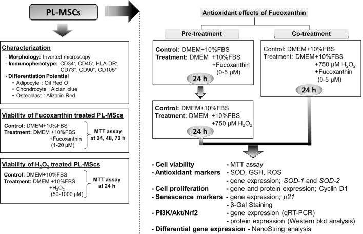
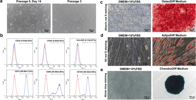
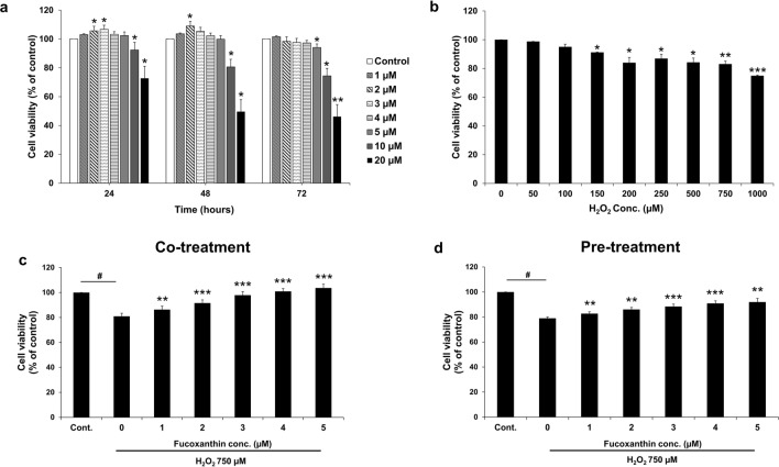
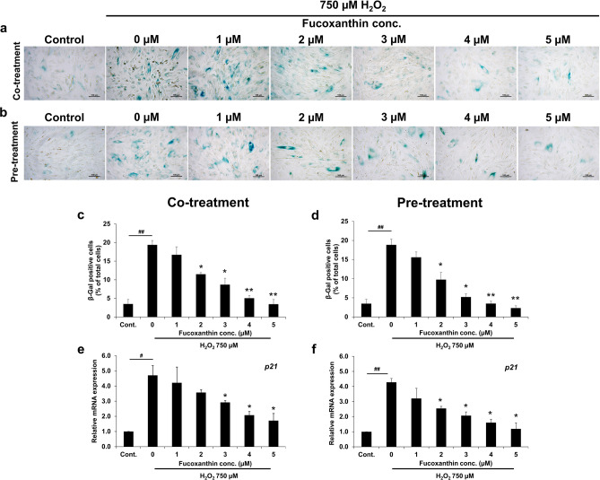
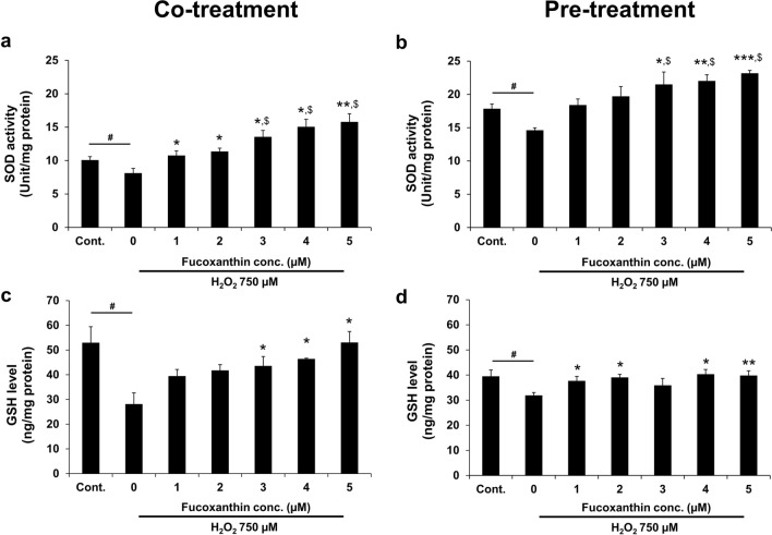
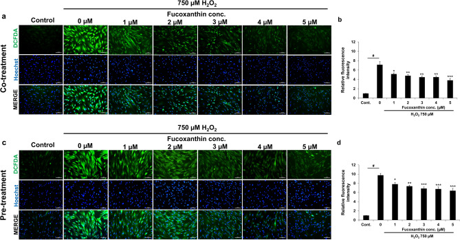
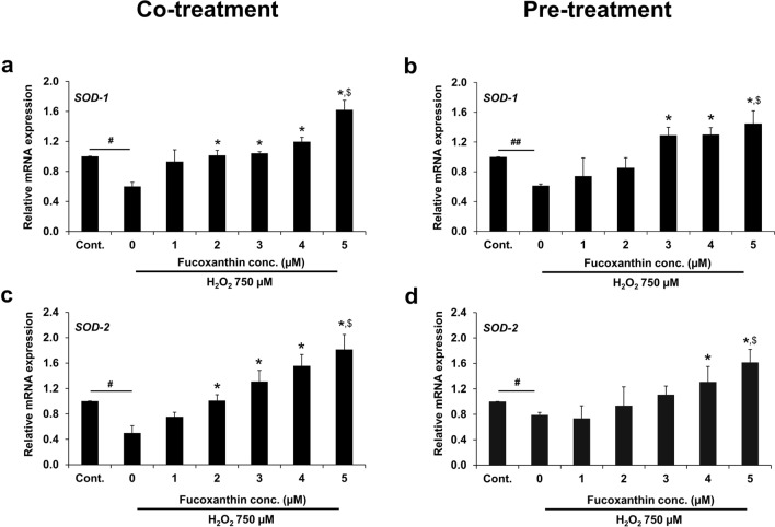
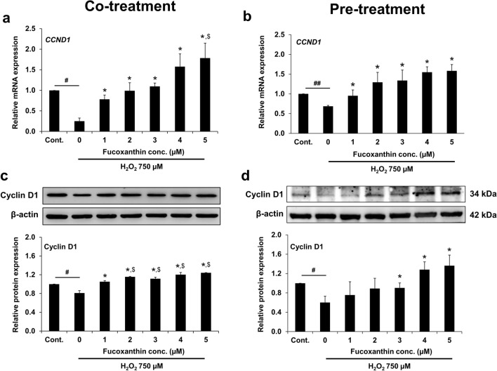
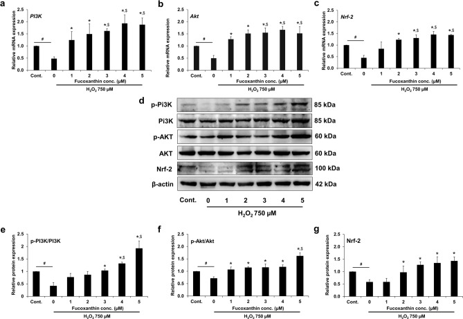
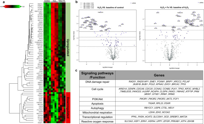
Placenta-derived mesenchymal stem cells (PL-MSCs) have therapeutic potential in various clinical contexts due to their regenerative and immunomodulatory properties. However, with increasing age or extensive in vitro culture, their viability and function are gradually lost, thus restricting their therapeutic application. The primary cause of this deterioration is oxidative injury from free radicals. Therefore, enhancing cell viability and restoring cellular repair mechanisms of PL-MSCs in an oxidative stress environment are crucial in this context. Fucoxanthin, a carotenoid derived from brown seaweed, demonstrates antioxidant activity by increasing the production of antioxidant enzymes and lowering the levels of reactive oxygen species (ROS). This study aimed to determine whether fucoxanthin protects PL-MSCs from hydrogen peroxide (H2O2)-induced oxidative stress. After characterization, PL-MSCs were co-treated with fucoxanthin and H2O2 for 24 h (co-treatment) or pre-treated with fucoxanthin for 24 h followed by H2O2 for 24 h (pre-treatment). The effects of fucoxanthin on cell viability and proliferation were examined using an MTT assay. The expression of antioxidant enzymes, PI3K/Akt/Nrf-2 and intracellular ROS production were investigated in fucoxanthin-treated PL-MSCs compared to the untreated group. The gene expression and involvement of specific pathways in the cytoprotective effect of fucoxanthin were investigated by high-throughput NanoString nCounter analysis. The results demonstrated that co-treatment and pre-treatment with fucoxanthin restored the viability and proliferative capacity of PL-MSCs. Fucoxanthin treatment increased the expression of antioxidant enzymes in PL-MSCs cultured under oxidative stress conditions and decreased intracellular ROS accumulation. Markedly, fucoxanthin treatment could restore PI3K/Akt/Nrf-2 expression in H2O2-treated PL-MSCs. High-throughput analysis revealed up-regulation of genes involved in cell survival pathways, including cell cycle and proliferation, DNA damage repair pathways, and down-regulation of genes in apoptosis and autophagy pathways. This study demonstrated that fucoxanthin protects and rescues PL-MSCs from oxidative stress damage through the PI3K/Akt/Nrf-2 pathway. Our data provide the supporting evidence for the use of fucoxanthin as an antioxidant cytoprotective agent to improve the viability and proliferation capacity of PL-MSCs both in vitro and in vivo required to increase the effectiveness of MSC expansion for therapeutic applications.


Conflict of interest statement

The authors declare no competing interests.

Figures

Figure 1
Figure 1
A schematic diagram illustrates the overall experimental schedule.
Figure 2
Figure 2
Characterization of human placenta-derived mesenchymal stem cells (PL-MSCs). (a) The spindle shape morphology of PL-MSCs cultured in DMEM supplemented with 10% fetal bovine serum on day 7 after removal of non-adherent cells (left) and in passages 3 (right). (b) Flow cytometric analysis of surface marker expression in PL-MSCs showing positive expression of MSC markers (CD73, CD90, CD105) and negative expression of hematopoietic markers (CD34, CD45, HLA-DR). (c) Brilliant orange-red staining of alizarin red S in PL-MSCs on day 28 of their osteogenic differentiation. (d) Positive signal of Oil Red O staining in PL-MSCs on day 28 on their adipogenic differentiation. (e) Chondrogenic differentiation potential of PL-MSCs demonstrated by Alcian positive blue color staining of positive colonies (right) that develop in the presence of chondrogenic differentiation media. Differentiated colonies were obtained from cells of all 5 donor placentas. (a) and (c) were captured with 10X magnification. Scale bar = 100 μm. (d) was captured with 40X magnification. Scale bar = 50 μm. (e) was captured with 20X magnification. Scale bar = 100 μm.
Figure 3
Figure 3
Effects of fucoxanthin on the viability of H2O2-treated PL-MSCs. (a) MTT assay showed the viability of PL-MSCs treated with increasing concentrations of fucoxanthin for 24–72 h. (b) MTT assay showed dose-dependent decreased viability of PL-MSCs treated with H2O2 for 24 h. (c) The viability of PL-MSCs after 24 h of co-treatment with 750 μM H2O2 and increasing concentrations (up to 5 μM) of fucoxanthin. (d) The viability of PL-MSCs pretreated with fucoxanthin for 24 h before H2O2 treatment for 24 h. (a) and (b) are presented as mean±SEM, n=3. The statistical significance was tested using the pair T-test. *p ≤ 0.05, **p ≤ 0.01, and ***p ≤ 0.001 vs. PL-MSCs cultured in completed DMEM medium. (c) and (d) are presented as mean ± SEM, n = 5. The statistical significance was tested using the pair T-test. #p ≤ 0.001 vs. PL-MSCs cultured in completed DMEM medium. *p ≤ 0.05, **p ≤ 0.01, and ***p ≤ 0.001 vs. H2O2-treated PL-MSCs without fucoxanthin.
Figure 4
Figure 4
Effects of fucoxanthin on replicative senescence of H2O2-treated PL-MSCs. (a) The representative images of β-Gal staining of PL-MSCs after 24 h of co-treatment with 750 μM H2O2 and 1–5 μM fucoxanthin. (b) β-Gal staining of PL-MSCs after pre-treated with fucoxanthin for 24 h, followed by treatment with 750 μM H2O2 for 24 h. (c) and (d) Quantitation of β-Gal positive cells (% of β-Gal positive cells) of co-treated or pre-treated with 1–5 μM fucoxanthin and 750 μM H2O2 for 24 h, respectively. (e) Quantitative real-time RT-PCR showed the expression of p21 in PL-MSCs after co-treatment with 750 μM H2O2 and 1–5 μM fucoxanthin for 24 h. (f) Quantitative real-time RT-PCR showed the expression of p21 in PL-MSCs pre-treated with fucoxanthin for 24 h followed by treatment with H2O2 for 24 h. Data are presented as mean ± SEM, n = 3. Statistical significance was tested using the pair T-test. #p ≤ 0.05, ##p ≤ 0.01 vs. PL-MSCs culture in completed DMEM medium. *p ≤ 0.05, **p ≤ 0.01 vs. H2O2-treated PL-MSCs without fucoxanthin. (a) and (b) were captured with 20X magnification. Scale bar = 100 μm.
Figure 5
Figure 5
Effects of fucoxanthin on SOD activity and GSH level in H2O2-treated PL-MSCs. (a) SOD activity of PL-MSCs after 24 h of co-treatment with 750 μM H2O2 and fucoxanthin. (b) SOD activity of PL-MSCs pre-treated with fucoxanthin for 24 h followed by treatment with 750 μM H2O2 for 24 h. (c) GSH level of PL-MSCs after 24 h co-treatment with 750 μM H2O2 and fucoxanthin. (d) GSH level of PL-MSCs after fucoxanthin treatment for 24 h, followed by H2O2 treatment for 24 h. Data are presented as mean ± SEM, n = 4. Statistical significance was tested using the pair T-test. #p ≤ 0.05 vs. PL-MSCs cultured in the completed DMEM medium. *p ≤ 0.05, **p ≤ 0.01, and ***p ≤ 0.001 vs. H2O2-treated PL-MSCs without fucoxanthin. $p ≤ 0.05 vs. PL-MSCs cultured in completed DMEM medium.
Figure 6
Figure 6
Effects of fucoxanthin on intracellular ROS production in H2O2-treated PL-MSCs. (a) Fluorescent micrograph illustrating the intracellular ROS content in PL-MSCs after 24 h of co-treatment with 750 μM H2O2 and fucoxanthin. (b) Relative fluorescence intensity of the intracellular ROS content in PL-MSCs after 24 h of co-treatment with H2O2 and fucoxanthin. (c) Fluorescent micrographs illustrated the intracellular ROS content in PLMSCs that were pre-treated with fucoxanthin for 24 h, followed by treatment with 750 μM H2O2 for 24 h. (d) Relative fluorescence intensity of the intracellular ROS content in PL-MSCs pre-treatment with fucoxanthin for 24 h, followed by treatment with H2O2 for 24 h. Data are presented as mean ± SEM, n = 3. Statistical significance was tested using the pair T-test. #p ≤ 0.001 vs. PL-MSCs cultured in completed DMEM medium. *p ≤ 0.05, **p ≤ 0.01, and ***p ≤ 0.001 vs. H2O2-treated PL-MSCs without fucoxanthin. Green = DCF-DA, Blue = Hoechst33342.
Figure 7
Figure 7
Quantitative real-time RT-PCR showed the expression of SOD-1 and SOD-2 in H2O2-treated PL-MSCs. (a) The expression of SOD-1 in PL-MSCs after 24 h of co-treatment with 750 μM H2O2 and fucoxanthin. (b) The expression of SOD-1 in PL-MSCs pre-treated with fucoxanthin for 24 h followed by treatment with 750 μM H2O2 for 24 h. (c) The expression of SOD-2 in PL-MSCs after 24 h of co-treatment with 750 μM H2O2 and fucoxanthin. (d) The expression of SOD-2 in PL-MSCs pre-treated with fucoxanthin for 24 h, followed by treatment with 750 μM H2O2 for 24 h. Data are presented as mean ± SEM, n = 3. Statistical significance was tested using the pair T-test. #p ≤ 0.01, ##p ≤ 0.001 vs. PL-MSCs cultured in the completed DMEM medium. *p ≤ 0.05 vs. H2O2-treated PL-MSCs without fucoxanthin. $p ≤ 0.05 vs. PL-MSCs cultured in the completed DMEM medium.
Figure 8
Figure 8
Effects of fucoxanthin on cyclin D1 expression in H2O2-treated PL-MSCs. (a) Quantitative real-time RT-PCR showed the expression of cyclin D1 in PL-MSCs after 24 h of co-treatment with H2O2 and fucoxanthin. (b) Quantitative real-time RT-PCR showed the expression of cyclin D1 in PL-MSCs pre-treated with fucoxanthin for 24 h followed by treated with H2O2 for 24 h. (c) Western blot analysis showed the expression of cyclin D1 in PL-MSCs after 24 h of co-treatment with H2O2 and fucoxanthin. (d) Western blot analysis showed the expression of cyclin D1 in PL-MSCs after pre-treatment with fucoxanthin for 24 h followed by H2O2-treatment for 24 h. The blotted membranes were cropped before hybridization with primary antibodies. The original blots of (c) and (d) are shown in Supplementary Fig. 2 and 3, respectively. Data are presented as mean ± SEM, n = 3. Statistical significance was tested using the pair T-test. #p ≤ 0.05, ##p ≤ 0.01 vs. PL-MSCs culture in the completed DMEM medium. *p ≤ 0.05 vs. H2O2-treated PL-MSCs without fucoxanthin. $p ≤ 0.05 vs. PL-MSCs cultured in the completed DMEM medium.
Figure 9
Figure 9
Effects of fucoxanthin on the expression of PI3K/Akt/Nrf-2 in H2O2-treated PL-MSCs. Quantitative real-time RT-PCR showed the expression of PI3K (a), Akt (b), and Nrf-2 (c) in PL-MSCs after 48 h of co-treatment with 750 μM H2O2 and fucoxanthin. (d) Western blot analysis showed the expression of PI3K/Akt/Nrf-2. The blotted membranes were cropped before hybridization with primary antibodies. The original blots of (d) are shown in Supplementary Fig. 4–6. (e)–(g) Relative protein expression level of p-PI3K/PI3K, p-Akt/Akt, and Nrf-2 in PL-MSCs after 48 h of co-treatment with 750 μM H2O2 and fucoxanthin. Data are presented as mean ± SEM, n = 3. Statistical significance was tested using the pair T-test. #p ≤ 0.001 vs. PL-MSCs cultured in the completed DMEM medium. *p ≤ 0.05 vs. H2O2-treated PL-MSCs without fucoxanthin. $p ≤ 0.05 vs. PL-MSCs cultured in the completed DMEM medium.
Figure 10
Figure 10
NanoString analysis of differential gene expression in PL-MSCs under oxidative stress conditions. (a) Heatmap of differentially expressed genes in 750 μM H2O2-treated PLMSCs and PL-MSCs treated with 750 μM H2O2 together with 5 μM fucoxanthin. (b) The volcano plot presents the distribution of differentially expressed genes (DE). (c) Summary of genes belonging to various metabolic pathways that showed statistical significance between treatment groups.

Similar articles

Cited by

References

    1. Dominici M, Le-Blanc K, Mueller I, Slaper-Cortenbach I, Marini F, Krause D, et al. Minimal criteria for defining multipotent mesenchymal stromal cells. The International Society for Cellular Therapy position statement. Cytotherapy. 2006;8(4):315–317. doi: 10.1080/14653240600855905. - DOI - PubMed
    1. Le Blanc K, Rasmusson I, Sundberg B, Götherström C, Hassan M, Uzunel M, et al. Treatment of severe acute graft-versus-host disease with third party haploidentical mesenchymal stem cells. Lancet. 2004;363(9419):1439–1441. doi: 10.1016/S0140-6736(04)16104-7. - DOI - PubMed
    1. Baker N, Boyette LB, Tuan RS. Characterization of bone marrow-derived mesenchymal stem cells in aging. Bone. 2015;70:37–47. doi: 10.1016/j.bone.2014.10.014. - DOI - PubMed
    1. Dufrane D. Impact of age on human adipose stem cells for bone tissue engineering. Cell Transplant. 2017;26(9):1496–1504. doi: 10.1177/0963689717721203. - DOI - PMC - PubMed
    1. Wu W, Niklason L, Steinbacher DM. The effect of age on human adipose-derived stem cells. Plast. Reconstr. Surg. 2013;131(1):27–37. doi: 10.1097/PRS.0b013e3182729cfc. - DOI - PubMed

MeSH terms