. 2023 Dec 27;13(1):22967.
doi: 10.1038/s41598-023-50108-1.

Trichostatin A enhances the titanium rods osseointegration in osteoporotic rats by the inhibition of oxidative stress through activating the AKT/Nrf2 pathway

Affiliations

Trichostatin A enhances the titanium rods osseointegration in osteoporotic rats by the inhibition of oxidative stress through activating the AKT/Nrf2 pathway

Zhi Zhou et al. Sci Rep. .

Abstract

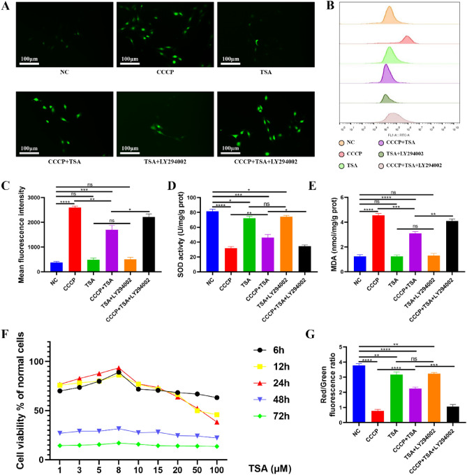
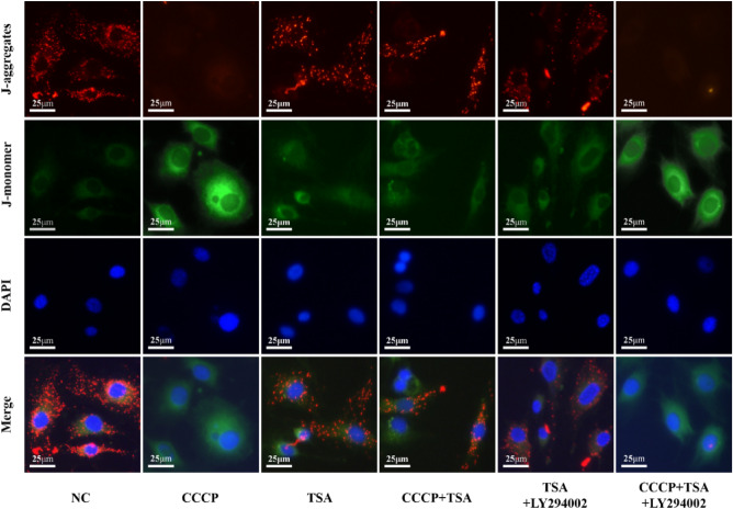
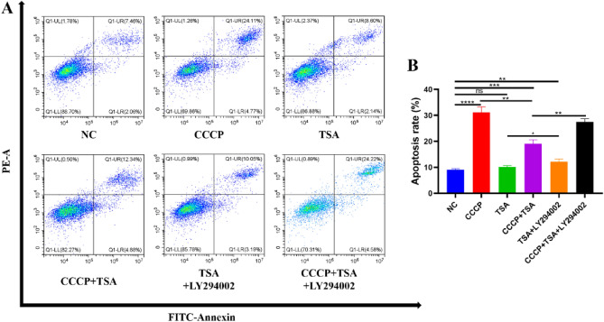
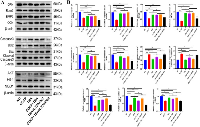
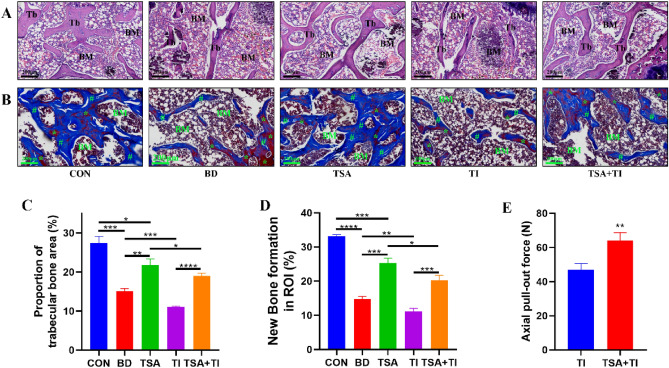
The use of titanium implants as fixed supports following fractures in patients with OP can often result in sterile loosening and poor osseointegration. Oxidative stress has been shown to play a particularly important role in this process. While TSA has been reported to facilitate in vivo osteogenesis, the underlying mechanisms remain to be clarified. It also remains unclear whether TSA can improve the osseointegration of titanium implants. This study investigated whether TSA could enhance the osseointegration of titanium rods by activating AKT/Nrf2 pathway signaling, thereby suppressing oxidative stress. MC3T3-E1 cells treated with CCCP to induce oxidative stress served as an in vitro model, while an OVX-induced OP rat model was employed for in vivo analysis of titanium rod implantation. In vitro, TSA treatment of CCCP-treated MC3T3-E1 cells resulted in the upregulation of osteogenic proteins together with increased AKT, total Nrf2, nuclear Nrf2, HO-1, and NQO1 expression, enhanced mitochondrial functionality, and decreased oxidative damage. Notably, the PI3K/AKT inhibitor LY294002 reversed these effects. In vivo, TSA effectively enhanced the microstructural characteristics of distal femur trabecular bone, increased BMSCs mineralization capacity, promoted bone formation, and improved the binding of titanium implants to the surrounding tissue. Finally, our results showed that TSA could reverse oxidative stress-induced cell damage while promoting bone healing and improving titanium rods' osseointegration through AKT/Nrf2 pathway activation.


Conflict of interest statement

The authors declare that they have no competing interests.

Figures

Figure 1
Figure 1
Effects of different treatments on oxidative stress indexes of MC3T3-E1 cells. (A) ROS images were detected by fluorescence microscope, scale bar = 100 μm; (B) ROS was detected by flow cytometry; (C) The average fluorescence intensity of ROS was determined by flow cytometry; (D) Quantitative analysis of SOD activity; (E) Quantitative analysis of MDA content; (F) CCK8 cell activity detection; (G) JC-1 fluorescence quantitative analysis (*P < 0.05, **P < 0.01, ***P < 0.001, ****P < 0.0001).
Figure 2
Figure 2
Effects of different treatments on MMP of MC3T3-E1 cells. (JC-1 staining image, scale bar = 25 μm).
Figure 3
Figure 3
Effects of different treatments on apoptosis rate of MC3T3-E1 cells. (A) Results of cell apoptosis rate detected by flow cytometry; (B) analysis of the results of apoptosis rate (*P < 0.05, **P < 0.01, ***P < 0.001, ****P < 0.0001).
Figure 4
Figure 4
Effects of different treatments on protein expression of MC3T3-E1 cells. (A) Representative bands of osteogenesis-related proteins (OPN, Runx2, BMP2, and OPN), apoptosis-related proteins (Caspase3, Bcl2, Bax, and Cleaved Caspase3), and some pathway-related proteins (AKT, HO-1, and NQO1); (B) Relative expression levels of related proteins in each group compared with NC group. Original blots are presented in Supplementary Panel A (*P < 0.05, **P < 0.01, ***P < 0.001, ****P < 0.0001).
Figure 5
Figure 5
The effects of TSA on Nrf2 and the effects of different treatments on protein expression of MC3T3-E1 cells after adding titanium powder were detected by nuclear cytoplasmic separation. (A) Representative expression bands of Nrf2 in nucleus, cytoplasm and total Nrf2; (B) Histogram of relative expression levels of Nrf2 in nucleus, cytoplasm and total Nrf2; (C) Representative bands of osteogenesis-related proteins (OPN, Runx2, BMP2, and OPN), apoptosis-related proteins (Caspase3, Bcl2, Bax, and Cleaved Caspase3), and some pathway-related proteins (AKT, HO-1, and NQO1) in MC3T3-E1 cells after adding titanium powder; (D) Relative expression levels of related proteins in each group compared with NC group. Original blots are presented in Supplementary Panel B and C (*P < 0.05, **P < 0.01, ***P < 0.001, ****P < 0.0001).
Figure 6
Figure 6
Effects of different treatments on osteogenic ability of MC3T3-E1 cells after adding titanium powder and results of MC3T3-E1 cells attached on a titanium sheet. (A) Results of ALP staining, scale bar = 100 μm, scale bar = 50 μm; (B) ALP activity diagram; (C) Images of electron microscopy; (D) Proportion of MC3T3-E1 cell adhesion area (*P < 0.05, **P < 0.01, ***P < 0.001, ****P < 0.0001).
Figure 7
Figure 7
Verification of rat OP model establishment. (A) Micro-CT images at femur metaphysis in Sham group and OVX group; (B) HE staining images at femur metaphysis in Sham group and OVX group, Scale bar = 200 μm; (C) Quantitative analysis of BMD at femur metaphysis; (D) Proportion of trabecular bone area in ROI. Tb: Trabecular bone. BM: bone marrow (**P < 0.01, ****P < 0.0001).
Figure 8
Figure 8
Results of micro-CT examination. (A) Three-dimensional reconstruction images of Titanium rods and surrounding tissues at femur metaphysis; (B) Quantitative analysis of ROI at femur Metaphysis (*P < 0.05, **P < 0.01, ***P < 0.001, ****P < 0.0001).
Figure 9
Figure 9
Results of HE and Masson's trichrome staining. (A) HE staining images at femur metaphysis. Scale bar = 200 μm; (B) Masson's trichrome staining images at femur metaphysis. Scale bar = 200 μm. (C) Proportion of trabecular bone area in ROI. (D) Histogram of new bone formation rate; (E) Axial pull-out force of titanium rods. Tb: trabecular bone. BM: bone marrow. Asterisk: mature bone. Pound sign: new bone and collagen fibers (*P < 0.05, **P < 0.01, ***P < 0.001, ****P < 0.0001).
Figure 10
Figure 10
Results of immunohistochemistry staining. (A) Results of AKT immunohistochemistry staining, scale bar = 50 μm; (B) Results of OCN immunohistochemistry staining, scale bar = 50 μm; (C,D) Immunohistochemical quantitative analysis of the differences in AKT and OCN expression among the five groups (*P < 0.05, **P < 0.01, ***P < 0.001, ****P < 0.0001).
Figure 11
Figure 11
Effects of different treatments on alkaline phosphatase and osteogenic mineralization function of rat BMSCs. (A) Results of ALP staining, scale bar = 100 μm; (B) Results of ARS staining, scale bar = 200 μm; (C) ALP activity diagram; (D) Absorbance histogram of ARS at 570 nm (*P < 0.05, **P < 0.01, ***P < 0.001, ****P < 0.0001).
Figure 12
Figure 12
Mechanism of TSA activation of PI3K/AKT/Nrf2 pathway.

References

    1. Rachner TD, Khosla S, Hofbauer LC. Osteoporosis: now and the future. The Lancet. 2011;377(9773):1276–1287. doi: 10.1016/S0140-6736(10)62349-5. - DOI - PMC - PubMed
    1. Aspray TJ, Hill TR. Osteoporosis and the ageing skeleton. Subcell Biochem. 2019;91:453–476. doi: 10.1007/978-981-13-3681-2_16. - DOI - PubMed
    1. Kimball JS, Johnson JP, Carlson DA. Oxidative stress and osteoporosis. J. Bone Joint Surg. Am. 2021;103(15):1451–1461. doi: 10.2106/JBJS.20.00989. - DOI - PubMed
    1. Liu HD, Ren MX, Li Y, Zhang RT, Ma NF, Li TL, et al. Melatonin alleviates hydrogen peroxide induced oxidative damage in MC3T3-E1 cells and promotes osteogenesis by activating SIRT1. Free Radic. Res. 2022;56(1):63–76. doi: 10.1080/10715762.2022.2037580. - DOI - PubMed
    1. Deng S, Dai G, Chen S, Nie Z, Zhou J, Fang H, et al. Dexamethasone induces osteoblast apoptosis through ROS-PI3K/AKT/GSK3beta signaling pathway. Biomed. Pharmacother. 2019;110:602–608. doi: 10.1016/j.biopha.2018.11.103. - DOI - PubMed