. 2023 Dec 27;13(1):22991.
doi: 10.1038/s41598-023-50390-z.

Protein N-myristoylation plays a critical role in the mitochondrial localization of human mitochondrial complex I accessory subunit NDUFB7

Affiliations

Protein N-myristoylation plays a critical role in the mitochondrial localization of human mitochondrial complex I accessory subunit NDUFB7

Haruna Harada et al. Sci Rep. .

Abstract

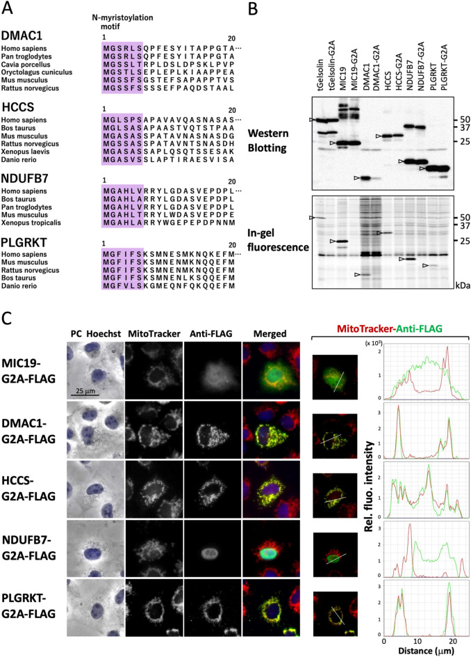
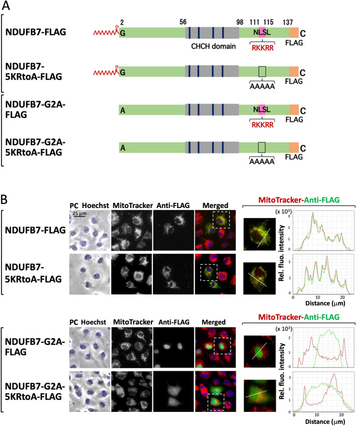
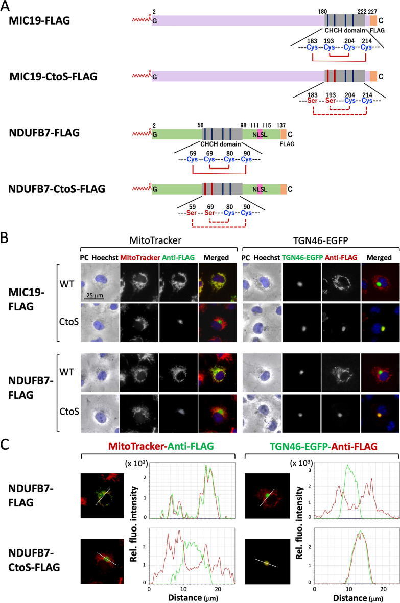
The present study examined human N-myristoylated proteins that specifically localize to mitochondria among the 1,705 human genes listed in MitoProteome, a mitochondrial protein database. We herein employed a strategy utilizing cellular metabolic labeling with a bioorthogonal myristic acid analog in transfected COS-1 cells established in our previous studies. Four proteins, DMAC1, HCCS, NDUFB7, and PLGRKT, were identified as N-myristoylated proteins that specifically localize to mitochondria. Among these proteins, DMAC1 and NDUFB7 play critical roles in the assembly of complex I of the mitochondrial respiratory chain. DMAC1 functions as an assembly factor, and NDUFB7 is an accessory subunit of complex I. An analysis of the intracellular localization of non-myristoylatable G2A mutants revealed that protein N-myristoylation occurring on NDUFB7 was important for the mitochondrial localization of this protein. Furthermore, an analysis of the role of the CHCH domain in NDUFB7 using Cys to Ser mutants revealed that it was essential for the mitochondrial localization of NDUFB7. Therefore, the present results showed that NDUFB7, a vital component of human mitochondrial complex I, was N-myristoylated, and protein N-myrisotylation and the CHCH domain were both indispensable for the specific targeting and localization of NDUFB7 to mitochondria.


Conflict of interest statement

The authors declare no competing interests.

Figures

Figure 1
Figure 1
Identification of N-myristoylated mitochondrial proteins from human genes listed in the MitoProteome protein database. Twelve candidate genes (BCAP31, CLN3, DMAC1/TMEM261, FLAD1, HCCS, LRRC10, MARC1/MTARC1, ME3, NDUFB7, NOL3, PLGRKT and TUSC3) were selected as N-myristoylated mitochondrial proteins from the human genes listed in MitoProteome, a mitochondrial protein database, and their susceptibility to protein N-myristoylation was evaluated by in vivo metabolic labeling in transfected COS-1 cells using fusion proteins in which the 10 N-terminal amino acid residues were fused to the FLAG-tagged model protein, tGelsolin. (A) Strategy to identify N-myristoylated mitochondrial proteins from human genes listed in the MitoProteome protein database. (B) Schematic representation of the generation of X(N10)-tGelsolin-FLAG with 10 N-terminal amino acids of the candidate protein X at its N terminus. (C) Analysis of the protein N-myristoylation of X(N10)-tGelsolin-FLAGs by metabolic labeling in transfected COS-1 cells. cDNAs coding X(N10)-tGelsolin-FLAGs were transfected into COS-1 cells. The expression of proteins was evaluated by Western blotting using an anti-FLAG antibody (upper panels). Protein N-myristoylation was evaluated by metabolic labeling with a myristic acid analog followed by click chemistry, as described in the Methods (lower panels). Images obtained by Western blotting or metabolic labeling were cropped. Uncropped full-length raw image data are shown in Supplementary Figure S1.
Figure 2
Figure 2
DMAC1, HCCS, NDUFB7, and PLGRKT are N-myristoylated mitochondrial proteins. Seven proteins (CLN3, DMAC1, HCCS, MARC1, NDUFB7, NOL3, and PLGRKT) were selected from proteins in which protein N-myristoylation was detected in X(N10)-tGelsolin-FLAG, and their protein N-myristoylation and intracellular localization were evaluated using C-terminally FLAG-tagged full-length proteins. (A) Analysis of the protein N-myristoylation of CLN3, DMAC1, HCCS, MARC1, NDUFB7, NOL3, and PLGRKT expressed in transfected COS-1 cells by metabolic labeling. cDNAs coding C-terminally FLAG-tagged full-length proteins were transfected into COS-1 cells and cells were then labeled with a myristic acid analog. The expression of proteins was evaluated by Western blotting using an anti-FLAG antibody (left panel). Protein N-myristoylation was evaluated by metabolic labeling followed by click chemistry, as described in the Methods (right panel). Arrows indicate the position of the expressed proteins. The images obtained by Western blotting or metabolic labeling were cropped. Uncropped full-length raw image data are shown in Supplementary Figure S2. (B) Analysis of the intracellular localization of CLN3, DMAC1, HCCS, MARC1, NDUFB7, NOL3, and PLGRKT by an immunofluorescence microscopic analysis. The intracellular localization of seven proteins was assessed by an immunofluorescence analysis of COS-1 cells transfected with cDNA coding C-terminally FLAG-tagged full-length proteins using an anti-FLAG antibody. MIC19-FLAG was used as a control mitochondrial N-myristoylated protein. Hoechst and MitoTracker Red were used as organelle markers for the nucleus and mitochondria, respectively. Experiments were repeated 3 times and similar results were obtained. Representative data are shown. Abbreviation used: PC, phase contrast image. The results of the line profile analysis are shown. In the line profile analysis, corresponding line-scan graphs of the relative fluorescence intensities of green anti-FLAG fluorescence (green line) and red MitoTracker red fluorescence (red line) along the white line indicated in the merged images are shown.
Figure 3
Figure 3
Protein N-myristoylation plays a critical role in the mitochondrial localization of NDUFB7. To clarify the role of protein N-myristoylation in the mitochondrial localization of DMAC1, HCCS, NDUFB7, and PLGRKT, non-myristoylatable G2A mutants were generated and their susceptibility to protein N-myristoylation and intracellular localization were evaluated. (A) Interspecies alignment of the N-terminal sequences of DMAC1, HCCS, NDUFB7, and PLGRKT. N-myristoylation motifs are shown in purple in N-terminal sequences. (B) Analysis of protein N-myristoylation of the WT and non-myristoylatable G2A mutants of DMAC1, HCCS, NDUFB7, and PLGRKT. cDNAs coding the WT and non-myristoylatable G2A mutants of four proteins were transfected into COS-1 cells. The expression of proteins was evaluated by Western blotting using an anti-FLAG antibody (upper panel). Protein N-myristoylation was evaluated by metabolic labeling with a myristic acid analog followed by click chemistry, as described in the Methods (lower panel). Arrows indicate the position of the expressed proteins. The images obtained by Western blotting or metabolic labeling were cropped. Uncropped full-length raw image data are shown in Supplementary Figure S4. (C) Analysis of the intracellular localization of the non-myristoylatable G2A mutant of DMAC1, HCCS, NDUFB7, and PLGRKT. The intracellular localization of the G2A mutant of the four proteins was assessed by an immunofluorescence analysis of COS-1 cells transfected with cDNA coding the C-terminally FLAG-tagged G2A mutant using an anti-FLAG antibody. MIC19-G2A-FLAG was used as a control non-myristoylatable G2A mutant. Hoechst and MitoTracker Red were used as organelle markers for the nucleus and mitochondria, respectively. Experiments were repeated 3 times and similar results were obtained. Representative data are shown. Abbreviation used: PC, phase contrast image. The results of the line profile analysis are shown. In the line profile analysis, corresponding line-scan graphs of the relative fluorescence intensities of green anti-FLAG fluorescence (green line) and red MitoTracker red fluorescence (red line) along the white line indicated in the merged images are shown.
Figure 4
Figure 4
Structure of NDUFB7. Schematic representation of the structures of MIC19 and NDUFB7. Interspecies alignments of the N-myristoylation motif, CHCH domain, and nuclear localization signal-like (NLSL) sequence of NDUFB7 are shown. N-myristoylation motifs were boxed. Four conserved Cys residues in the CHCH domain were indicated by the blue characters in the amino acid sequences. Conserved RKKRR sequences in the NLSL sequence were indicated by the red characters in the amino acid sequences.
Figure 5
Figure 5
Positive charge cluster localized in the C-terminal region functions as a nuclear localization signal of the non-myristoylatable G2A mutant of NDUFB7. To establish whether the positive charge cluster at positions 111 to 115 of NDUFB7 function as a nuclear localization signal, two mutants in which amino acids at this positions of NDUFB7-FLAG and NDUFB7-G2A-FLAG were changed to Ala (NDUFB7-5KRtoA-FLAG and NDUFB7-G2A-5KRtoA-FLAG, respectively) were generated (Fig. 5A), and their susceptibility to protein N-myristoylation and their intracellular localization were evaluated. The results obtained on protein expression and protein N-myristoylation from a Western blotting analysis and metabolic labeling experiments, respectively, are shown in Supplementary Figure S6. (A) Structure of NDUFB7 mutants lacking the C-terminal positive charge cluster. (B) Analysis of the intracellular localization of NDUFB7 mutants lacking the C-terminal positive charge cluster. The intracellular localization of NDUFB7-FLAG, NDUFB7-5KRtoA-FLAG, NDUFB7-G2A-FLAG, and NDUFB7-G2A-5KRtoA-FLAG was assessed by an immunofluorescence analysis of COS-1 cells transfected with cDNA coding these four proteins using an anti-FLAG antibody. Hoechst and MitoTracker Red were used as organelle markers for the nucleus and mitochondria, respectively. Experiments were repeated 3 times and similar results were obtained. Representative data are shown. Abbreviation used: PC, phase contrast image. The results of the line profile analysis are shown. In the line profile analysis, corresponding line-scan graphs of the relative fluorescence intensities of green anti-FLAG fluorescence (green line) and red MitoTracker red fluorescence (red line) along the white line indicated in the merged images are shown.
Figure 6
Figure 6
The CHCH domain plays a critical role in the mitochondrial localization of NDUFB7. To elucidate the role of the CHCH domain in NDUFB7 in the mitochondrial localization of this protein, two mutants in which Cys residues at positions 59 and 69 in the wild type and G2A mutant of NDUFB7 were changed to Ser (NDUFB7-CtoS-FLAG and NDUFB7-G2A-CtoS-FLAG, respectively) were generated (Fig. 6A and Supplementary Fig. S8A) and their susceptibility to protein N-myristoylation and their intracellular localization were evaluated. As controls for this analysis, two MIC19 mutants in which the corresponding Cys residues at 183 and 193 in the wild type and G2A mutant of MIC19-FLAG were changed to Ser (MIC19-CtoS-FLAG and MIC19-G2A-CtoS-FLAG, respectively) were generated and the same experiments were performed. The results obtained on protein expression and protein N-myristoylation from a Western blotting analysis and metabolic labeling experiments, respectively, are shown in Supplementary Figure S9. (A) Structures of MIC19-FLAG, MIC19-CtoS-FLAG, NDUFB7-FLAG, and NDUFB7-CtoS-FLAG. (B) Analysis of the intracellular localization of MIC19-FLAG, MIC19-CtoS-FLAG, NDUFB7-FLAG, and NDUFB7-CtoS-FLAG. The intracellular localization of MIC19-FLAG, MIC19-CtoS-FLAG, NDUFB7-FLAG, and NDUFB7-CtoS-FLAG was assessed by an immunofluorescence analysis of COS-1 cells transfected with cDNA coding these four proteins using an anti-FLAG antibody. Hoechst, MitoTracker Red, and TGN-46-EGFP were used as organelle markers for the nucleus, mitochondria, and TGN, respectively. Experiments were repeated 3 times and similar results were obtained. Representative data are shown. Abbreviation used: PC, phase contrast image. (C) A line profile analysis of the intracellular localization of NDUFB7-FLAG and NDUFB7-CtoS-FLAG. The results of a line profile analysis are shown. In the line profile analysis, corresponding line-scan graphs of the relative fluorescence intensities of green anti-FLAG or TGN46-EGFP fluorescence (green line) and red MitoTracker red or anti-FLAG fluorescence (red line) along the white line indicated in the merged images are shown.
Figure 7
Figure 7
Schematic representation of possible roles of protein N-myristoylation and the CHCH domain in the mitochondrial targeting and localization of NDUFB7. NDUFB7 was synthesized by cytosolic ribosomes in the cytoplasm, targeted to the mitochondrial outer membrane, and then translocated across the outer membrane through outer membrane-localized TOM translocase (TOM complex) by a protein N-myristoylation-dependent mechanism. The Mia40/CHCHD4 import machinery then mediated the oxidation-dependent retention of NDUFB7 in the intermembrane space. NDUFB7 functioned as an accessory subunit of complex I of the mitochondrial respiratory chain (Complex I). In this mechanism, protein N-myristoylation and CHCH-domain were both required for the proper mitochondrial localization of NDUFB7.

Similar articles

Cited by

References

    1. Meinnel T, Dian C, Giglione C. Myristoylation, an ancient protein modification mirroring eukaryogenesis and evolution. Trends Biochem. Sci. 2020;45:619–632. doi: 10.1016/j.tibs.2020.03.007. - DOI - PubMed
    1. Wang B, et al. Protein N-myristoylation: Functions and mechanisms in control of innate immunity. Cell. Mol. Immunol. 2021;18:878–888. doi: 10.1038/s41423-021-00663-2. - DOI - PMC - PubMed
    1. Jiang H, et al. Protein lipidation: Occurrence, mechanisms, biological functions, and enabling technologies. Chem. Rev. 2018;118:919–988. doi: 10.1021/acs.chemrev.6b00750. - DOI - PMC - PubMed
    1. Udenwobele DI, et al. Myristoylation: An important protein modification in the immune response. Front Immunol. 2017;8:751. doi: 10.3389/fimmu.2017.00751. - DOI - PMC - PubMed
    1. Zha J, et al. Posttranslational N-myristoylation of BID as a molecular switch for targeting mitochondria and apoptosis. Science. 2000;290:1761–1765. doi: 10.1126/science.290.5497.1761. - DOI - PubMed

LinkOut - more resources