. 2024 Jan;5(1):66-84.
doi: 10.1038/s43018-023-00699-5. Epub 2023 Dec 27.

Small-molecule inhibition of kinesin KIF18A reveals a mitotic vulnerability enriched in chromosomally unstable cancers

Affiliations

Small-molecule inhibition of kinesin KIF18A reveals a mitotic vulnerability enriched in chromosomally unstable cancers

Marc Payton et al. Nat Cancer. 2024 Jan.

Abstract

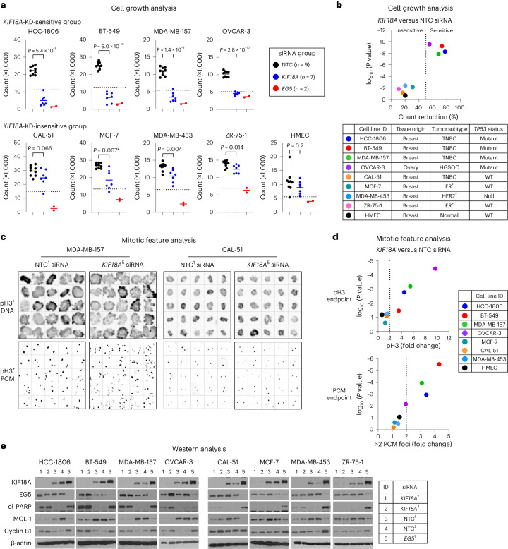
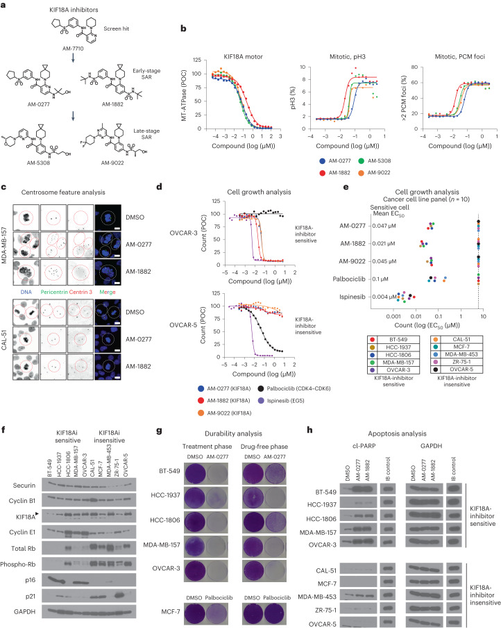
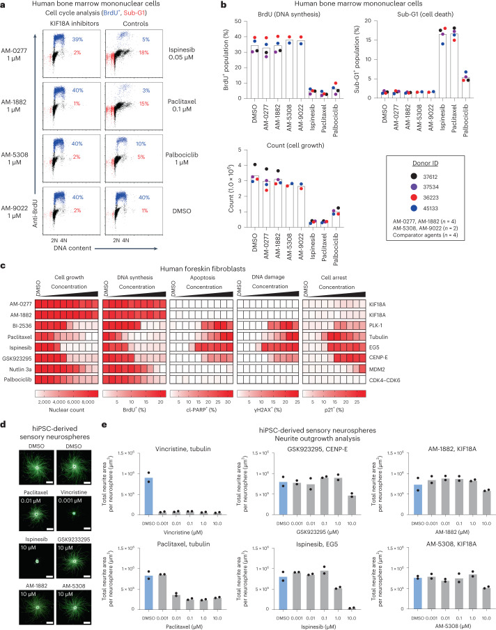
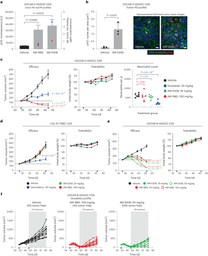
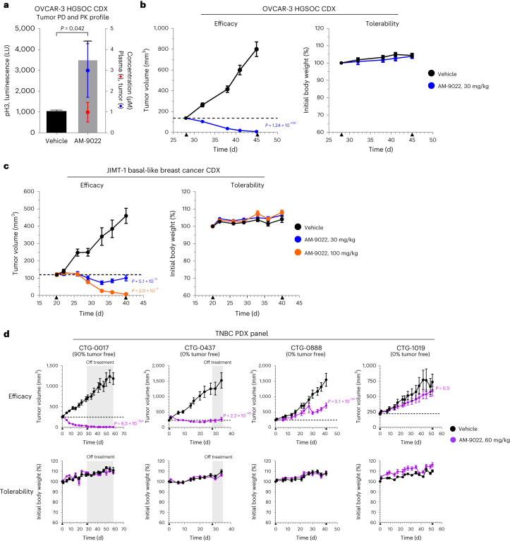
Chromosomal instability (CIN) is a hallmark of cancer, caused by persistent errors in chromosome segregation during mitosis. Aggressive cancers like high-grade serous ovarian cancer (HGSOC) and triple-negative breast cancer (TNBC) have a high frequency of CIN and TP53 mutations. Here, we show that inhibitors of the KIF18A motor protein activate the mitotic checkpoint and selectively kill chromosomally unstable cancer cells. Sensitivity to KIF18A inhibition is enriched in TP53-mutant HGSOC and TNBC cell lines with CIN features, including in a subset of CCNE1-amplified, CDK4-CDK6-inhibitor-resistant and BRCA1-altered cell line models. Our KIF18A inhibitors have minimal detrimental effects on human bone marrow cells in culture, distinct from other anti-mitotic agents. In mice, inhibition of KIF18A leads to robust anti-cancer effects with tumor regression observed in human HGSOC and TNBC models at well-tolerated doses. Collectively, our results provide a rational therapeutic strategy for selective targeting of CIN cancers via KIF18A inhibition.


Conflict of interest statement

M.P., B.B., K.H., J.M., K.C., J.D.M., G.C., M.S.N., J.S., R.M., S.C., S.-M.H., R.J.M.K., K.Z.E., U.P.D., T.W., S.W., P.J.B., J.C., S.M., M.P.B., J.R.A., A.C., N.A.T. and P.E.H. are current or former employees and shareholders of Amgen. A.S.B., M.G.R., M.M.R. and J.A.R. declare no conflict of interest.

Figures

Fig. 1
Fig. 1. Characterization of KIF18A gene dependency in human cell lines.
a, Cell growth analysis was performed on cell lines (n = 9) treated for 96 h with individual siRNA species for KIF18A (n = 7), EG5 (n = 2) or NTC (n = 9). Scatterplots show cell counts for individual siRNA species with group means; the dotted line denotes 50% cell count reduction (n = 2 independent experiments in duplicate). Statistical significance was determined for KIF18A and NTC siRNA groups by unpaired two-tailed t-test (*Welch’s correction) and is shown as P values. b, Scatterplot shows cell count reduction (%) for each cell line and associated P values for KIF18A siRNA species relative to NTC siRNA species; the dotted line denotes 50% cell count reduction. Color-coded cell line identification (ID), tissue of origin, tumor subtype (TNBC, HGSOC, ER, human epidermal growth factor receptor 2 (HER2)) and TP53 status are shown. c,d, Mitotic imaging analysis was performed on the cell line panel (n = 8) treated for 48 h with individual siRNA species for KIF18A (n = 5) or NTC (n = 4). Cells were stained to detect DNA, pH3 and PCM (pericentrin), and images were captured by laser scanning cytometry with a ×40 objective (n = 1 or 2 independent experiments in four to eight replicate wells). Color-coded cell line identification is shown. c, Representative images of MDA-MB-157 and CAL-51 pH3+ cells for NTC (siRNA ID = NTC_1) and KIF18A (siRNA ID = hKIF18A_5) siRNA species (5 × 5 square gallery). d, Scatterplots show cell line pH3+ and PCM foci count fold change and associated P values for KIF18A siRNA species relative to NTC siRNA species; the dotted line denotes twofold change. e, WBA was performed on cell lines (n = 8) treated for 48 h with individual siRNA species for KIF18A (n = 2), EG5 (n = 1) or NTC (n = 2). Protein levels were determined for KIF18A, EG5, cl-PARP, MCL-1, cyclin B1 and β-actin (n = 1 experiment). Immunoblot protein size information is found in the source data. See supporting data (Extended Data Fig. 1 and Supplementary Tables 1, 2 and 7). Source data
Fig. 2
Fig. 2. In vitro characterization of potent and selective KIF18A inhibitors.
a, Structures of AM-7710-series analogs (AM-0277, AM-1882, AM-5308, AM-9022), denoted as early or late SAR stage. b, Graph (left) shows compound profiles in a KIF18A motor assay presented as MT-ATPase luminescence signal relative to the percentage of DMSO control (POC), as assessed by ADP-Glo (n = 2 or 4 independent experiments). Graphs (center, right) show compound profiles in a 24-h mitotic feature assay for pH3 and PCM foci in MDA-MB-157 cells (n = 2 independent experiments). c, Centrosome feature analysis in MDA-MB-157 and CAL-51 cells treated for 24 h with DMSO, AM-0277 (0.5 µM) or AM-1882 (0.05 µM) and stained to detect DNA, PCM (pericentrin) and centrioles (centrin 3). Representative images were captured with a ×60 objective (scale bars, 8 µm); dashed lines silhouette mitotic objects (n = 1 experiment). d,e, Cell growth analysis in a panel of cancer cell lines (n = 10) treated with DMSO, AM-0277, AM-1882, AM-9022, palbociclib or ispinesib. d, Representative concentration–response profiles are presented as count relative to the percentage of the DMSO control. e, Graph shows compound count EC50 values reported for each cell line and mean EC50 values for the sensitive cell lines. The dotted line indicates that >50% cell count reduction was not reached at 6 µM (n = 2 independent experiments in duplicate). f, WBA across the cancer cell lines (n = 10). Protein levels were determined for securin, cyclin B1, KIF18A, cyclin E1, total Rb, phospho-Rb (serine 807 and 811), p16, p21 and glyceraldehyde-3-phosphate dehydrogenase (GAPDH). The arrowhead indicates the KIF18A protein band (n = 1 experiment). KIF18A inhibitor (KIF18Ai). g, Durability analysis in KIF18A-inhibitor-sensitive cancer cell lines (n = 5); cells were treated for 6 d with DMSO or AM-0277 (0.5 µM). MCF-7 cells were treated with palbociclib (1 µM) as a cytostatic control. After the treatment phase (left), cells were replated in drug-free medium and cultured until the DMSO control reached confluence (right), dishes were stained with crystal violet, and images were captured with a digital scanner (n = 1 experiment). h, Western blot apoptosis analysis of cancer cell lines (n = 10) treated for 48 h with DMSO, AM-0277 (0.5 µM) or AM-1882 (0.1 µM). HCC-1806 cells were treated with ispinesib (0.05 µM) as an immunoblot (IB) control. Protein levels were determined for cl-PARP and GAPDH (n = 1 experiment). Immunoblot protein size information is found in the source data. See supporting data (Extended Data Figs. 2–4 and Supplementary Table 7). Source data
Fig. 3
Fig. 3. In vitro characterization of KIF18A-inhibitor effects on normal somatic cells.
a,b, Cell cycle (48-h) and cell growth (96-h) analysis of human bone marrow mononuclear cells from unaffected donors (n = 2 or 4) treated with DMSO, KIF18A inhibitors (AM-1882, AM-0277, AM-5308, AM-9022 at 1 µM), ispinesib (0.05 µM), paclitaxel (0.1 µM) or palbociclib (1 µM). a, Cell cycle analysis; scatterplots show BrdU+ (blue) and sub-G1 (red) populations and percentages for donor 36223. b, Graphs show cell cycle (BrdU+ and sub-G1 population percentages) and cell growth (count) summaries for individual donors with group means (n = 2 or 4 independent experiments). c, Multiparametric image analysis of human foreskin fibroblast cells treated for 48 h with DMSO, AM-0277 and AM-1882 (KIF18A), BI-2536 (Polo Like Kinase 1, PLK-1), paclitaxel (tubulin), ispinesib (EG5), GSK923295 (CENP-E), nutlin 3a (Mouse Double Minute 2, MDM2) or palbociclib (CDK4–CDK6). Cells were stained to detect DNA, BrdU, cl-PARP, γH2AX and p21. Heatmaps show nuclear count (growth) and percentage of counts that stain positive for BrdU (DNA synthesis), cl-PARP (apoptosis), phosphorylated γH2AX (DNA damage) and p21 (cell arrest); scales are indicated below each heatmap (n = 1 experiment). d,e, Neurite outgrowth analysis of hiPSC-derived sensory neurospheres treated for 24 h with DMSO, vincristine, paclitaxel, ispinesib, GSK923295, AM-1882 or AM-5308 at the indicated concentrations. Neurospheres were stained to detect DNA (blue) and β3-tubulin protein (green). d, Representative images of neurite outgrowth (scale bars, 500 µm) captured with a ×20 objective. e, Concentration–response graphs presented as total neurite area (µm2) per neurosphere with group means (n = 2 independent experiments in duplicate or triplicate). See supporting data (Extended Data Fig. 5). Source data
Fig. 4
Fig. 4. AM-1882 phenocopies KIF18A gene dependency across a panel of DNA-barcoded cancer cell lines.
af, Pooled cancer cell lines were treated with AM-1882 in a 5-d cell growth assay; the relative abundance of unique barcodes estimates cell viability (n = 1 experiment in triplicate). AM-1882 AUC values were determined for cancer cell lines (n = 631). a, Heatmap of the AM-1882 concentration–response profile; viability values for AM-1882 are presented as fold change relative to the DMSO control; a scale is shown on the right, with viability scoring as more sensitive (blue) or less sensitive (red). b, Violin plots show AM-1882 AUC distribution by tumor type (n = 24); numbers of cell lines per type are denoted on the x axis. The dotted line indicates an AUC value of 0.65, representing the lower-quartile (LQ) cutoff. c, AM-1882 AUC versus genome-wide gene dependency scores from RNAi KD (n ≤ 447 cell lines) or CRISPR KO (n ≤ 439 cell lines). Volcano plots show Pearson correlation scores and q values for 10,000 genes. KIF18A gene KD or KO was scored as the top-ranked correlation with AM-1882 sensitivity; other genes with positive or negative correlations with AM-1882 sensitivity are denoted in blue or red, respectively. d,e, AM-1882 AUC versus gene mutation (n = 629 cell lines). d, Volcano plot shows Pearson correlation scores and P values for 10,000 genes. TP53 gene mutation was scored as the top-ranked correlation with AM-1882 sensitivity. e, Scatterplot shows AM-1882 AUC versus TP53-WT or TP53-mutant group with mean AUC values for each group. The dashed line indicates an AUC value of 0.65. Statistical significance was determined for the TP53-WT group relative to the TP53-mutant group by unpaired two-tailed t-test (*Welch’s correction) and is shown as P values. f, AM-1882 AUC versus CIN features (WGD, DNA ploidy, AS) in breast and ovarian cancer cell lines (n = 58). Scatterplots show AM-1882 AUC versus TP53 status plus CIN features with mean AUC values for each group. Dashed lines indicate an AUC value of 0.65. Statistical significance was determined for CIN features in the TP53-mutant group by unpaired two-tailed t-test (*Welch’s correction) and is shown as P values. See supporting data (Extended Data Fig. 6 and Supplementary Tables 3–5). Source data
Fig. 5
Fig. 5. Characterization of KIF18A inhibition in vivo.
a, PD and PK profiles of two KIF18A compounds in the OVCAR-3 HGSOC CDX tumor model. Mice were administered a single i.p. dose of vehicle, AM-1882 (100 mg per kg) or AM-5308 (50 mg per kg). Tumor and blood were collected 24 h after treatment and analyzed for pH3 signal in tumor and compound PK in tumor and plasma. Graph shows pH3 luminescence signal (LU) with mean + s.d. (bar, left axis) and tumor (blue) and plasma (red) concentrations with mean ± s.d. (right axis) for each treatment group (n = 3 mice per group). Statistical significance was determined for treatment groups relative to the vehicle by one-way ANOVA with Dunnett’s multiplicity adjustment and is shown as P values. b, PD imaging analysis of AM-5308 in the OVCAR-3 HGSOC CDX tumor model. Mice were administered an i.p. dose of vehicle or AM-5308 (25 mg per kg) for 2 d. Tumors were collected 24 h after treatment. Tumors were stained to detect DNA, pH3 and α-tubulin; representative images were captured with a ×60 objective (scale bars, 12 µm). Graph shows pH3+ nucleus count per tissue area with mean + s.d. from three image fields per tumor (n = 3 mice per group). Statistical significance was determined for AM-5308 relative to the vehicle by unpaired two-tailed t-test (*Welch’s correction) and is shown as P values. c,d, AM-1882 and AM-5308 efficacy and tolerability analysis in OVCAR-3 HGSOC (c) and CAL-51 TNBC (d) CDX tumor models. Mice were administered an i.p. dose of vehicle, AM-1882 (100 mg per kg) or AM-5308 (25 mg per kg) daily for 18 d. Mice were administered an i.p. dose of docetaxel (20 mg per kg) once weekly (c) or gemcitabine (120 mg per kg) twice weekly (d) as positive controls. Graphs show tumor volume and mouse body weight measurements as mean ± s.e.m. versus time (d) (n = 10 mice per group); dashed line indicates 100% TGI or tumor stasis. Treatment start and stop (▴) are indicated on the x axis. c, Graph shows end-of-study neutrophil counts from the OVCAR-3 study (n = 6 mice per group). Statistical significance was determined for neutrophil counts for treatment groups relative to the vehicle by one-way ANOVA with Dunnett’s multiplicity adjustment and is shown as P values. e,f, AM-1882 and AM-5308 efficacy, tolerability and durability analysis in the OVCAR-8 HGSOC CDX tumor model. Mice were administered an i.p. dose of vehicle, AM-1882 (50 or 100 mg per kg) or AM-5308 (25 or 50 mg per kg) daily for 18 d. e, Graphs show tumor volume and body weight measurements as mean ± s.e.m. versus time (d) (n = 10 mice per group); the dashed line indicates 100% TGI. Treatment start and stop (▴) are indicated on the x axis. f, After the cessation of treatment (shaded gray area), tumor durability analysis was performed until day 81. Graphs show tumor volume measurements versus time (d) for individual mice. Mice with no measurable tumor are indicated as tumor free. Statistical significance was determined for tumor efficacy for treatment groups relative to the vehicle by a linear mixed-effect analysis model with Dunnett’s multiplicity adjustment and is shown as P values. See supporting data (Extended Data Fig. 7). Source data
Fig. 6
Fig. 6. Oral candidate AM-9022 induces TR in vivo.
a, PD and PK profiles of AM-9022 in the OVCAR-3 HGSOC CDX tumor model. Mice were administered a single oral (p.o.) dose of vehicle or AM-9022 (30 mg per kg). Tumor and blood were collected 24 h after treatment and analyzed for pH3 signal in tumor and compound PK in tumor and plasma. Graph shows pH3 luminescence signal with mean + s.d. (bar, left axis) and tumor (blue) and plasma (red) concentrations with mean ± s.d. (right axis) for each treatment group (n = 3 mice per group). Statistical significance was determined for AM-9022 relative to the vehicle by unpaired two-tailed t-test (Welch’s correction) and is shown as P values. b, AM-9022 efficacy and tolerability analysis in the OVCAR-3 HGSOC CDX tumor model. Mice were administered a p.o. dose of vehicle or AM-9022 (30 mg per kg) daily for 18 d. Graphs show tumor volume and mouse body weight measurements as mean ± s.e.m. versus time (d) (n = 10 mice per group); the dashed line indicates 100% TGI. Treatment start and stop (▴) are indicated on the x axis. c, AM-9022 efficacy and tolerability analysis in the JIMT-1 basal-like breast cancer CDX tumor model. Mice were administered a p.o. dose of vehicle or AM-9022 (30 or 100 mg per kg) daily for 21 d. Graphs show tumor volume and mouse body weight measurements as mean ± s.e.m. versus time (d) (n = 10 mice per group); the dashed line indicates 100% TGI. Treatment start and stop (▴) are indicated on the x axis. d, AM-9022 efficacy and tolerability analysis in four low-passage TNBC PDX tumor models (CTG-0017, CTG-0437, CTG-0888, CTG-1019). Mice were administered a p.o. dose of vehicle or AM-9022 (60 mg per kg) daily for ≥28 consecutive days. Graphs show tumor volume (top) and mouse body weight (bottom) measurements as mean ± s.e.m. versus time (d) (n = 10 mice per group); dashed lines indicate 100% TGI. Treatment start and stop (▴) are indicated on the x axis. After the cessation of treatment (shaded gray area), tumor durability analysis was performed for CTG-0017 and CTG-0437 models until day 58 and day 34, respectively. Mice with no measurable tumor are indicated as tumor free. Statistical significance was determined for tumor efficacy for the treatment group(s) relative to the vehicle by the linear mixed-effect analysis model with Dunnett’s multiplicity adjustment and is shown as P values. See supporting data (Extended Data Fig. 8 and Supplementary Table 6). Source data
Extended Data Fig. 1
Extended Data Fig. 1. KIF18A gene perturbation effects on human cell lines.
a, WBA was performed on HeLa, BT-549, MCF-7, and HMEC cell lines treated for 48 h with individual siRNA species for KIF18A (n = 7) or non-targeting control (NTC) (n = 1). HeLa cells were treated with nocodazole (NOC) as a mitotic control. Protein levels were determined for KIF18A and β-Actin (n = 1 experiment). b and c, Mitotic image analysis was performed by laser scanning cytometry (LSC). Cell lines were treated for 48 h with individual siRNA species for KIF18A (n = 5) or NTC (n = 4). Imaging data was collected for DNA, phospho-histone H3 (serine-10) (pH3), and PCM (pericentrin) channels with x40 objective. b, MDA-MB-157 cells treated with KIF18A siRNA (siRNA ID = hKIF18A_5) or NTC siRNA (siRNA ID = NTC_1), showing the LSC segmentation scheme (1) Integral versus Max Pixel (DAPI) to establish DNA content profiles, (2) Integral (pH3) versus Integral (DAPI) to gate pH3+ mitotic population, (3) pH3+ PCM foci count scored as >2 PCM foci (red, R5) or ≤ 2 PCM foci (blue, R6). c, Scatter plot of each cell line (n = 8) shows individual siRNA species for pH3+ and >2 PCM foci count percentages with group means and associated significance (n = 1 or 2 independent experiments in 4 to 8 replicate wells). Cell lines grouped as KIF18A knockdown (KD) sensitive or insensitive based on 96-h cell growth assay (see Fig. 1a). d, Mitotic image analysis was performed by ArrayScan VTi. MDA-MB-157 Cas9 cells were treated for 48 h with tracrRNA complexed to individual or pool crRNA species for KIF18A (n = 5), NTC (n = 5), or Eg5 (n = 5). Imaging data was collected for DNA, pH3, and PCM channels with x20 objective. Scatter plots show individual and pool crRNA species for pH3 + , and >2 PCM foci count percentages with group means and associated significance (n = 2 independent experiments in triplicate). Statistical significance was determined for KIF18A and NTC siRNA or crRNA groups by unpaired two-tailed t-test (*Welch’s correction) and is shown as P values. Immunoblot protein size information is found in the source data. See supporting data (Supplementary Tables 1–2, 7). Source data
Extended Data Fig. 2
Extended Data Fig. 2. Discovery of potent and selective KIF18A inhibitors.
a, AM-7710 series analogs were assessed in kinesin-8 microtubule (MT)-ATPase motor assays and 24-h mitotic image assay in MDA-MB-157 cells. Scatter plots show MT-ATPase IC50 values for KIF18A versus KIF19A motors (n = 97 analogs), KIF18A versus KIF18B motors (n = 77 analogs), or cellular EC50 values for pH3 versus PCM foci endpoints (n = 150 analogs) (n ≥ 1 experiment (s) per analog). b and c, KIF18A compounds were assessed in a panel of kinesin MT-ATPase motor assays and 24-h mitotic image assay in MDA-MB-157 cells. b, Graphs show compound profiles for each motor presented as MT-ATPase luminescence signal relative to the percentage of DMSO control (POC), as assessed by ADP-Glo (n = 2 or 4 independent experiments). Ispinesib and GSK923295 were included as motor inhibitor controls. c, Upper table shows compound MT-ATPase IC50 values for KIF18A, KIF18B, KIF19A, Eg5, CENP-E, and KIFC1 motors. Middle table shows compound (1 µM) MT-ATPase POC values for a panel of motors (n = 8), as assessed by enzyme-linked inorganic phosphate assay (ELIPA), results presented as a percentage of DMSO control (POC) (n = 1 or 2 independent experiments in duplicate). Lower table shows the compound cellular EC50 values for mitotic image assay (n = 2 independent experiments). d, AM-0277 and AM-1882 were assessed in KIF18A ATPase motor assays (± MTs) or varied ATP or MT concentrations, as assessed by ADP-Glo. Graphs show compound profiles in KIF18A motor assays presented as ATPase luminescence signal relative to the percentage of DMSO control (POC) (± MTs n = 2 independent experiments in duplicate, varied ATP, or MT concentrations n = 1 experiment). Source data
Extended Data Fig. 3
Extended Data Fig. 3. In vitro characterization of potent and selective KIF18A inhibitors.
a, KIF18A compounds (1 µM) were assessed across a panel of kinases (n = 96) by a competitive binding assay. Graph presented as percentage of control (POC); dashed line indicates POC value of 30 (n = 1 experiment). b, Tubulin polymerization analysis. Profile of KIF18A compounds (10 µM) relative to DMSO, paclitaxel (5 µM, tubulin stabilizer), or nocodazole (5 µM, tubulin destabilizer). Graphs presented as tubulin fluorescence signal at 440 nm expressed as arbitrary units (a.u.) versus time (min) with area-under-the-curve (AUC) values (n = 1 or 3 independent experiments). c, Mitotic spindle image analysis. MDA-MB-157 cells were treated for 24 h with DMSO, AM-1882 (0.2 µM), or AM-5308 (0.5 µM) and stained to detect DNA, α-tubulin, and pericentrin. Representative images were captured with x40 objective (scale bar = 10 µm) (n = 1 experiment). d-f, KIF18A compound effects on HeLa cells. d, Cells were treated for 96 h with DMSO or KIF18A compounds at the indicated concentrations. KIF18A compound concentration-response profiles presented as count relative to the percentage of DMSO control (POC) with count EC50 values (n = 2 independent experiments in duplicate). e, KIF18A protein localization in cells after treatment for 6 hours with DMSO or AM-1882 (0.05 µM). Cells stained to detect DNA, KIF18A, and centrin-3. Representative images of mitotic cells (n = 3 per group) were captured with x60 objective (scale bar = 5 µm) and graphs of line scan measurements were obtained for DNA, KIF18A, and centrin-3 channels expressed as fluorescence intensity (a.u.) versus distance (µm) (n = 1 experiment). f, Mitotic cell fate imaging analysis. HeLa Kyoto cells co-expressing α-tubulin-EGFP and H2B-mCherry proteins released from a G1S block in media containing DMSO or AM-1882 (0.2 µM). Analysis was performed on cells entering the first mitosis (n = 40 cells per group). Graph presented as time in mitosis (h) versus mitotic cell fate, either complete cell division (CCD) or death in mitosis (DiM). Classified cells exiting mitosis by the number of daughter cells and cell death in early interphase (n = 1 experiment). Images were captured every 15 min for 48 h with x20 objective (see Supplementary Videos 1, 2). Source data
Extended Data Fig. 4
Extended Data Fig. 4. In vitro effects of KIF18A inhibitors on sensitive cancer cell lines.
a, Six-day cell growth analysis. KIF18A inhibitor-sensitive cancer cell lines were treated with DMSO or AM-0277 (0.5 µM). MCF-7 cells were treated with palbociclib (1 µM) as a cytostatic control. Graphs presented as cell count from 100-mm dish (n = 1 experiment). b, WBA of synchronized OVCAR-3 cells released from a G1S block in growth media containing DMSO or AM-0277 (0.5 µM). Cell lysates were prepared at 4, 8, 10, 12, 14, 24 h post-release. Asynchronous cells were treated with DMSO or AM-0277 (0.5 µM) as a control. Protein levels were determined for cl-PARP, cyclin B1, BubR1, KIF18A, MCL-1, cyclin E1, and β-Actin. Arrowheads indicate BubR1 and KIF18A protein band doublets (n = 1 experiment). c-e, KIF18A compound effects on BT-549 cells. c and d, Cells were treated for 48 h with DMSO, AM-0277 (0.5 µM), or AM-1882 (0.1 µM). Ispinesib (0.05 µM) was included as cytotoxic control. c, Cell growth profile presented as cell confluency (%) versus time (h) were captured by live-cell imaging, arrow indicates treatment phase (n = 1 experiment in triplicate). d, WBA shows protein levels determined for phospho-γ-H2AX (serine-139) (γH2AX) and GAPDH (n = 1 experiment). e, Images of cells treated for 48 h with DMSO or AM-1882 (0.2 µM) and stained to detect DNA, γH2AX, and cGAS. Representative images were captured with x40 objective (scale bar = 10 µm) (n = 1 experiment). f-h, KIF18A compound effects on parental and ADRRES OVCAR-8 cells. f, Determined P-gp protein expression levels versus isotype antibody control by flow cytometry. g, Cell growth analysis of OVCAR-8 paired cell lines treated for 96 h with DMSO, AM-1882, or AM-9022 at the indicated concentrations with and without P-gp inhibitor at GF120918 at 1 µM). Paclitaxel and doxorubicin were included as controls. Graphs of compound concentration-response profiles presented as count relative to the percentage of DMSO control (POC) with count EC50 values (n = 2 independent experiments). h, WBA of OVCAR-8 paired cells treated for 48 h with DMSO, AM-0277 (0.5 µM), or AM-1882 (0.1 µM). Protein levels were determined for cl-PARP and GAPDH (n = 1 experiment). Immunoblot protein size information is found in the source data. See supporting data (Supplementary Table 7). Source data
Extended Data Fig. 5
Extended Data Fig. 5. In vitro effects of KIF18A inhibitors on normal somatic cells.
a, Cell-cycle (48-h) and cell growth (96-h) analysis of human bone marrow mononuclear cells from normal donors (n = 2 or 4) treated with DMSO, KIF18A compounds (AM-1882, AM-0277, AM-5308, AM-9022 at 1 µM), ispinesib (0.05 µM), paclitaxel (0.1 µM), or palbociclib (1 µM) (see Fig. 3b). Each donor assessed in independent experiments. Table shows statistical significance for treatment groups relative to DMSO control by One-way ANOVA with Dunnett’s multiplicity adjustment and is shown as P values. b, Human mammary epithelial cell (HMEC) BrdU incorporation analysis by flow cytometry. Cells were treated for 48 h with DMSO, AM-0277, ispinesib, or palbociclib at the indicated concentrations (n = 2 independent experiments). Graphs show concentration-response profiles presented as the percentage of BrdU+ cells for each treatment condition. c, Activated human T cells 3H-thymidine incorporation analysis. Cells from two normal donors were treated for 48 h with DMSO, AM-1882, AM-0277, ispinesib, or palbociclib at the indicated concentrations (n = 1 experiment in triplicate). Graphs show concentration-response profiles presented as 3H-thymidine incorporation relative to the percentage of DMSO control (POC) for each donor with mean EC50 values. Source data
Extended Data Fig. 6
Extended Data Fig. 6. AM-1882 PRISM association analysis and combinability potential of KIF18A and PARP inhibition.
a-d, Pooled cancer cell lines treated with AM-1882 in a 5-d cell growth assay; the relative abundance of unique barcodes estimate cell viability (n = 1 experiment in triplicate). AM-1882 area-under-the-curve (AUC) values were determined for cancer cell lines (n = 631). a, Scatterplots show the pan-cancer AM-1882 AUC distribution versus KIF18A KD or KO Gene Effect scores and Pearson correlation coefficient values. KIF18A KD versus KO Gene Effect scores were included as a reference. b-d, AM-1882 PRISM association analysis in breast and ovarian cancer cell lines (n = 58). The dotted line indicates an AUC value of 0.65. b, Scatterplot shows AM-1882 AUC versus TP53 status grouped by wildtype (WT) or mutant (MUT) cell lines with mean AUC indicated for each group. c, Scatterplots show AM-1882 AUC versus CCNE1 or RB1 copy number. Solid line indicates either a log2 copy number value of 2 (CCNE1) or 0.5 (RB1), black circle indicates a copy number >2 or < 0.5, respectively. Red circle indicates RB1 mutant cell lines. d, Scatterplot shows AM-1882 AUC versus BRCA1 status, cell lines grouped by WT or altered (MUT as black circle, promoter methylated (PM) as red circle) with mean AUC values indicated for each group. b and d, Statistical significance was determined for the BRCA1 groups by unpaired two-tailed t-test (*Welch’s correction) and is shown as P values. e, WBA to detect full-length (FL) BRCA1 protein in HCC-1937 (BRCA1 MUT), OVCAR-5 (BRCA1 WT), OVCAR-8 (BRCA1 PM), and CAL-51 (BRCA1 WT) cell lines. CAL-51 cells were treated for 48 h with pooled BRCA1 or NTC siRNAs as knockdown controls. Protein levels were determined for BRCA1 using n-terminal or c-terminal antibodies and GAPDH. Arrowhead indicates FL BRCA1 protein band (n = 1 experiment). f and g, BRCA1-altered cell lines were treated with AM-1882 alone, olaparib alone, or in combination. f, WBA to detect apoptosis. HCC-1937 cells were treated for 48 h with DMSO, AM-1882 (0.01 µM), olaparib (20 µM), or in combination. OVCAR-8 cells were treated with DMSO, AM-1882 (0.03 µM), olaparib (5 µM), or in combination. Protein levels were determined for cl-PARP and GAPDH. g, Durability analysis after drug withdrawal. HCC-1937 or OVCAR-8 cells were treated for 6 d or 4 d, respectively, with DMSO, AM-1882, olaparib, or in a 3 × 2 combination matrix at the indicated concentrations. After the treatment phase, cell were re-plated in drug-free growth media and cultured until DMSO control reached confluence, plates were stained with crystal violet, and images were captured by digital scanner (n = 1 experiment). Immunoblot protein size information is found in the source data. See supporting data (Supplementary Tables 1, 3, 5, 7). Source data
Extended Data Fig. 7
Extended Data Fig. 7. Characterization of intraperitoneal candidates AM-1882 and AM-5308.
a, Mitotic pH3 and pharmacokinetic (PK) analysis. OVCAR-3 cells were treated for 24 h with DMSO, AM-1882, or AM-5308 at indicated concentrations. Cells were stained to detect DNA and pH3. Graph of compound concentration-response profiles presented as pH3+ percentage and EC50 values (n = 3 independent experiments in triplicate). Mouse plasma PK area-under-the-curve (AUC) values (µM·h) were determined after a single intraperitoneal (i.p.) dose of AM-1882 (100 mg per kg) or AM-5308 (100 mg per kg) (n = 3 mice per group). b, Amino acid sequence alignment of human and mouse KIF18A motor domains. Protein sequences Q8NI77 (human) and Q91WD7 (mouse), motor domain, and nucleotide binding site are highlighted in yellow and green, respectively. c, Human and mouse KIF18A motor MT-ATPase assays performed for AM-1882 and AM-5308, as assessed by ADP-Glo. Graph of compound concentration-response profiles presented as MT-ATPase luminescence signal relative to the percentage of DMSO control (POC) and IC50 values (n = 2 independent experiments). d, End of study mouse blood counts from OVCAR-3 CDX tumor model. Mice were administered vehicle, AM-1882 (100 mg per kg), or AM-5308 (25 mg per kg) daily for 18 d. Mice were administered docetaxel (20 mg per kg) once weekly as a positive control. Blood samples were collected after the last dose (n = 6 mice per group). Scatterplots show individual and mean blood counts for reticulocytes, red blood cells (RBC), lymphocytes, and white blood cells (WBC). Statistical significance was determined for treatment groups relative to vehicle by One-way ANOVA with Dunnett’s multiplicity adjustment and is shown as P values. e, End of study mouse PK profiles from OVCAR-3, CAL-51, and OVCAR-8 CDX tumor models. Scatterplots show mouse PK profiles presented as mean plasma concentration versus time (n = 2 mice per time point) with AUC values (µM·h). Source data
Extended Data Fig. 8
Extended Data Fig. 8. Characterization of oral candidate AM-9022.
a, Mitotic pH3 and pharmacokinetic (PK) analysis. OVCAR-3 cells were treated for 24 h with DMSO, AM-1882, or AM-9022 at indicated concentrations. Cells were stained to detect DNA and pH3. Graphs of compound concentration-response profiles presented as pH3 percentage and EC50 values (n = 2 independent experiments in duplicate). Mouse plasma PK area-under-the-curve (AUC) values (µM·h) were determined after a single oral (p.o.) dose of AM-1882 (100 mg per kg) or AM-9022 (10 mg per kg) (n = 3 mice per group). b, Human and mouse KIF18A motor MT-ATPase assays performed for AM-9022, as assessed by ADP-Glo. Graph of AM-9022 concentration-response profiles presented as MT-ATPase luminescence signal relative to percentage of DMSO control (POC) and IC50 values (n = 2 independent experiments). c, End of study mouse plasma PK profile from OVCAR-3 CDX tumor model. Mice were administered a p.o. dose of AM-9022 (30 mg per kg) for 18 d. Scatterplot shows the mouse PK profile presented as mean plasma concentration versus time (n = 2 mice per time point) and AUC values (µM·h). d, Mitotic pH3 imaging analysis. JIMT-1 cells were treated for 24 h with DMSO, AM-0277, AM-1882, AM-5308, or AM-9022 at the indicated concentrations. Cells were stained to detect DNA and pH3. Graphs of compound concentration-response profiles presented as pH3 percentage and EC50 values (n = 2 independent experiments in duplicate). e, End of study mouse plasma PK profile from JIMT-1 CDX tumor model. Mice were administered a p.o. dose of AM-9022 (30 or 100 mg per kg) for 21 d. Scatterplot shows the mouse PK profile presented as mean plasma concentration versus time (n = 2 mice per time point) and AUC values (µM·h). Source data

Comment in

References

    1. Dumontet C, Jordan MA. Microtubule-binding agents: a dynamic field of cancer therapeutics. Nat. Rev. Drug Discov. 2010;9:790–803. doi: 10.1038/nrd3253. - DOI - PMC - PubMed
    1. Beck A, et al. Strategies and challenges for the next generation of antibody–drug conjugates. Nat. Rev. Drug Discov. 2017;16:315–337. doi: 10.1038/nrd.2016.268. - DOI - PubMed
    1. Zasadil LM, et al. Cytotoxicity of paclitaxel in breast cancer is due to chromosome missegregation on multipolar spindles. Science Transl. Med. 2014;6:229ra43. doi: 10.1126/scitranslmed.3007965. - DOI - PMC - PubMed
    1. Komlodi-Pasztor E, Sackett DL, Fojo AT. Inhibitors targeting mitosis: tales of how great drugs against a promising target were brought down by a flawed rationale. Clin. Cancer Res. 2012;18:51–63. doi: 10.1158/1078-0432.CCR-11-0999. - DOI - PubMed
    1. Mitchison TJ. The proliferation rate paradox in antimitotic chemotherapy. Mol. Biol. Cell. 2012;23:1–6. doi: 10.1091/mbc.e10-04-0335. - DOI - PMC - PubMed