

. 2023 Dec 13:2023:10.17912/micropub.biology.001022.
doi: 10.17912/micropub.biology.001022.
eCollection 2023.
Split-GFP lamin as a tool for studying C. elegans LMN-1 dynamics in vivo
Affiliations
- PMID: 38152058
- PMCID: PMC10751582
- DOI: 10.17912/micropub.biology.001022
Item in Clipboard
Split-GFP lamin as a tool for studying C. elegans LMN-1 dynamics in vivo
MicroPubl Biol.
.
Abstract
We engineered a fluorescent fusion protein of C. elegans lamin, by fusing the eleventh beta strand of GFP to the N-terminus of LMN-1 at the endogenous lmn-1 locus. When co-expressed with GFP1-10, GFP11::LMN-1 was observed at the nuclear periphery of a wide variety of somatic cells. Homozygous gfp11::lmn-1 animals had normal numbers of viable embryos. However, the gfp11::lmn-1 animals had a mild swimming defect. While not completely functional, the GFP11::LMN-1 strain is more healthy than other published fluorescent LMN-1 lines, making it a valuable reagent for studying lamins.
Copyright: © 2023 by the authors.
Conflict of interest statement
The authors declare that there are no conflicts of interest present.
Figures
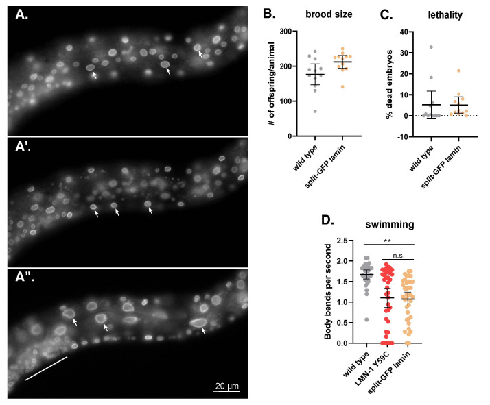
(A) GFP fluorescence is shown in three different focal planes of an L4 larvae. A lateral view is shown with ventral down and anterior to the left. The scale bar is 20 mm. GFP localizes to the periphery of most somatic cells, including cells from the ventral hypodermis (arrows in A), body wall muscles (arrows in A’), and intestine (arrows in A”). GFP can also be seen in many other cells, including the developing vulva (above the line in A”). (B) The brood size and, (C) the percent of embryonic lethality in wild type and GFP11::LMN-1 (split-GFP lamin) animals. Each data point represents the brood from a single adult. The means are shown with error bars of 95% CI. Significance was calculated using student’s t-test. (D) The swimming rate, measured in body bends per second, of wild type,
lmn-1
(Y59C)
and split-GFP lamin in L4 larvae. Means and error bars of 95% CI are shown. n=40. Significance was calculated using a one-way ANOVA and corrected for multiple comparisons by Tukey HSD. **p≤0.01; n.s.= not significant.
Similar articles
-
Application of Split-GFP Reassembly Assay to Study Myogenesis and Myofusion In Vitro.Methods Mol Biol. 2017;1668:127-134. doi: 10.1007/978-1-4939-7283-8_9. Methods Mol Biol. 2017. PMID: 28842906
-
Tissue-Specific Split sfGFP System for Streamlined Expression of GFP Tagged Proteins in the Caenorhabditis elegans Germline.G3 (Bethesda). 2019 Jun 5;9(6):1933-1943. doi: 10.1534/g3.119.400162. G3 (Bethesda). 2019. PMID: 30992318 Free PMC article.
-
A Split-GFP Gateway Cloning System for Topology Analyses of Membrane Proteins in Plants.PLoS One. 2017 Jan 13;12(1):e0170118. doi: 10.1371/journal.pone.0170118. eCollection 2017. PLoS One. 2017. PMID: 28085941 Free PMC article.
-
Electron microscopy of lamin and the nuclear lamina in Caenorhabditis elegans.Methods Cell Biol. 2008;88:411-29. doi: 10.1016/S0091-679X(08)00421-4. Methods Cell Biol. 2008. PMID: 18617045 Review.
-
Fluorescence protein complementation in microscopy: applications beyond detecting bi-molecular interactions.Methods Appl Fluoresc. 2018 Nov 20;7(1):012001. doi: 10.1088/2050-6120/aaef01. Methods Appl Fluoresc. 2018. PMID: 30457122 Review.
Cited by
-
N-terminal tags impair the ability of lamin A to provide structural support to the nucleus.J Cell Sci. 2024 Aug 15;137(16):jcs262207. doi: 10.1242/jcs.262207. Epub 2024 Aug 23. J Cell Sci. 2024. PMID: 39092499
References
-
- Breimann Laura, Preusser Friedrich, Preibisch Stephan. Light-microscopy methods in C. elegans research. Current Opinion in Systems Biology. 2019 Feb 1;13:82–92. doi: 10.1016/j.coisb.2018.11.004. - DOI
Grants and funding
LinkOut - more resources
Full Text Sources
Research Materials
Miscellaneous